Hubbry Logo
Aneurysmal bone cystAneurysmal bone cystMain
Open search
Aneurysmal bone cyst
Community hub
Aneurysmal bone cyst
logo
8 pages, 0 posts
0 subscribers
Be the first to start a discussion here.
Aneurysmal bone cyst
from Wikipedia
Not found
Revisions and contributorsEdit on WikipediaRead on Wikipedia
from Grokipedia
An aneurysmal bone cyst (ABC) is a benign, non-malignant, tumor-like vascular of characterized by multiple blood-filled cystic spaces separated by fibrous septa containing tissue and osteoclast-like giant cells, which can expand rapidly, cause local bone destruction, and lead to pathologic fractures. These lesions are rare, accounting for 1% to 2% of all primary tumors, with an incidence of approximately 1 in 100,000 individuals annually, and they predominantly affect children and adolescents under 20 years of age, showing a slight predominance (-to-male of 1:1 to 1.3:1). ABCs most commonly arise in the metaphysis of long bones such as the and (about 67% of cases), followed by the spine (15%) and (9%), though they can occur in any bone. The etiology remains unclear, but evidence suggests they may develop as primary lesions due to genetic abnormalities like the t(16;17)(q22;p13) translocation involving the USP6 gene in up to 69% of cases, or as secondary reactive processes to trauma, other benign bone tumors (e.g., chondroblastoma or tumor), or vascular malformations. Clinically, patients often present with insidious onset of localized pain over weeks to months, swelling, or deformity; acute severe pain may signal a , while spinal involvement can cause neurologic symptoms such as or due to compression. 
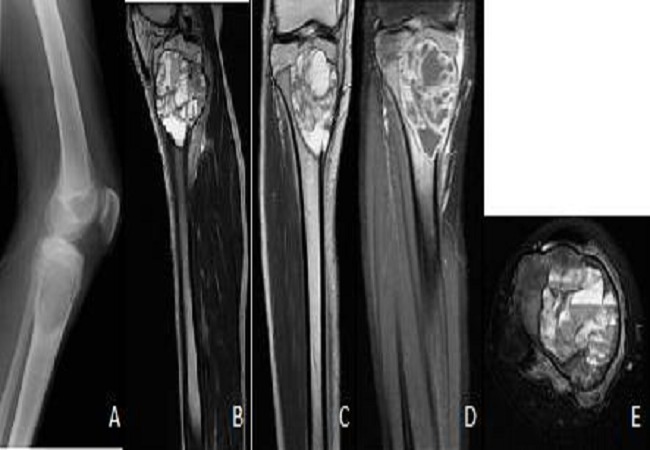
Diagnosis typically involves radiographic imaging, where ABCs appear as expansile, lytic lesions with thinned "eggshell" cortical borders and internal septations; advanced modalities like MRI reveal characteristic fluid-fluid levels from blood sedimentation, aiding differentiation from malignant mimics such as telangiectatic osteosarcoma. Histopathologic confirmation via is often required, showing cavernous blood-filled spaces without endothelial lining and the presence of spindle cells, multinucleated giant cells, and reactive formation. Treatment is primarily surgical, with and as the standard approach to decompress the lesion and restore stability, though adjuncts like phenol or reduce recurrence rates (which approach 25%); alternatives include percutaneous , selective arterial , or for unresectable cases, particularly in the spine.

Overview

Definition

An aneurysmal bone cyst (ABC) is a benign, non-malignant, tumor-like vascular of characterized by expansile, lytic destruction and the formation of blood-filled cystic spaces separated by fibrous . Histologically, ABCs consist of numerous blood-filled channels and cystic cavities lined by fibrous tissue, often featuring osteoclastic giant cells, fibroblasts, and areas of reactive woven formation within the . The cystic spaces may contain blood, , and fibrinoid material, contributing to the lesion's characteristic hemorrhagic appearance. Despite their benign histology, ABCs demonstrate aggressive local growth that can weaken the surrounding structure, potentially leading to fractures, though they possess no metastatic potential.

Classification

Aneurysmal bone cysts (ABCs) are classified into primary and secondary types based on their origin and association with underlying , which influences diagnostic approaches and strategies. Primary ABCs arise de novo without an associated preexisting and account for approximately 70% of cases. These s are considered true neoplasms and typically exhibit characteristic genetic alterations, aiding in their distinction from other entities during histopathological evaluation. In contrast, secondary ABCs develop in conjunction with other primary tumors or conditions, comprising about 30% of cases, and represent reactive cystic changes within the preexisting . Common associations include , chondroblastoma, fibrous dysplasia, osteoblastoma, and occasionally post-traumatic or post-radiation changes, which can complicate diagnosis as the cystic components may obscure the underlying on . The classification carries prognostic implications, as primary ABCs are uniformly benign with a focus on local control, while secondary ABCs' outcomes depend on the of the associated tumor, potentially requiring more aggressive intervention if the primary lesion is malignant or recurrent. Both types share a benign expansile but can demonstrate locally aggressive behavior, such as cortical thinning and soft tissue extension, which may mimic more aggressive processes on radiographs. Accurate subclassification often relies on correlation between clinical history, imaging features like fluid-fluid levels on MRI, and confirmation to guide treatment and predict recurrence risk, which is higher in aggressive presentations. Rare variants of ABC include the solid variant, which is less cystic and more fibrous in composition, comprising 3-8% of cases and often lacking the typical blood-filled spaces. This subtype can present with aggressive radiographic features, such as rapid growth and bone destruction, frequently mimicking malignancy like telangiectatic osteosarcoma or giant cell tumor, leading to potential misdiagnosis without histopathological review. Additionally, some ABCs exhibit unusually aggressive behavior regardless of variant, with extensive periosteal reaction or invasion, further emphasizing the need for multidisciplinary evaluation to differentiate from sarcomas.

Epidemiology

Incidence and Prevalence

Aneurysmal bone cysts (ABCs) are rare benign tumors, accounting for approximately 1% to 6% of all primary tumors. This range reflects their infrequent occurrence within the spectrum of osseous neoplasms, with some studies reporting a narrower proportion of 1.4% to 2.3%. The annual incidence of primary ABCs is estimated at about 0.14 per 100,000 individuals, based on population-based epidemiologic analyses. In pediatric populations, ABCs represent roughly 1% to 2% of biopsied primary neoplasms, underscoring their relative rarity even among younger patients where the condition predominantly arises.

Demographics

Aneurysmal bone cysts primarily affect children and adolescents, with approximately 80% of cases diagnosed before the age of 20 years and the peak incidence occurring in the 5- to 15-year age group. The condition is rare in adults, with most occurrences limited to individuals under 30 years old. A slight female predominance is observed, with reported female-to-male ratios ranging from 1:1 to 1.3:1 across various studies. No strong ethnic or geographic predispositions have been identified in the literature on aneurysmal cysts.

Clinical Presentation

Signs and Symptoms

Aneurysmal cysts typically present with an insidious onset of localized that develops over weeks to months, often worsening at night or with physical activity. This is the most common symptom, reported in approximately 80% of cases, and is usually confined to the affected . Swelling or a palpable over the site is also frequent, occurring in about 37% of patients, reflecting the expansile nature of the . Pathologic fractures arise due to progressive bone weakening and represent the initial presentation in 10-20% of cases, often manifesting as sudden, severe pain following minor trauma or even spontaneously. These fractures are more likely in long tubular s where the cyst's cystic spaces erode the cortex, leading to structural compromise. When the spine is involved, patients may experience neurological symptoms such as , weakness, or paraparesis due to compression of the or nerve roots. In cases affecting long bones, joint dysfunction, stiffness, or deformity can develop from the lesion's expansion, limiting range of motion in the adjacent joint.

Anatomic Sites

Aneurysmal bone cysts (ABCs) exhibit a broad distribution across the but preferentially affect certain anatomic sites, with approximately 67% occurring in the of . The most frequently involved are the , , and , where lesions typically arise in the metaphyseal region and can extend into the . These cysts are characteristically eccentric and cortical-based, often expanding the cortex and extending into adjacent soft tissues, which contributes to their expansile nature. Involvement of the spine accounts for 15-20% of cases, predominantly affecting the posterior elements such as the vertebral arches and spinous processes, though the vertebral bodies may also be involved in some instances. The is affected in about 9% of cases, typically involving the ilium, , or pubis. ABCs in flat bones, such as the ribs, , or , occur less commonly, representing a smaller proportion of overall cases. Lesions in the small bones of the hands and feet are rare, comprising fewer than 5% of reported instances, and often present unique diagnostic challenges due to their atypical location.

Etiology and Pathogenesis

Causes

The exact cause of aneurysmal bone cysts (ABCs) remains unknown, though they are widely hypothesized to represent a reactive process arising from intraosseous vascular malformations or hemorrhage that leads to cystic expansion. Hemodynamic disturbances are implicated in this , with proposed mechanisms including arteriovenous shunting or venous blockage that elevates intraosseous pressure, thereby promoting bone and resorption through the formation of blood-filled cavities. A history of preceding trauma or is reported in some cases, potentially serving as a trigger for initial hemorrhage and subsequent lesion development. Secondary ABCs account for 23-32% of all cases and arise in association with underlying primary bone tumors, such as chondroblastoma or tumor, or other conditions including fibrous , where the ABC-like changes represent a reactive component to the primary . In contrast, primary ABCs are characterized by specific genetic rearrangements that support their neoplastic nature, though non-genetic factors like those described above may contribute to their initiation.

Genetic Factors

Aneurysmal bone cysts (ABCs) are classified into primary and secondary forms based on their genetic profiles, with primary ABCs characterized by specific chromosomal rearrangements involving the USP6 . The most common alteration is the t(16;17)(q22;p13) translocation, which fuses the promoter region of the 11 (CDH11) on 16q22 to the entire coding sequence of the ubiquitin-specific protease 6 (USP6, also known as TRE17) on 17p13, resulting in USP6 upregulation through promoter swapping. This fusion drives neoplastic transformation in primary ABCs by enhancing USP6 transcription, a mechanism first identified in seminal studies on ABC . USP6 rearrangements occur in approximately 69% of primary ABCs, as demonstrated by (FISH) analysis targeting USP6 and CDH11 loci, while variant fusions with other partners (e.g., COL1A1, ZNF9) account for additional cases. Overexpression of USP6 promotes osteolysis, the hallmark bone destruction in ABCs, by disrupting osteoblastic maturation through autocrine dysregulation of bone morphogenetic proteins (BMPs) and enhancing activity via stimulation of production and inflammatory signaling pathways. These molecular effects underscore the neoplastic nature of primary ABCs, distinguishing them from reactive processes. In contrast, secondary ABCs—those arising in association with other bone tumors such as tumor or chondroblastoma—typically lack USP6 rearrangements, though they may exhibit genetic alterations inherited from the underlying primary . This absence supports the view that secondary ABCs represent reactive cystic changes rather than independent . The detection of USP6 rearrangements holds significant diagnostic utility, as or (PCR)-based assays can confirm primary ABC and differentiate it from mimics like telangiectatic , which may show overlapping radiographic and histologic features but lack USP6 fusions. Such molecular testing is particularly valuable in challenging cases, enabling precise classification and guiding management.

Histopathology

Aneurysmal bone cysts (ABCs) are characterized grossly by a well-demarcated, expansile, spongy, and hemorrhagic composed of multiloculated cystic spaces filled with and friable tissue. These spaces range in size from less than 1 mm to several centimeters and are separated by thin, fibrous , often surrounded by a delicate "eggshell" shell of reactive . The may exhibit irregular borders and variable amounts of solid components, particularly in certain anatomic sites like the digits, with overall dimensions varying based on location and duration. deposits from chronic hemorrhage contribute to the dark, mottled appearance of the cut surface. Microscopically, ABCs feature blood-filled cystic cavities lacking an endothelial or epithelial lining, instead separated by cellular fibrous composed of bland fibroblasts, osteoclast-like multinucleated giant cells, and reactive woven trabeculae. The often contain areas of hemorrhage, hemosiderin-laden macrophages, and mixed inflammatory cells, with clusters of giant cells concentrated near the cystic spaces. Mitotic figures are common in the stromal cells but lack , and is rare. A distinctive feature is the presence of calcified, basophilic, reticulated chondroid-like material within the . In the solid variant of ABC, cystic spaces are reduced, with a predominance of spindle-shaped stromal cells and more prominent fibro-osseous tissue, though giant cells and reactive persist. Immunohistochemically, the multinucleated giant cells express , consistent with their osteoclast-like nature, while the cystic walls are negative for endothelial markers such as and , confirming the absence of a true vascular lining. Stromal cells may show positivity for , and giant cells for , supporting the lesion's reactive osteoclastic activity, though no specific panel is diagnostic for ABC. Genetic confirmation can be achieved through detection of USP6 gene rearrangements in approximately 65% of cases via .

Diagnosis

Imaging Characteristics

Aneurysmal bone cysts (ABCs) typically present on plain radiographs as eccentric, expansile lytic lesions located in the of long bones, with a characteristic "blown-out" appearance due to cortical thinning and a thinned sclerotic rim. Internal septations may be visible, imparting a multiloculated or soap-bubble appearance, while aggressive lesions can show periosteal reaction or pathologic fractures. These features reflect the lesion's vascular and cystic nature, often without matrix mineralization unless secondary changes are present. Magnetic resonance imaging (MRI) is the modality of choice for characterizing ABCs, revealing multiloculated cystic spaces with fluid-fluid levels due to sedimentation of blood products, a characteristic finding seen in a majority of cases and highly suggestive of the diagnosis. On T1-weighted images, the lesion shows variable signal intensity from hemorrhage, while T2-weighted sequences demonstrate hyperintense cystic components with a hypointense rim; post-contrast enhancement occurs in the thin septa and nodular solid areas, aiding in assessing lesion extent and soft-tissue involvement. Surrounding bone marrow and soft-tissue edema may also be evident, indicating reactive changes. Computed tomography (CT) complements radiography and MRI by better delineating cortical thinning, internal fluid levels, and subtle matrix mineralization in expansile lesions, particularly in complex sites like the spine or pelvis. It highlights the geographic, lytic nature with a well-defined sclerotic margin and can identify periosteal reactions or fractures not fully appreciated on plain films. No significant soft-tissue mass is typically seen unless associated with fracture. Bone scintigraphy demonstrates increased radiotracer uptake in ABCs, reflecting active bone turnover, hyperemia, and osteoblastic activity, often with a peripheral "doughnut" pattern of intense rim uptake and central photopenia corresponding to the cystic spaces. This nonspecific finding helps assess lesion activity but is less diagnostic than MRI. While imaging strongly suggests ABC, histological confirmation via is generally required for definitive .

Biopsy Findings

Biopsy is indicated for the definitive of aneurysmal bone cyst (ABC), particularly to exclude malignant mimics such as telangiectatic , when imaging findings are suggestive but not conclusive. image-guided core needle is the preferred initial method due to its minimally invasive nature, high diagnostic yield, and safety for most primary bone tumors, including ABC; open with frozen section analysis may be employed if the needle is nondiagnostic or yields insufficient tissue. Histological examination of biopsy specimens reveals characteristic blood-filled cystic cavities separated by fibrous septa, lacking an endothelial lining and thereby confirming the lesion as a non-vascular malformation rather than a true or . The septa typically contain fibroblasts, spindle cells, multinucleated giant cells, and areas of reactive formation, with mitotic activity present but no atypical cells or significant pleomorphism. Due to the highly vascular nature of ABC, biopsy carries a risk of significant hemorrhage, which can complicate the procedure and potentially affect limb salvage. Preoperative selective arterial embolization may be performed to reduce intraoperative bleeding and facilitate safer tissue sampling. Molecular testing for USP6 rearrangements on tissue is integrated into the diagnostic workup to distinguish primary ABC, which harbors these alterations in approximately 65-70% of cases, from secondary ABC or other cystic lesions lacking such clonal changes.

The of aneurysmal bone cyst (ABC) includes several benign and malignant bone lesions that may present with expansile lytic changes on plain . Distinguishing ABC from these mimics relies on imaging features, such as the presence of fluid-fluid levels on MRI, which are characteristic of ABC due to its blood-filled cystic spaces, and for USP6 rearrangements, found in approximately 70% of primary ABC cases but absent in most differentials. Giant cell tumor of bone (GCTB) is a key benign mimic, typically occurring in the epiphysis of long bones in adults aged 20-40 years, presenting as a more solid, eccentric lytic lesion without the multiloculated cystic spaces or fluid-fluid levels seen in ABC on MRI. Unlike ABC, GCTB harbors the H3-3A gene mutation (H3.3 pGly34Trp), which can be confirmed via immunohistochemistry to differentiate the two. Telangiectatic osteosarcoma (TOS), a rare malignant variant of , closely resembles ABC clinically and radiographically, affecting similar age groups () and sites (long bones), with both showing aggressive, expansile lytic destruction and cystic components. However, TOS exhibits permeative bone margins, extension, and lacks USP6 rearrangements; definitive distinction requires revealing anaplastic sarcomatous cells and production, absent in ABC. Unicameral bone cyst (UBC), also known as simple bone cyst, is a benign primarily in the or of long bones in children, appearing as a non-expansile, unilocular lytic defect with thin walls and content, lacking the internal septations, blood-filled cavities, and fluid-fluid levels of ABC on cross-sectional imaging. UBC typically does not cause cortical thinning or pathologic fractures as aggressively as ABC. Fibrous dysplasia (FD) may mimic ABC in its monostotic form, presenting as an expansile with ground-glass matrix opacity on due to irregular bony trabeculae in a fibrous stroma, contrasting with the purely cystic, radiolucent appearance of ABC. FD often affects a broader age range and lacks USP6 fusions, though secondary ABC changes can occur in up to 10% of FD cases, requiring histologic correlation to identify the fibro-osseous component. Chondroblastoma, a rare benign tumor predominantly in the of long bones in adolescents, can resemble ABC when secondary aneurysmal changes develop (in 10-20% of cases), but primary chondroblastoma features chondroid matrix and calcifications on imaging, with histologic evidence of chondroblasts, distinguishing it from the vascular, spindle-cell stroma of ABC.

Management

Surgical Treatments

Surgical treatments for aneurysmal bone cysts (ABCs) primarily involve operative interventions aimed at removing the while preserving bone function, with intralesional serving as the cornerstone approach for most cases. This method is preferred due to its relative preservation of surrounding bone stock compared to more radical procedures. Adjuvant therapies are commonly incorporated to enhance local control by targeting residual tumor cells. Intralesional curettage involves scraping out the cystic contents and fibrous septa under direct visualization, often using high-speed burrs to extend the margins into healthy bone. Adjuvants such as phenol for chemical cauterization, cryotherapy with liquid nitrogen to induce cellular necrosis, or polymethylmethacrylate (PMMA) cement for thermal ablation and structural support are applied to reduce the risk of local recurrence, particularly in aggressive lesions. This technique is suitable for accessible sites like long bones and is the standard for initial management. Following , the resulting bone defect is typically filled with autogenous bone grafts from the or rib, allografts, or synthetic materials like PMMA to restore structural integrity and promote healing, especially in areas. These fillers help prevent pathological fractures and support early mobilization. For highly aggressive, recurrent, or spinal ABCs where margins are inadequate, en bloc resection provides complete excision of the lesion with a margin of normal bone, often requiring and instrumentation for stability. This approach is technically demanding and reserved for cases where preservation of function is feasible, such as select mobile spine locations. Preoperative selective arterial is frequently employed to devascularize the lesion, using agents like particles or N-2-butyl-cyanoacrylate to minimize intraoperative blood loss, particularly in hypervascular tumors of the or spine. It is performed 24-48 hours prior to via catheter access to feeding arteries. In young patients, these surgical strategies are essential to address the lesion's potential for rapid growth while minimizing long-term skeletal deformities.

Non-Surgical Options

Non-surgical options for aneurysmal bone cysts (ABCs) are particularly valuable in cases involving difficult surgical access, such as spinal or pelvic locations, or when preserving bone growth in pediatric patients is prioritized. These approaches aim to induce cyst obliteration, inhibit lesion progression, or monitor stable lesions without intervention, often serving as alternatives or adjuncts to surgical curettage, which remains the first-line treatment for most accessible ABCs. Percutaneous sclerotherapy involves the image-guided injection of sclerosing agents directly into the cyst to promote and endothelial damage, leading to gradual obliteration of the cystic spaces. Commonly used agents include , a sclerosant that achieves cyst consolidation through multiple outpatient sessions, with reported cure rates up to 97% in long-term follow-up. , another effective agent, is injected percutaneously to induce and sclerosis, demonstrating low recurrence rates of approximately 5% after more than 24 months in treated cases. This typically requires 2–6 sessions spaced 4–6 weeks apart, under fluoroscopic or CT guidance, and is well-tolerated with minimal complications such as transient pain or skin irritation. Radiotherapy is reserved for inoperable ABCs, such as those in the spine or , or for recurrent lesions where surgery poses high risk. It delivers targeted to arrest lesion growth and promote , achieving local control rates exceeding 90% when used as an adjuvant or primary modality with doses of 26–30 Gy in fractionated regimens. However, its use is limited due to risks including secondary malignancies, such as radiation-induced , and potential growth plate disturbances in children. Medical therapy with , a that inhibits to suppress activity, offers a targeted option for unresectable or recurrent ABCs, particularly in axial skeletons. Administered subcutaneously at a weight-based dose (e.g., 70 mg/m² monthly after loading doses) in pediatric patients, it has shown radiographic evidence of lesion sclerosis and volume reduction within 3–6 months, with clinical improvement in pain and function. This approach is especially beneficial in pediatric cases where surgical morbidity is a concern; recent reports (as of 2024) also support its use for recurrent lesions via re-challenge. However, long-term data on durability remain limited, and side effects include during treatment as well as severe rebound hypercalcemia upon discontinuation, necessitating careful monitoring and potential bridging with bisphosphonates. For small, ABCs in children, active with serial (e.g., MRI or radiographs every 3–6 months) allows monitoring for spontaneous involution, which occurs in some cases without intervention. This conservative strategy is supported by reports of post-biopsy alone in active lesions, avoiding unnecessary treatment in stable presentations. Progression or symptom development prompts escalation to other therapies.

Prognosis

Outcomes

Aneurysmal bone cysts (ABCs) exhibit an excellent overall prognosis following appropriate treatment, with cure rates ranging from 90% to 95% in most cases. These benign lesions carry a low mortality risk, as they do not metastasize. However, if left untreated, ABCs can lead to significant morbidity, primarily through fractures, , and disruption of normal skeletal growth. In pediatric patients, who comprise the majority of cases, preservation of the growth plate during intervention is crucial to prevent limb length discrepancies and angular deformities. Successful outcomes are influenced by factors such as the completeness of lesion excision and whether the ABC is primary or secondary to another bone pathology. Primary ABCs generally demonstrate better prognoses compared to secondary forms, particularly in spinal locations among adults. Recent pharmacologic options, such as followed by bisphosphonates, have shown promise in improving outcomes for aggressive or recurrent cases, potentially lowering recurrence rates. Long-term expectations include restored function and minimal complications when these elements are addressed effectively.

Recurrence and Monitoring

Aneurysmal bone cysts exhibit a recurrence rate of 10-20% following initial treatment, with rates approaching 19% in cases of incomplete excision or . Most recurrences manifest within the first year post-intervention, though delayed cases beyond two years have been documented. Several factors elevate the risk of recurrence, including incomplete during surgery, patient age under 10 years, lesion size exceeding 5 cm, and involvement of the spine or . These elements contribute to higher local persistence due to the lesion's aggressive, expansile nature and anatomical challenges in certain sites. Recommended monitoring protocols emphasize serial imaging to facilitate early detection. This includes MRI or plain X-rays performed every 3-6 months for the initial two years after treatment, transitioning to annual assessments up to five years to track potential regrowth or complications. Recurrent lesions are generally managed through repeat , such as extended with adjuvants or , depending on location and extent; remains exceedingly rare, occurring in fewer than 1% of cases.

References

Add your contribution
Related Hubs
User Avatar
No comments yet.