Recent from talks
Ciliary body
Knowledge base stats:
Talk channels stats:
Members stats:
Ciliary body
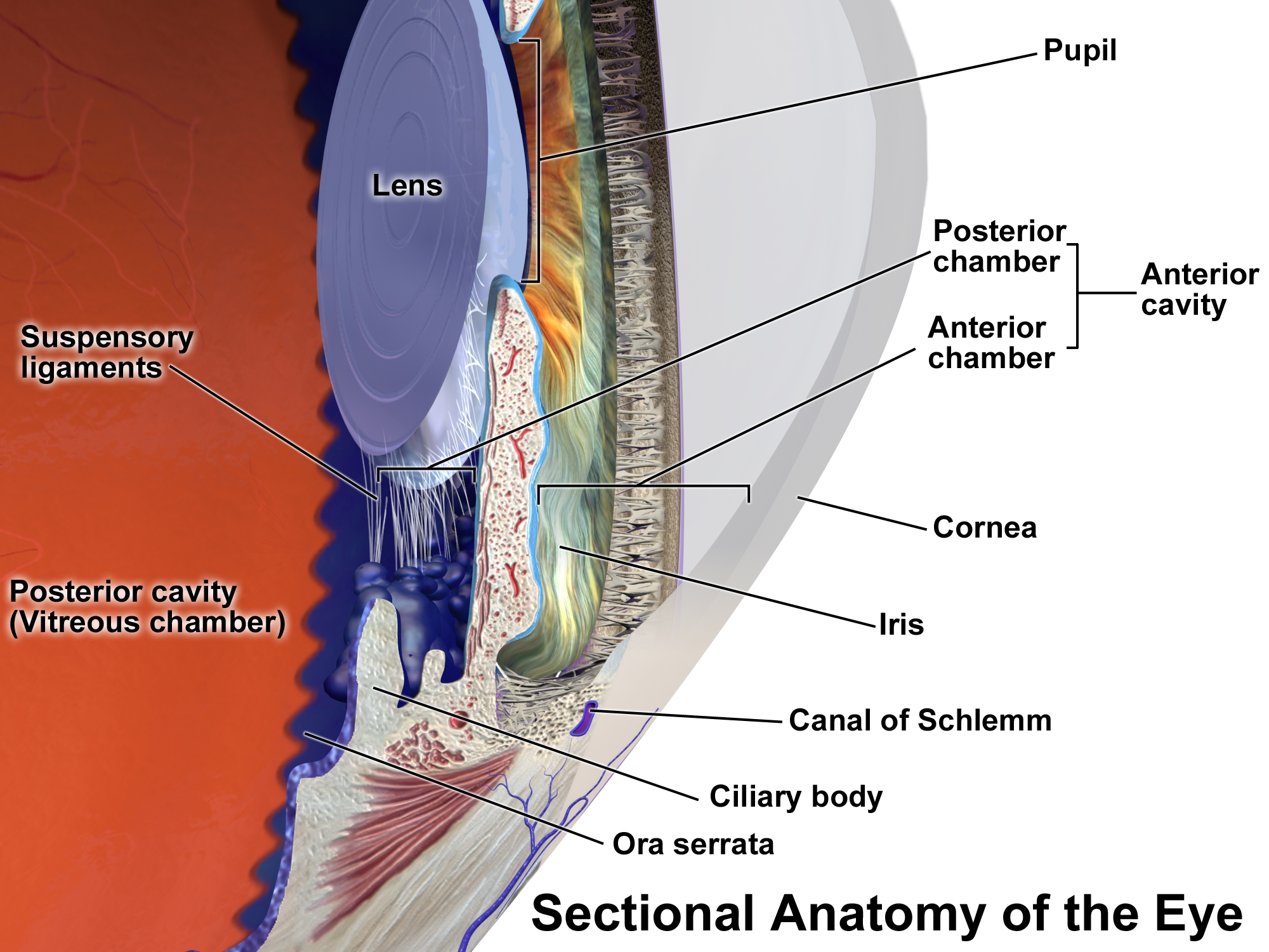
The ciliary body is a part of the eye that includes the ciliary muscle, which controls the shape of the lens, and the ciliary epithelium, which produces the aqueous humor. The aqueous humor is produced in the non-pigmented portion of the ciliary body. The ciliary body is part of the uvea, the layer of tissue that delivers oxygen and nutrients to the eye tissues. The ciliary body joins the ora serrata of the choroid to the root of the iris.
The ciliary body is a ring-shaped thickening of tissue inside the eye that divides the posterior chamber from the vitreous body. It contains the ciliary muscle, vessels, and fibrous connective tissue. Folds on the inner ciliary epithelium are called ciliary processes, and these secrete aqueous humor into the posterior chamber. The aqueous humor then flows through the iris into the anterior chamber.
The ciliary body is attached to the lens by connective tissue called the Zonule of Zinn (fibers of Zinn). Relaxation of the ciliary muscle puts tension on these fibers and changes the shape of the lens in order to focus light on the retina.
The inner layer is transparent and covers the vitreous body, and is continuous from the neural tissue of the retina. The outer layer is highly pigmented, continuous with the retinal pigment epithelium, and constitutes the cells of the dilator muscle. This double membrane is often considered continuous with the retina and a rudiment of the embryological correspondent to the retina. The inner layer is unpigmented until it reaches the iris, where it takes on pigment. The retina ends at the ora serrata.
The space between the ciliary body and the base of the iris is the ciliary sulcus.
The parasympathetic innervation of the ciliary body is the most clearly understood. Presynaptic parasympathetic signals that originate in the Edinger-Westphal nucleus are carried by cranial nerve III (the oculomotor nerve) and travel through the ciliary ganglion. Postsynaptic fibers from the ciliary ganglion form the short ciliary nerves. Parasympathetic activation of the M3 muscarinic receptors causes ciliary muscle contraction, the effect of contraction is to decrease the diameter of the ring of ciliary muscle. The parasympathetic tone is dominant when a higher degree of accommodation of the lens is required, such as reading a book.
The ciliary body is also known to receive sympathetic innervation via long ciliary nerves. When test subjects are startled, their eyes automatically adjust for distance vision.
The ciliary body has three functions: accommodation, aqueous humor production, and resorption, and maintenance of the lens zonules for the purpose of anchoring the lens in place.
Hub AI
Ciliary body AI simulator
(@Ciliary body_simulator)
Ciliary body
The ciliary body is a part of the eye that includes the ciliary muscle, which controls the shape of the lens, and the ciliary epithelium, which produces the aqueous humor. The aqueous humor is produced in the non-pigmented portion of the ciliary body. The ciliary body is part of the uvea, the layer of tissue that delivers oxygen and nutrients to the eye tissues. The ciliary body joins the ora serrata of the choroid to the root of the iris.
The ciliary body is a ring-shaped thickening of tissue inside the eye that divides the posterior chamber from the vitreous body. It contains the ciliary muscle, vessels, and fibrous connective tissue. Folds on the inner ciliary epithelium are called ciliary processes, and these secrete aqueous humor into the posterior chamber. The aqueous humor then flows through the iris into the anterior chamber.
The ciliary body is attached to the lens by connective tissue called the Zonule of Zinn (fibers of Zinn). Relaxation of the ciliary muscle puts tension on these fibers and changes the shape of the lens in order to focus light on the retina.
The inner layer is transparent and covers the vitreous body, and is continuous from the neural tissue of the retina. The outer layer is highly pigmented, continuous with the retinal pigment epithelium, and constitutes the cells of the dilator muscle. This double membrane is often considered continuous with the retina and a rudiment of the embryological correspondent to the retina. The inner layer is unpigmented until it reaches the iris, where it takes on pigment. The retina ends at the ora serrata.
The space between the ciliary body and the base of the iris is the ciliary sulcus.
The parasympathetic innervation of the ciliary body is the most clearly understood. Presynaptic parasympathetic signals that originate in the Edinger-Westphal nucleus are carried by cranial nerve III (the oculomotor nerve) and travel through the ciliary ganglion. Postsynaptic fibers from the ciliary ganglion form the short ciliary nerves. Parasympathetic activation of the M3 muscarinic receptors causes ciliary muscle contraction, the effect of contraction is to decrease the diameter of the ring of ciliary muscle. The parasympathetic tone is dominant when a higher degree of accommodation of the lens is required, such as reading a book.
The ciliary body is also known to receive sympathetic innervation via long ciliary nerves. When test subjects are startled, their eyes automatically adjust for distance vision.
The ciliary body has three functions: accommodation, aqueous humor production, and resorption, and maintenance of the lens zonules for the purpose of anchoring the lens in place.
Recent media