Hubbry Logo
DroshaDroshaMain
Open search
Drosha
Community hub
Drosha
logo
8 pages, 0 posts
0 subscribers
Be the first to start a discussion here.
Drosha
Drosha
from Wikipedia

Crystal structure of Drosha and DGCR8, which form the core of the microprocessor complex.
DROSHA
Available structures
PDBOrtholog search: PDBe RCSB
Identifiers
AliasesDROSHA, ETOHI2, HSA242976, RANSE3L, RN3, RNASE3L, RNASEN, drosha ribonuclease III
External IDsOMIM: 608828; MGI: 1261425; HomoloGene: 8293; GeneCards: DROSHA; OMA:DROSHA - orthologs
Orthologs
SpeciesHumanMouse
Entrez
Ensembl
UniProt
RefSeq (mRNA)

NM_001100412
NM_013235
NM_001382508

NM_001130149
NM_026799

RefSeq (protein)

NP_001093882
NP_037367
NP_001369437

NP_001123621
NP_081075

Location (UCSC)Chr 5: 31.4 – 31.53 MbChr 15: 12.82 – 12.94 Mb
PubMed search[3][4]
Wikidata
View/Edit HumanView/Edit Mouse

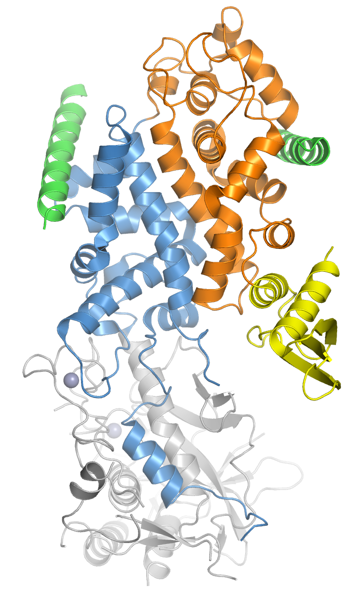
Drosha is a Class 2 ribonuclease III enzyme[5] that in humans is encoded by the DROSHA (formerly RNASEN) gene.[6][7][8] It is the primary nuclease that executes the initiation step of miRNA processing in the nucleus. It works closely with DGCR8 and in correlation with Dicer. It has been found significant in clinical knowledge for cancer prognosis.[9] and HIV-1 replication.[10]

History

[edit]

Human Drosha was cloned in 2000 when it was identified as a nuclear dsRNA ribonuclease involved in the processing of ribosomal RNA precursors.[11] The other two human enzymes that participate in the processing and activity of miRNA are the Dicer and Argonaute proteins. Recently, Drosha has been associated with cancer [9] and childhood acute lymphoblastic leukemia prognosis,[12] and HIV-1 replication.[10]

Function

[edit]

Members of the ribonuclease III superfamily of double-stranded (ds) RNA-specific endoribonucleases participate in diverse RNA maturation and decay pathways in eukaryotic and prokaryotic cells.[13] The RNase III Drosha is the core nuclease that executes the initiation step of microRNA (miRNA) processing in the nucleus.[8][11]

The microRNAs thus generated are short RNA molecules that regulate a wide variety of other genes by interacting with the RNA-induced silencing complex (RISC) to induce cleavage of complementary messenger RNA (mRNA) as part of the RNA interference pathway. MicroRNA molecules are synthesized as long RNA primary transcripts known as a pri-miRNAs, which are cleaved by Drosha to produce a characteristic stem-loop structure of about 70 base pairs long, known as a pre-miRNA.[11] Pre-miRNAs, when associated with EXP5, are stabilized due to removal of the 5' cap and 3' poly(A) tail.[14] Drosha exists as part of a protein complex called the Microprocessor complex, which also contains the double-stranded RNA binding protein DGCR8 (called Pasha in D. melanogaster and C. elegans).[15] DGCR8 is essential for Drosha activity and is capable of binding single-stranded fragments of the pri-miRNA that are required for proper processing.[16] The Drosha complex also contains several auxiliary factors such as EWSR1, FUS, hnRNPs, p68, and p72.[17]

Both Drosha and DGCR8 are localized to the cell nucleus, where processing of pri-miRNA to pre-miRNA occurs. These two proteins homeostatically control miRNA biogenesis by an auto-feedback loop.[17] A 2nt 3' overhang is generated by Drosha in the nucleus recognized by Dicer in the cytoplasm, which couples the upstream and downstream processing events. Pre-miRNA is then further processed by the RNase Dicer into mature miRNAs in the cell cytoplasm.[11][17] There also exists an isoform of Drosha that does not contain a nuclear localization signal, which results in the generation of c-Drosha.[18][19] This variant has been shown to localize to the cell cytoplasm rather than the nucleus, but the effects on pri-miRNA processing are yet unclear.

Both Drosha and Dicer also participate in the DNA damage response.[20]

Certain miRNAs have been found to deviate from conventional biogenesis pathways and do not necessarily require Drosha or Dicer, which is because they do not require the processing of pri-miRNA to pre-miRNA.[17] Drosha-independent miRNAs derive from mirtrons, which are genes that encode for miRNAs in their introns and make use of splicing to bypass Drosha cleavage. Simtrons are mirtron-like, splicing-independent, and do require Drosha mediated cleavage, although they do not require most proteins in the canonical pathway such as DGCR8 or Dicer.[10]

Clinical significance

[edit]

Drosha and other miRNA processing enzymes may be important in cancer prognosis.[9] Both Drosha and Dicer can function as master regulators of miRNA processing and have been observed to be down-regulated in some types of breast cancer.[21] The alternative splicing patterns of Drosha in The Cancer Genome Atlas have also indicated that c-drosha appears to be enriched in various types of breast cancer, colon cancer, and esophageal cancer.[19] However, the exact nature of the association between microRNA processing and tumorigenesis is unclear,[22] but its function can be effectively examined by siRNA knockdown based on an independent validation.[23]

Drosha and other miRNA processing enzymes may also be important in HIV-1 replication. miRNAs contribute to the innate antiviral defense. This can be shown by the knockdown of two important miRNA processing proteins, Drosha and Dicer, which leads to a significant enhancement of viral replication in PBMCs from HIV-1-infected patients. Thus, Drosha, in conjunction with Dicer, seem to have a role in controlling HIV-1 replication.[10]

References

[edit]

Further reading

[edit]
[edit]
Revisions and contributorsEdit on WikipediaRead on Wikipedia
from Grokipedia
Drosha is a nuclear III (RNase III) that initiates the canonical (miRNA) biogenesis pathway by cleaving primary miRNA transcripts (pri-miRNAs) into precursor miRNAs (pre-miRNAs). This processing step occurs in the nucleus and is essential for generating mature miRNAs, which are small non-coding RNAs that regulate post-transcriptionally by targeting messenger RNAs for degradation or translational repression. Drosha forms the core of the microprocessor complex, a ∼650 kDa multiprotein assembly that includes the double-stranded DGCR8 (also known as in ). Structurally, Drosha is a ∼160 kDa protein featuring two tandem RNase III domains (A and B) and a ds-binding domain, which together enable it to recognize the stem-loop structure of pri-miRNAs and perform coordinated cleavage on both strands of the double-stranded stem. DGCR8 enhances substrate specificity by binding to the pri-miRNA and facilitating accurate positioning for cleavage, which generates a ∼60–70 pre-miRNA with a characteristic 2-nucleotide 3′ overhang. Beyond its canonical role, Drosha participates in non-canonical miRNA processing pathways, such as the maturation of endogenous short hairpin RNAs (shRNAs) in a Dicer-independent manner, often in collaboration with 2 (AGO2). Additionally, Drosha has been implicated in mRNA splicing regulation and other RNA processing events, underscoring its broader influence on cellular metabolism. Dysfunctions in Drosha activity are associated with developmental disorders and diseases, including various cancers, where altered miRNA profiles contribute to oncogenesis.

Discovery and History

Initial Identification

Drosha was first identified as a member of the RNase III enzyme family in the early 2000s through a combination of cloning efforts, sequence homology searches, and functional studies in both human and Drosophila models. In 2000, the human DROSHA gene was cloned from a liver cDNA library, revealing it encodes a 1,374-amino-acid protein with an approximate molecular weight of 160 kDa, featuring N-terminal proline- and serine/arginine-rich domains alongside a C-terminal RNase III domain. The gene was mapped to the cytogenetic location 5p13.3. Concurrently, the Drosophila ortholog of Drosha was cloned as a cDNA sequence, demonstrating evolutionary conservation and suggesting a role in double-stranded RNA processing. Between 2001 and 2003, genetic screens in identified key components of the miRNA pathway, while biochemical assays in cells established Drosha as an essential RNase III member for the initial steps of miRNA biogenesis, distinguishing it from other RNases involved in . The pivotal functional characterization occurred in 2003, when Drosha was shown to reside in the nucleus and execute the first cleavage event on primary miRNA transcripts (pri-miRNAs), generating precursor miRNAs (pre-miRNAs) as 60–70 nucleotide hairpin structures. This nuclear processing step was demonstrated through immunopurification of epitope-tagged Drosha from cell extracts, followed by assays confirming its nuclease activity on pri-miRNAs, and experiments that led to pri-miRNA accumulation and reduced mature miRNA levels upon depletion. Unlike the cytoplasmic RNase III enzyme , which processes pre-miRNAs into mature miRNAs, Drosha's nuclear localization and substrate specificity underscored its unique role in the miRNA maturation pathway.

Key Developments

In 2004, parallel studies independently identified DGCR8 as the essential double-stranded RNA-binding cofactor for Drosha, forming the core complex responsible for the initial nuclear processing of primary microRNAs (pri-miRNAs). These discoveries utilized biochemical techniques, including co-immunoprecipitation of endogenous proteins followed by identification, and demonstrated that DGCR8 stabilizes Drosha and enhances its substrate specificity for pri-miRNA hairpins, without which Drosha exhibits negligible processing activity. This breakthrough shifted understanding from Drosha as a solitary RNase III to a heterodimeric complex, with DGCR8 providing structural scaffolding via its dsRNA-binding domains. A pivotal structural advance came in 2016 with the determination of the crystal structure of the minimal catalytic core of human Drosha in complex with the C-terminal α-helix of DGCR8, resolved at 3.2 Å resolution. This revealed that Drosha adopts a pseudodimeric , with its two RNase III domains forming an intramolecular dimer, and features two distinct DGCR8-binding sites: one at the C-terminal end of the RIIIDb domain and another at the N-terminal extension, facilitating heterodimerization and allosteric activation. The structure elucidated how the DGCR8 helix inserts into a hydrophobic groove on Drosha, rigidifying its flexible regions and positioning the catalytic sites for precise cleavage ~11 nucleotides from the single-stranded RNA-duplex junction of pri-miRNAs. In 2025, research uncovered non-canonical localization of Drosha to cytoplasmic compartments, including the Golgi apparatus in human cancer cells, independent of DGCR8 and miRNA biogenesis activity, suggesting novel -independent functions potentially linked to cellular stress responses. Concurrently, studies highlighted the role of an intrinsically disordered region (IDR) in Drosha's N-terminal domain in selectively promoting processing of specific pri-miRNAs, such as those in C. elegans, by facilitating dynamic interactions that enhance Microprocessor condensate formation without relying on tissue-specific factors. These findings expand Drosha's functional repertoire beyond the nucleus, emphasizing regulatory flexibility in miRNA maturation.

Molecular Structure

Protein Domains

Drosha is an endoribonuclease belonging to the class II RNase III family, characterized by a modular domain organization that enables its role in RNA processing. The protein has an overall molecular weight of approximately 160 in humans. The N-terminal region features a proline-rich domain spanning 1–212, which is highly disordered and enriched with prolines (comprising about 32% of the first 200 residues), facilitating protein-protein interactions essential for its regulatory functions. Adjacent to this is an arginine/serine-rich (RS-rich) domain ( 219–316), which contains serine/arginine repeats and contributes to additional protein interactions, including a brief association with the cofactor DGCR8. This N-terminal segment also includes acidic regions that influence structural flexibility and stability. At the core of the protein lies the RNase III domain (RIIID), comprising two tandem catalytic subunits: RIIIDa and RIIIDb. These domains form an intramolecular pseudodimer, with each containing conserved Rsr1 and Rsr2 motifs that harbor the catalytic DDE/H triad—consisting of two acidic aspartate or glutamate residues and a —for coordinating magnesium ions and executing endonucleolytic cleavage of double-stranded substrates. The RIIID architecture ensures precise recognition and processing of structured elements. The C-terminal double-stranded RNA-binding domain (dsRBD) is responsible for substrate recognition by binding to double-stranded features, such as mismatched motifs in pri-miRNA hairpins, thereby positioning the RNA for cleavage by the RIIID. This domain adopts a conserved α-β-β-β-α fold, enhancing the specificity of Drosha's enzymatic activity. The domain architecture of Drosha is evolutionarily conserved across metazoans, with the RIIID and dsRBD showing high ; for instance, the human protein shares approximately 50–60% identity with its homolog, particularly in the catalytic regions (65% for RIIIDa and 45% for RIIIDb). This conservation underscores the fundamental role of these domains in biogenesis throughout animal .

Complex Formation

Drosha assembles into the functional complex primarily through interactions with DGCR8, forming a heterotrimeric unit consisting of one Drosha polypeptide and two DGCR8 molecules. This architecture is mediated by specific binding interfaces on Drosha's RNase III domains (RIIIDs), where the C-terminal tails of DGCR8 anchor to distinct sites: one at the junction between the RIIIDa and RIIIDb domains and another near the C-terminal region of Drosha's catalytic core. These interactions stabilize the complex and position the catalytic centers for substrate recognition, with biochemical and structural analyses confirming that disruption of these sites abolishes complex formation and pri-miRNA processing activity.00558-9) DGCR8 contributes to complex stability and RNA substrate binding via its double-stranded RNA-binding domains (dsRBDs) and dimerization domain within the heme-binding region (also known as the RNA-binding heme domain or Rhed). The dsRBDs clamp onto the double-stranded stem of pri-miRNA hairpins, measuring approximately one helical turn to ensure precise positioning, while the dimerization domain bridges the two DGCR8 molecules and enhances affinity for the apical loop structure, promoting overall hairpin stabilization. Heme binding to DGCR8 further refines this recognition by inducing conformational changes that favor productive RNA engagement.00643-2) The DEAD-box RNA helicase p68 (DDX5) serves as an accessory factor that integrates into the Microprocessor complex, enhancing its dynamics without being essential for core assembly. p68 promotes ATP-dependent remodeling of RNA secondary structures, facilitating efficient pri-miRNA loading and cleavage while preventing off-target interactions; its absence reduces processing efficiency for certain substrates in reconstitution assays. Recent cryo-EM and biochemical studies from 2025 have revealed that intrinsically disordered regions (IDRs) in Drosha's N-terminal domain and DGCR8's N-terminus play critical roles in selective cofactor recruitment, such as the enhancer of rudimentary homolog (ERH), enabling context-dependent modulation of complex activity and tissue-specific miRNA production.

Function in miRNA Biogenesis

Processing Mechanism

Drosha, functioning within the Microprocessor complex alongside DGCR8, performs the initial processing of primary microRNAs (pri-miRNAs) in the nucleus, where the is predominantly localized through its N-terminal nuclear localization signal. Pri-miRNAs are recognized by the complex based on their characteristic stem-loop structures, which feature a double-stranded (dsRNA) stem of typically 20-40 base pairs (including bulges), flanked by single-stranded (ssRNA) regions on both the 5' and 3' sides. These flanking ssRNA segments are essential for efficient binding, as DGCR8 primarily interacts with the dsRNA stem while the ssRNA extensions help position the substrate for cleavage. The cleavage reaction is catalyzed by Drosha's two RNase III domains, which execute an asymmetric endonucleolytic cut approximately 11 nucleotides from the ssRNA-dsRNA junction at the base of the pri-miRNA stem, releasing a ~60-70 nucleotide hairpin precursor (pre-miRNA) with a characteristic 2-nucleotide 3' overhang. This precise positioning ensures the mature miRNA sequence is retained within the pre-miRNA stem. The reaction requires divalent cations, specifically Mg^{2+} ions, which coordinate within the active sites of the RNase III domains to facilitate phosphodiester bond hydrolysis. In vitro assays demonstrate that substituting Mg^{2+} with Mn^{2+} can support activity, but Mg^{2+} is the physiological cofactor. The core Drosha-mediated cleavage is an energy-independent process driven solely by the RNase III catalytic mechanism. However, processing efficiency is significantly enhanced by the RNA helicase p68 (also known as DDX5), which associates with the and uses to remodel the pri-miRNA secondary structure, unwinding internal bulges or stabilizing the stem for optimal substrate presentation. This ATP-stimulated remodeling is particularly important for pri-miRNAs with complex folds that might otherwise hinder access to the cleavage site. Overall processing fidelity and rate are further modulated by the intrinsic secondary of the pri-miRNA, with more stable stems or suboptimal loops reducing under limiting Drosha conditions, as evidenced by genome-wide analyses showing structure-dependent variations in cleavage yields. For instance, pri-miRNAs with extensive base-pairing in the basal stem exhibit slower processing compared to those with flexible flanking regions, highlighting how structural features fine-tune the enzymatic output without altering the fundamental cleavage .

Substrate Recognition

Drosha recognizes primary (pri-miRNA) substrates through specific structural features of the RNA , primarily the length and thermodynamic stability of the double-stranded stem region. Optimal occurs with stems approximately 22-25 base pairs in length, as longer or shorter helices reduce cleavage by altering the positioning of the basal single-stranded to double-stranded RNA junction relative to the enzyme's . Thermodynamic stability of the stem, typically around ΔG ≈ -25 kcal/mol, serves as a key selector, balancing sufficient rigidity for binding with flexibility to accommodate mismatches and bulges characteristic of pri-miRNAs; overly stable stems (more negative ΔG) hinder unwinding for precise cleavage, while unstable ones fail to engage the complex effectively. Sequence motifs within the pri-miRNA further enhance substrate specificity, particularly the UGUG motif located in the 5' region of the apical loop or flanking sequences, which increases binding affinity by interacting with accessory factors or stabilizing the hairpin conformation. This motif is present in about 26% of pri-miRNAs and boosts processing fidelity, as mutations disrupt recognition and reduce efficiency in vitro and in vivo assays. The motif's uracil-rich nature facilitates base-specific contacts, distinguishing pri-miRNAs from other structured RNAs. The double-stranded RNA-binding domains (dsRBDs) of Drosha and its cofactor DGCR8 play complementary roles in scanning and measuring pri-miRNA features. DGCR8's dsRBDs survey the RNA for structural motifs, such as the basal junction and apical loop, by binding dsRNA segments and sensing overall geometry to initiate complex assembly. In contrast, Drosha's dsRBD acts as a molecular , gauging the distance from the Drosha-binding site (often termed the dD-box or basal recognition element) to ensure cleavage occurs ~11 from the ss-ds , thereby enforcing substrate length specificity. Recent cryo-EM structures (as of 2024) reveal that these domains undergo conformational changes upon pri-miRNA binding, locking the complex in a cleavage-competent state. Drosha discriminates against non-canonical substrates, such as ribosomal RNAs (rRNAs) or viral RNAs, which often lack the precise architecture or flanking sequences required for efficient binding, resulting in minimal . Recent 2025 studies highlight how intrinsically disordered regions (IDRs) in Drosha, particularly the N-terminal IDR1, bias recognition toward canonical pri-miRNAs by sensing flanking disordered RNA elements; deletion of IDR1 impairs of specific pri-miRs while sparing non-canonical mimics, suggesting these regions provide an additional layer of selectivity through dynamic interactions that favor miRNA biogenesis over off-target cleavage of abundant cellular or viral transcripts.

Regulation

Expression Control

The expression of the DROSHA gene is tightly controlled at both transcriptional and post-transcriptional levels to ensure appropriate levels of the Drosha protein in different cellular contexts. involves transcription factors that bind to the DROSHA promoter, including , which induces Drosha expression to enhance miRNA production in response to stress signals. A notable feedback loop involves miR-103 and miR-107, where Drosha processes the primary transcripts of these miRNAs, which in turn regulate components of the miRNA biogenesis pathway, including indirect effects on Drosha activity through modulation, creating a self-regulatory circuit. Post-transcriptional control of DROSHA mRNA stability is mediated by the HuR, which binds directly to AU-rich elements in the 3'- (3'-UTR) of the Drosha transcript, preventing its degradation and thereby increasing Drosha protein levels to support miRNA processing. This mechanism is particularly important in cells with high miRNA demands, such as those undergoing rapid proliferation. Drosha expression exhibits developmental stage-specific patterns, with protein levels translationally upregulated during oocyte maturation in laevis, where poly(A) tail extension of the Drosha mRNA boosts translation upon transition to eggs, coinciding with the onset of embryonic proliferation; levels are low in quiescent oocytes but rise dramatically to facilitate early embryonic development. In mammals, Drosha is highly expressed in proliferating cells, including embryonic tissues and certain tumors, where elevated levels correlate with increased miRNA output to regulate growth and differentiation. Tissue-specific variations in Drosha expression are well-documented, with notably higher levels in the compared to other organs, reflecting the role of miRNAs in neuronal function and development. In contrast, expression is lower in the liver and salivary glands, potentially adapting miRNA profiles to metabolic and secretory demands. Recent studies have linked epigenetic mechanisms to Drosha regulation, including TET2 and TET3-mediated (5hmC) deposition across the DROSHA body, which promotes its expression in immune cells; disruptions in this pathway can lead to silencing and altered miRNA landscapes. Although histone deacetylases (HDACs) influence miRNA processing through interactions with the Drosha complex, their in direct epigenetic silencing of the DROSHA remains under investigation. These regulatory mechanisms ensure that Drosha abundance aligns with cellular needs, ultimately influencing global miRNA output and control.

Activity Modulation

Drosha's enzymatic activity is modulated through various post-translational modifications that influence its stability, localization, and interaction with the DGCR8 cofactor in the Microprocessor complex. at serine residues 300 and 302 (S300/S302) promotes Drosha's nuclear retention and enhances its processing efficiency of primary (pri-miRNA) transcripts. This modification, mediated by (GSK3β), strengthens the association between Drosha and DGCR8, thereby facilitating stable complex formation and increased miRNA biogenesis. In contrast, under cellular stress conditions such as oxidative damage or heat shock, p38 (MAPK) phosphorylates Drosha at multiple N-terminal sites, including S300 and S355, which disrupts its interaction with DGCR8, promotes nuclear export, and leads to calpain-mediated degradation, thereby inhibiting activity. Ubiquitination serves as a key mechanism for regulating Drosha protein levels by targeting it for proteasomal degradation. The E3 MDM2 directly ubiquitinates Drosha in response to nutrient deprivation signals via the pathway, reducing Drosha abundance and suppressing miRNA production to adapt cellular metabolism. Additionally, N-terminal of Drosha competes with ubiquitination sites, stabilizing the protein and preventing degradation, which maintains baseline activity levels. A critical feedback loop autoregulates Drosha activity through its cleavage of DGCR8 mRNA. The Microprocessor complex recognizes and processes stem-loop structures within the 3' untranslated region of DGCR8 mRNA, leading to its destabilization and reduced DGCR8 protein synthesis when complex levels are high, thus limiting further pri-miRNA processing and preventing overproduction of miRNAs. Environmental factors, such as iron availability, also modulate activity via heme binding to the double-stranded RNA-binding domain of DGCR8; under iron-depleted conditions, apo-DGCR8 exhibits diminished pri-miRNA binding affinity, inhibiting the complex's catalytic function and reducing overall miRNA maturation rates. Recent studies highlight dynamic relocalization of Drosha as a stress-responsive modulation mechanism. In 2025 research on infection in lung epithelial cells, viral-induced cellular stress triggers proteolytic cleavage of Drosha, resulting in its translocation from the nucleus to the , which limits access to nuclear pri-miRNA substrates and attenuates canonical miRNA biogenesis while potentially enabling non-canonical functions. This relocalization is independent of and is associated with decreased upon Drosha loss, underscoring its role in antiviral stress responses.

Physiological and Pathological Roles

Non-canonical Functions

Beyond its canonical role in pri-miRNA processing, Drosha exhibits non-canonical functions involving the cleavage of alternative RNA substrates in the . Originally identified for its involvement in (rRNA) biogenesis, Drosha cleaves the 48S pre-rRNA precursor into the 12S rRNA intermediate, contributing to the maturation of ribosomal components. A significant fraction of nuclear Drosha translocates to the during the of the , facilitating this activity. Additionally, the Drosha/DGCR8 complex processes a subset of small Cajal body-specific RNAs (scaRNAs), which are nucleolus-associated and function in modification; this processing generates smaller RNA fragments with potential regulatory roles distinct from canonical miRNA pathways. Drosha also participates in cytoplasmic RNA turnover, particularly under cellular stress conditions. Stress signals, such as heat shock or , activate p38 MAPK, which phosphorylates Drosha at its , disrupting its interaction with DGCR8 and promoting nuclear export to the . In the cytoplasm, alternatively spliced isoforms of Drosha localize and retain endonucleolytic activity to process pri-miRNAs in a DGCR8-dependent manner, contributing to cytoplasmic miRNA biogenesis. In the context of mRNA stability regulation, Drosha indirectly influences transcript levels through the biogenesis of specific miRNAs that target mRNAs for decay. For instance, in models of neurodegeneration, Drosha-dependent miRNAs modulate FUS mRNA expression, preventing excessive cytoplasmic accumulation of FUS protein and mitigating associated motor and survival defects. Depletion of Drosha exacerbates FUS-mediated , highlighting its role in fine-tuning mRNA stability via miRNA-mediated mechanisms.

Dysregulation in Diseases

In , levels of the Drosha protein are significantly reduced in postmortem human brain tissues and in transgenic rat models, primarily through p38 MAPK-mediated nuclear exclusion and degradation triggered by amyloid-β accumulation. This downregulation impairs pri-miRNA processing, leading to widespread miRNA deficits that exacerbate neuronal toxicity and degeneration by disrupting metabolism and stress responses. Recent 2023 Drosophila models further illustrate how FUS protein sequesters Drosha into cytoplasmic inclusions, reducing its nuclear availability and amplifying miRNA biogenesis deficits, which in turn promotes neuronal loss and motor impairments relevant to neurodegenerative pathologies including those overlapping with Alzheimer's features. Haploinsufficiency of the DGCR8 gene, resulting from deletions in the 22q11.2 locus characteristic of , disrupts the Drosha-DGCR8 microprocessor complex essential for canonical miRNA maturation. This genetic alteration leads to impaired endothelial cell differentiation, migration, and tubular network formation in patient-derived (iPSC) models, manifesting as and compromised vascular integrity. Consequently, defects arise, contributing to cardiovascular malformations observed in the syndrome, such as conotruncal anomalies, due to dysregulated miRNA control over vascular cell proliferation, , and differentiation. Certain viruses exploit Drosha dysregulation to promote infection. HIV-1 infection reduces levels of Drosha and , suppressing the host miRNA-silencing pathway and thereby evading miRNA-mediated translational repression of viral transcripts. The viral Tat protein contributes to this suppression by inhibiting activity. This inhibition enhances HIV-1 production in infected cells, such as peripheral blood mononuclear cells, facilitating immune evasion and chronic persistence.

Clinical Significance

Implications in Cancer

Drosha plays a critical role in oncogenesis by dysregulating microRNA (miRNA) biogenesis, which influences tumor initiation, progression, and metastasis across various cancer types. In many solid tumors, including breast, ovarian, and lung cancers, Drosha expression is frequently downregulated, contributing to impaired miRNA processing and the derepression of oncogenic pathways. For instance, studies have reported decreased Drosha levels in approximately 51% of ovarian cancer cases, correlating with advanced disease stages and poorer patient outcomes. This downregulation often occurs via epigenetic mechanisms such as promoter hypermethylation, as observed in lung cancer where DROSHA promoter methylation serves as a potential biomarker for early tumor detection. The resulting global reduction in mature miRNA levels disrupts post-transcriptional regulation, leading to the upregulation of oncogenes like BCL2 and KRAS, thereby promoting cell proliferation, invasion, and resistance to apoptosis in cancers such as breast and colon. Paradoxically, Drosha can exhibit overexpression or aberrant activation in certain hematological malignancies, enhancing tumor survival signaling. In (AML), particularly in cases with FLT3-ITD mutations, cytoplasmic relocation and activation of Drosha drive non-canonical miRNA processing, which confers growth advantages to blasts and correlates with aggressive disease. This overexpression facilitates the maturation of pro-survival miRNAs, contributing to therapy resistance and relapse. Similarly, in some subtypes, elevated Drosha levels have been linked to altered miRNA profiles that support epithelial-to-mesenchymal transition and . In , Drosha dysregulation is associated with unfavorable . High miR-21 expression strongly correlates with tumor grade, invasion, and reduced overall survival in patients. Prognostic models incorporating miRNA signatures in liquid biopsies, such as circulating miRNAs from blood or , show promise for early detection and monitoring of glioma progression. These biomarkers enable non-invasive assessment of miRNA alterations, aiding in personalized risk stratification for high-grade gliomas.

Therapeutic Potential

Drosha, as a key in (miRNA) biogenesis, presents therapeutic opportunities in diseases characterized by its dysregulation, particularly cancers where reduced expression correlates with poor prognosis. Strategies to restore or modulate Drosha activity aim to reinstate mature miRNA production, thereby suppressing oncogenic pathways. For instance, in , low Drosha mRNA levels are associated with shorter progression-free and overall survival, suggesting that enhancing Drosha function could improve outcomes by normalizing miRNA-mediated tumor suppression. One approach involves small-molecule inhibitors targeting the miRNA machinery, including Drosha, to disrupt aberrant miRNA production in cancer cells. Although specific Drosha inhibitors remain under development, compounds that interfere with Drosha's enzymatic activity have shown promise in preclinical models by reducing levels and inhibiting tumor growth. For example, small molecules designed to bind Drosha's can block pri-miRNA cleavage, offering a targeted way to downregulate miRNAs that promote cancer progression. Complementing this, (CDK) inhibitors like roscovitine and dinaciclib, already in clinical trials for various cancers, have been investigated for their potential to maintain Drosha expression and amplify antitumor effects through restored miRNA biogenesis. CRISPR-based methods provide precise tools for modulating Drosha function in Drosha-deficient tumors. This approach has potential in contexts like pineoblastoma, where Drosha acts as a tumor suppressor. Indirect modulation via antisense oligonucleotides () targets Drosha-regulated miRNAs, offering a to Drosha dysfunction by sequestering specific oncomiRs. (LNA)-based have demonstrated efficacy in clinical trials for cancers with dysregulated miRNAs, such as miR-21 inhibition in , which indirectly leverages the Drosha pathway by altering miRNA availability and downstream signaling. In viral diseases, enhancing Drosha activity holds promise; Drosha cleaves viral RNAs in an interferon-independent manner, inhibiting HIV-1 replication in infected cells, and pharmacological upregulation could augment host antiviral defenses. Emerging delivery systems, including nanoparticles, address challenges in Drosha modulation by enabling tissue-specific targeting. Lipid-based nanoparticles for miRNA therapies have shown improved blood-brain barrier penetration in preclinical models of , paving the way for broader clinical translation despite hurdles like delivery efficiency and immune responses.

References

Add your contribution
Related Hubs
User Avatar
No comments yet.