Hubbry Logo
ElliptocyteElliptocyteMain
Open search
Elliptocyte
Community hub
Elliptocyte
logo
8 pages, 0 posts
0 subscribers
Be the first to start a discussion here.
Elliptocyte
Elliptocyte
from Wikipedia
Cigar cells in elliptocytosis
Blood smear showing elliptocytes
SpecialtyHematology
blood film in a patient with hereditary elliptocytosis: approximately 60% to 70% of the RBCs are elliptocytes.
Elliptocyte compared to other forms of poikilocytosis.

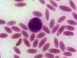
Elliptocytes, also known as ovalocytes or cigar cells, are abnormally shaped red blood cells that appear oval or elongated, from slightly egg-shaped to rod or pencil forms. They have normal central pallor with the hemoglobin appearing concentrated at the ends of the elongated cells when viewed through a light microscope. The ends of the cells are blunt and not sharp like sickle cells.[1]

Elliptocytes are commonly associated with hereditary elliptocytosis. However, they may also be seen in iron deficiency anemia, sepsis, malaria and other pathological states that decrease red blood cell turnover and or production.[2] In the case of iron deficiency anemia, microcytosis and hypochromia would also be expected.[3]

Causes

[edit]

Rare elliptocytes (less than 1%) on a peripheral blood smear are a normal finding.[citation needed]

These abnormal red blood cells are seen in higher numbers in the blood films of patients with blood disorders such as:[4]

References

[edit]
Revisions and contributorsEdit on WikipediaRead on Wikipedia
from Grokipedia
An elliptocyte, also known as an ovalocyte, is an abnormally shaped characterized by an elongated, oval, or elliptical form observed on a , typically resulting from defects in the . These cells often exhibit parallel sides with blunt ends and a central area of , resembling a or pencil shape, and measure approximately the same size as normal discocytes but with increased mechanical fragility. Elliptocytes are a hallmark feature of hereditary elliptocytosis (HE), a group of inherited heterogeneous disorders of the caused primarily by in genes encoding proteins such as α-spectrin (most common, affecting ~65% of cases), β-spectrin (~30%), protein 4.1 (~5%), band 3, or glycophorin C. These genetic alterations impair the vertical and horizontal interactions of the , leading to elliptical morphology that develops after maturation in the circulation. In HE, elliptocytes comprise 15% to 100% of circulating s, depending on the subtype (e.g., common HE, hereditary pyropoikilocytosis, spherocytic elliptocytosis, or Southeast Asian ovalocytosis), and the condition follows an autosomal dominant inheritance pattern in most cases, except for the recessive hereditary pyropoikilocytosis variant. Clinically, HE ranges from asymptomatic carrier states to severe , with manifestations including , , and gallstones in symptomatic individuals; the global prevalence is estimated at 1 in 2,000 to 4,000, with higher rates (up to 1-2%) in malaria-endemic regions like , where the mutation may confer partial resistance to infection. Elliptocytes can also appear in acquired conditions such as , , , or even as artifacts in certain smears, though they are less prominent and not diagnostic in these contexts. typically involves peripheral examination, osmotic fragility testing, and genetic confirmation, while management focuses on supportive care like transfusions or for severe cases, as no curative therapy exists for the underlying defect.

Definition and Morphology

Definition

Elliptocytes are elongated, oval-shaped s, or erythrocytes, that deviate from the typical biconcave disc morphology observed in normal erythrocytes. These cells appear as - or cigar-like forms with blunt ends, representing a variation in shape due to alterations in membrane structure. In healthy individuals, small numbers of elliptocytes, up to 5% of total s, may occur as a normal variation without . However, when elliptocytes exceed 25-40% of the population, this typically indicates an underlying pathological process, such as a membrane disorder. The term "elliptocyte" originates from the early 20th-century literature, with the condition first systematically described by Dresbach in 1904. Elliptocytes differ from similar abnormal red blood cell forms, such as ovalocytes, which are more rounded or egg-shaped, and sickle cells, which exhibit a rigid or sickle-like .

Physical Characteristics

Elliptocytes are elongated, oval-shaped s observable under light microscopy, often resembling cigars or pencils due to their uniform, parallel sides and rounded ends. Their typical dimensions feature a length that is 2 to 3 times the width, distinguishing them from the more circular normal erythrocytes. The central is usually reduced or shifted along the elongated axis, contributing to their distinctive morphology. In Wright-Giemsa stained peripheral blood smears, elliptocytes exhibit staining of the cytoplasm, appearing pink without unless associated with active in . This staining highlights their smooth, elongated contours without surface projections or irregularities in typical cases. Variations occur in severe presentations, where manifests as a mixture of shapes including fragmented forms, and red blood cell fragmentation produces microelliptocytes—small, elliptical remnants. Relative to normal biconcave discocytes, elliptocytes demonstrate reduced deformability stemming from their inherent elongated structure, which hinders passage through microvasculature; studies confirm decreased maximum elongation index in affected cells.

Associated Disorders

Hereditary Elliptocytosis

Hereditary elliptocytosis (HE) is a congenital disorder primarily characterized by the presence of elliptical-shaped erythrocytes, resulting from inherited defects that lead to varying degrees of . It follows an autosomal dominant pattern in most cases, with a global estimated at 1 in 2,000 to 4,000 individuals. The condition exhibits higher in malaria-endemic regions, such as West and , where rates can reach up to 2% (1 in 50), attributed to a conferring partial resistance to severe infection. Clinical manifestations of HE depend on severity, with many cases remaining . In mild forms, individuals often experience no symptoms or only subtle signs like and reduced exercise tolerance. Moderate cases involve compensated , featuring intermittent episodes of , , and , often with elevated counts. Homozygous or compound heterozygous forms, which are rarer, present severely in infancy with , persistent , , and complications such as pigmented gallstones due to chronic overload. HE encompasses several subtypes distinguished by clinical features and associated red cell morphology. Common HE, accounting for approximately 90% of cases, arises from mutations in spectrin genes and is often mild or , with 15% to 100% elliptocytes visible on peripheral smears. Spherocytic HE combines elliptocytes with spherocytes, leading to mild to moderate and more frequent symptoms like , predominantly observed in individuals of European descent. Hereditary pyropoikilocytosis (HPP), a severe recessive variant, features extreme red cell fragmentation into poikilocytes and microspherocytes, resulting in profound neonatal and . Southeast Asian ovalocytosis, a related variant prevalent in populations from , , and the (with frequencies up to 25%), typically causes minimal and is linked to resistance through a deletion in the SLC4A1 gene. Epidemiologically, HE was first described in 1904 by Dresbach, with its hereditary nature later confirmed in the early . Ethnic variations are notable, with elevated carrier rates in African (up to 3% in some West African groups) and Mediterranean populations due to historical selective pressures from , while Southeast Asian ovalocytosis represents a distinct but mechanistically similar entity in Oceanic and Asian communities.

Acquired Forms

Acquired elliptocytosis refers to the presence of elliptical red blood cells resulting from secondary, non-hereditary conditions that disrupt normal erythrocyte morphology, often transiently and reversible upon addressing the underlying cause. Unlike hereditary forms, which involve persistent genetic defects, acquired elliptocytosis typically manifests as a poikilocytotic feature in peripheral blood smears secondary to nutritional, inflammatory, or infiltrative processes. The most common cause is , where elliptocytes appear as a correlate of disease severity and are fully reversible with iron supplementation. In severe cases, the percentage of elliptocytes can increase alongside reductions in , , and , reflecting altered red cell production and membrane pliability due to iron scarcity. Beta-thalassemia minor also frequently associates with elliptocytosis, with affected individuals showing variable numbers of elliptocytes alongside microcytosis and target cells, though this persists as a trait rather than resolving completely. Other notable associations include myelophthisic anemias, such as those arising from infiltration by or myelodysplastic syndromes, where elliptocytes emerge due to disrupted hematopoiesis and premature release. Severe burns can induce elliptocytosis through thermal damage to cell membranes, leading to temporary shape abnormalities. Additionally, artifactual elliptocytes may appear in blood smears from improper preparation, such as prolonged storage or drying artifacts, mimicking true pathological changes. In nutritional deficiencies like iron lack, elliptocytes may be present, but this resolves with correction of the deficiency, in contrast to the lifelong presence in . Specific case examples highlight the transient nature of acquired forms; post-splenectomy states can lead to persistent but mild elliptocytosis due to loss of splenic filtering, resulting in irregular shapes like contracted elliptocytes. Elliptocytosis is rare in , occasionally noted in recovery phases after immune-mediated destruction subsides.

Pathophysiology

Genetic Mutations

Hereditary elliptocytosis (HE) is primarily caused by heterozygous mutations in genes encoding key components of the red blood cell membrane cytoskeleton, leading to structural weaknesses that manifest as elliptical erythrocytes. The most common genetic defects occur in the SPTA1 gene encoding α-spectrin, accounting for approximately 65% of cases, followed by mutations in the SPTB gene encoding β-spectrin at about 30%, and the EPB41 gene encoding protein 4.1R at roughly 5%. Mutations in SLC4A1, which encodes band 3 (anion exchanger 1), are rarer and typically associated with specific subtypes like Southeast Asian ovalocytosis rather than common HE. These mutations often involve missense substitutions, frameshift deletions or insertions, or alterations in mRNA splicing and processing, resulting in qualitative defects that impair spectrin dimer or tetramer self-association or lead to partial spectrin deficiency. For instance, many spectrin variants cluster in the tetramerization domain, disrupting the lateral interactions essential for membrane stability without completely abolishing protein function. A notable example is the low-expression αLELY in SPTA1, characterized by polymorphisms in 40 (αV/41) and 45 that cause partial skipping of 46, reducing α-spectrin synthesis by approximately 50% and functional output accordingly. Inheritance of HE is typically autosomal dominant, with affected individuals inheriting one mutant sufficient to produce the due to the or dominant-negative effects of the mutations. However, the severe variant known as hereditary pyropoikilocytosis (HPP) follows an autosomal recessive pattern, arising from —such as one structural HE-causing mutation in trans to the αLELY —or rarely homozygous mutations, which exacerbate spectrin deficiency. In these cases, total spectrin content is markedly reduced, often to about 70% of normal levels (a 30% deficit relative to band 3), though severe presentations can involve even lower effective functionality due to unstable heterodimers. The role of spectrin in integrity was first elucidated in the 1970s through biochemical studies revealing its filamentous network beneath the , with defects linked to elliptocytosis shortly thereafter. The first molecularly characterized in SPTB was a associated with αI/74 , identified in 1990, marking a key advance in understanding spectrin's genetic basis in HE.

Membrane Abnormalities

The membrane in elliptocytosis exhibits structural defects primarily in the spectrin-based , which maintains cellular integrity and deformability. Disruptions in vertical linkages between the and the underlying skeletal network weaken the membrane's , causing elliptical remodeling of erythrocytes under physiological . These abnormalities arise from impaired assembly of the spectrin lattice, where compromise the formation of stable heterodimers and higher-order structures essential for membrane stability. Key protein interactions are altered, leading to reduced binding affinity between spectrin and junctions, as well as between spectrin, protein 4.1, and the ankyrin-band 3 complex. Protein 4.1 deficiency, for instance, can reduce levels by up to 40%, destabilizing spectrin- associations and disrupting ankyrin-mediated anchoring of spectrin to band 3, an . These changes result in overall instability, with weakened horizontal and vertical interactions in the promoting fragmentation and loss of material. Mechanically, affected erythrocytes display increased fragility, as evidenced by osmotic fragility tests showing at higher saline concentrations (0.5-0.6%) compared to normal cells (0.3-0.45%). Under flow conditions, ektacytometry demonstrates reduced elongation index and a leftward shift in the deformability curve, indicating diminished ability to extend in response to shear forces. These properties reflect compromised and heightened susceptibility to mechanical stress. The pathogenic cascade begins with mutant spectrin tetramers that fail to self-associate effectively, weakening the skeletal network and initiating vesiculation or fragmentation under circulatory shear. This progressive loss reduces the surface area-to-volume ratio by 10-20%, altering cellular from biconcave disc to and exacerbating instability without affecting overall cell volume.

Diagnosis

Laboratory Detection

The primary laboratory method for detecting elliptocytes involves preparing and examining a peripheral blood smear using manual light microscopy after staining with Wright's or Wright-Giemsa stain. This technique allows visualization of red blood cell morphology, where elliptocytes appear as elongated, oval-shaped cells with central pallor. Sample preparation requires fresh venous blood collected in an anticoagulant such as EDTA to preserve cell integrity and prevent clotting. The blood should be smeared promptly on a clean glass slide using the wedge or coverslip method to achieve a thin, even monolayer of cells, followed by immediate air-drying and staining; prolonged storage or improper handling can induce artifacts mimicking elliptocytes, known as pseudoelliptocytosis. Quantification entails counting at least 200-500 red blood cells under (100x objective) and calculating the of elliptocytes relative to total red blood cells. A diagnostic threshold for is often set at greater than 25% elliptocytes, though values can range from 15% to 100% depending on severity. The peripheral smear reliably identifies elliptocytes when they exceed 15% of total red blood cells, but milder cases with lower may require additional testing. For more advanced detection, particularly in subtle or atypical cases, ektacytometry employs laser diffraction ektacytometry to evaluate deformability under controlled and osmotic gradients. This method, introduced in the early 1980s, generates an elongation index curve that reveals decreased maximum deformability in , often appearing as a trapezoidal pattern, making it preferable for confirming membrane defects when smear findings are equivocal. Flow cytometry, using eosin-5-maleimide (EMA) binding assays, can also assess membrane protein expression, such as band 3, which may show reduced in some variants due to protein deficiencies. This quantitative technique measures mean channel on labeled cells and aids in detecting subtle membrane abnormalities, though it is more commonly applied to differentiate related disorders.

Differential Diagnosis

Elliptocytosis must be differentiated from other anemias presenting with elliptical or fragmented red blood cells on peripheral smear, including , which features microcytic, hypochromic erythrocytes due to impaired synthesis. In contrast, often shows prominent target cells and variable microcytosis from globin chain imbalances, while is characterized by drepanocytes (sickled cells) triggered by deoxygenation. These conditions can mimic the mild seen in elliptocytosis, particularly when elliptocytes exceed 25% of red cells without severe . Clinical evaluation provides key clues for distinction: a positive family history of similar hemolytic features strongly suggests , often inherited autosomal dominantly, whereas acquired forms may correlate with nutritional deficiencies indicated by low serum levels. plays a role in hemoglobinopathies, with more prevalent in Mediterranean, African, or Southeast Asian populations and common in those of African descent. Diagnostic challenges arise from overlapping severity, where levels may fall below 110 g/L in both and , complicating initial assessment without additional context. Rare mimics include Southeast Asian ovalocytosis, a related disorder caused by a 27-base-pair deletion in the band 3 gene (SLC4A1), resulting in rigid, oval-shaped cells resistant to invasion by . Resolution involves targeted strategies such as trialing iron supplementation, where improvement in supports over elliptocytosis, which shows no response. confirms hereditary forms by identifying mutations in genes like SPTA1 or SPTB encoding spectrin. Per guidelines, first-line evaluation excludes common through assessment and basic indices before pursuing specialized testing.

Treatment and Management

Supportive Care

Supportive care for elliptocytosis focuses on alleviating symptoms of hemolysis and preventing complications through non-invasive measures, particularly in patients with moderate hereditary elliptocytosis (HE). Folic acid supplementation is standard for individuals experiencing increased due to , as it addresses the heightened folate requirements and helps maintain production. This intervention is especially beneficial in hemolytic states, where chronic turnover can deplete stores. Routine monitoring via (CBC) every 6 to 12 months is recommended for stable patients to track levels, counts, and signs of worsening , with more frequent assessments during periods of stress such as infections or . These strategies reduce the risk of megaloblastic crises arising from . Lifestyle guidance includes avoiding except in cases of severe, transfusion-dependent , as most patients manage well without surgical intervention. For those who undergo , prophylaxis against infections is critical, including administration of the to mitigate the elevated risk of . These approaches, supported by American Society of guidelines on red cell membrane disorders, promote long-term stability and quality of life without addressing underlying membrane defects.

Advanced Interventions

In severe cases of hereditary elliptocytosis (HE), particularly those involving transfusion-dependent or hereditary pyropoikilocytosis (HPP), is indicated to alleviate erythrocyte sequestration and markedly reduce hemolytic activity, though it does not address the underlying membrane defect. This procedure is reserved for approximately 5-20% of patients with clinically significant , often after the age of 5 years to minimize risks, and partial is preferred in children to preserve some splenic function while achieving similar reductions in and improvements in lifespan. Preoperative vaccinations against pneumococcus, meningococcus, and type b are essential due to the lifelong risk of (OPSI), with additional risks including . Post-splenectomy outcomes show improvement or stabilization of in affected individuals with hemolytic anemias, leading to decreased transfusion requirements and enhanced , though long-term monitoring for complications is required. Experimental therapies for HE target genetic and membrane defects, with preclinical efforts focusing on editing of spectrin genes like SPTB to correct mutations underlying elliptocyte formation, though no phase I human trials specific to HE have advanced as of 2025. These interventions hold promise for high-risk patients but require rigorous evaluation given the rarity of severe HE manifestations.

References

Add your contribution
Related Hubs
User Avatar
No comments yet.