Hubbry Logo
MicrogliaMicrogliaMain
Open search
Microglia
Community hub
Microglia
logo
8 pages, 0 posts
0 subscribers
Be the first to start a discussion here.
Microglia
Microglia
from Wikipedia
Microglia
Microglia in resting state from rat cortex before traumatic brain injury (lectin staining with HRP)
Microglia/macrophage – activated form from rat cortex after traumatic brain injury (lectin staining with HRP)
Details
PrecursorPrimitive yolk-sac derived macrophage
SystemCentral nervous system
Identifiers
MeSHD017628
THH2.00.06.2.00004, H2.00.06.2.01025
FMA54539
Anatomical terms of microanatomy

Microglia are a type of glial cell located throughout the brain and spinal cord of the central nervous system (CNS).[1] Microglia account for about around 5–10% of cells found within the brain.[2][3] As the resident macrophage cells, they act as the first and main form of active immune defense in the CNS.[4] Microglia originate in the yolk sac under tightly regulated molecular conditions.[5] These cells (and other neuroglia including astrocytes) are distributed in large non-overlapping regions throughout the CNS.[6][7] Microglia are key cells in overall brain maintenance – they are constantly scavenging the CNS for plaques, damaged or unnecessary neurons and synapses, and infectious agents.[8] Since these processes must be efficient to prevent potentially fatal damage, microglia are extremely sensitive to even small pathological changes in the CNS.[9] This sensitivity is achieved in part by the presence of unique potassium channels that respond to even small changes in extracellular potassium.[8] Recent evidence shows that microglia are also key players in the sustainment of normal brain functions under healthy conditions.[10] Microglia also constantly monitor neuronal functions through direct somatic contacts via their microglial processes, and exert neuroprotective effects when needed.[11][12]

The brain and spinal cord, which make up the CNS, are not usually accessed directly by pathogenic factors in the body's circulation due to a series of endothelial cells known as the blood–brain barrier, or BBB. The BBB prevents most infections from reaching the vulnerable nervous tissue. In the case where infectious agents are directly introduced to the brain or cross the blood–brain barrier, microglial cells must react quickly to decrease inflammation and destroy the infectious agents before they damage the sensitive neural tissue. Due to the lack of antibodies from the rest of the body (few antibodies are small enough to cross the blood–brain barrier), microglia must be able to recognize foreign bodies, swallow them, and act as antigen-presenting cells activating T-cells.

History

[edit]

The ability to view and characterize different neural cells including microglia began in 1880 when Nissl staining was developed by Franz Nissl. Franz Nissl and William Ford Robertson first described microglial cells during their histology experiments. The cell staining techniques in the 1880s showed that microglia are related to macrophages. The activation of microglia and formation of ramified microglial clusters was first noted by Victor Babeş while studying a rabies case in 1897. Babeş noted the cells were found in a variety of viral brain infections but did not know what the clusters of microglia he saw were.[13] The Spanish scientist Santiago Ramón y Cajal defined a "third element" (cell type) besides neurons and astrocytes.[14] Pío del Río Hortega, a student of Santiago Ramón y Cajal, first called the cells "microglia" around 1920. He went on to characterize microglial response to brain lesions in 1927 and note the "fountains of microglia" present in the corpus callosum and other perinatal white matter areas in 1932. After many years of research Rio Hortega became generally considered as the "father of microglia".[15][16] For a long period of time little improvement was made in our knowledge of microglia. Then, in 1988, Hickey and Kimura showed that perivascular microglial cells are bone-marrow derived, and express high levels of MHC class II proteins used for antigen presentation. This confirmed Pio Del Rio-Hortega's postulate that microglial cells functioned similarly to macrophages by performing phagocytosis and antigen presentation.[citation needed]

At the end of the 20th century, the experimental psychology group at Oxford University classified microglial cells into 3 types according to their morphology, tissue location and duration of phagocytic activity.[17] Today, many researchers around the world are trying to establish a relationship between microglial cell morphology and the levels of expression of immune mediators by microglial cells, using different software.[18].

Forms

[edit]
Rat microglia grown in tissue culture in green, along with nerve fiber processes shown in red.
Microglia in rat cerebellar molecular layer in red, stained with antibody to IBA1/AIF1. Bergmann glia processes are shown in green, DNA in blue.

Microglial cells are extremely plastic, and undergo a variety of structural changes based on location and system needs. This level of plasticity is required to fulfill the vast variety of functions that microglia perform. The ability to transform distinguishes microglia from macrophages, which must be replaced on a regular basis, and provides them the ability to defend the CNS on extremely short notice without causing immunological disturbance.[8] Microglia adopt a specific form, or phenotype, in response to the local conditions and chemical signals they have detected.[19] It has also been shown, that tissue-injury related ATP signalling plays a crucial role in the phenotypic transformation of microglia.[20]

Ramified

[edit]

This form of microglial cell is commonly found at specific locations throughout the entire brain and spinal cord in the absence of foreign material or dying cells. This "resting" form of microglia is composed of long branching processes and a small cellular body. Unlike the amoeboid forms of microglia, the cell body of the ramified form remains in place while its branches are constantly moving and surveying the surrounding area. The branches are very sensitive to small changes in physiological condition and require very specific culture conditions to observe in vitro.[19]

Unlike activated or ameboid microglia, ramified microglia do not phagocytose cells and secrete fewer immunomolecules (including the MHC class I/II proteins). Microglia in this state are able to search for and identify immune threats while maintaining homeostasis in the CNS.[21][22][23] Although this is considered the resting state, microglia in this form are still extremely active in chemically surveying the environment. Ramified microglia can be transformed into the activated form at any time in response to injury or threat.[19]

Reactive (activated)

[edit]

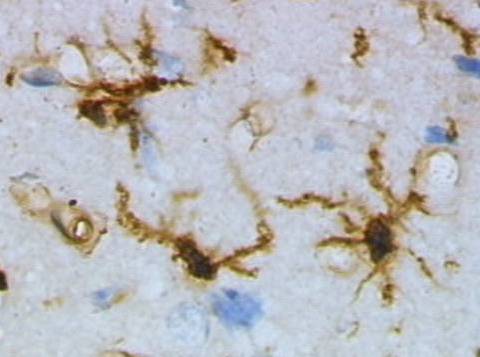
Although historically frequently used, the term "activated" microglia should be replaced by "reactive" microglia.[24] Indeed, apparently quiescent microglia are not devoid of active functions and the "activation" term is misleading as it tends to indicate an "all or nothing" polarization of cell reactivity. The marker Iba1, which is upregulated in reactive microglia, is often used to visualize these cells.[25]

Non-phagocytic

[edit]

This state is actually part of a graded response as microglia move from their ramified form to their fully active phagocytic form. Microglia can be activated by a variety of factors including: pro-inflammatory cytokines, cell necrosis factors, lipopolysaccharide, and changes in extracellular potassium (indicative of ruptured cells). Once activated the cells undergo several key morphological changes including the thickening and retraction of branches, uptake of MHC class I/II proteins, expression of immunomolecules, secretion of cytotoxic factors, secretion of recruitment molecules, and secretion of pro-inflammatory signaling molecules (resulting in a pro-inflammation signal cascade). Activated non-phagocytic microglia generally appear as "bushy", "rods", or small ameboids depending on how far along the ramified to full phagocytic transformation continuum they are. In addition, the microglia also undergo rapid proliferation in order to increase their numbers. From a strictly morphological perspective, the variation in microglial form along the continuum is associated with changing morphological complexity and can be quantitated using the methods of fractal analysis, which have proven sensitive to even subtle, visually undetectable changes associated with different morphologies in different pathological states.[8][21][22][26]

Phagocytic

[edit]

Activated phagocytic microglia are the maximally immune-responsive form of microglia. These cells generally take on a large, ameboid shape, although some variance has been observed. In addition to having the antigen presenting, cytotoxic and inflammation-mediating signaling of activated non-phagocytic microglia, they are also able to phagocytose foreign materials and display the resulting immunomolecules for T-cell activation. Phagocytic microglia travel to the site of the injury, engulf the offending material, and secrete pro-inflammatory factors to promote more cells to proliferate and do the same. Activated phagocytic microglia also interact with astrocytes and neural cells to fight off any infection or inflammation as quickly as possible with minimal damage to healthy brain cells.[8][21]

Amoeboid

[edit]

This shape allows the microglia free movement throughout the neural tissue, which allows it to fulfill its role as a scavenger cell. Amoeboid microglia are able to phagocytose debris, but do not fulfill the same antigen-presenting and inflammatory roles as activated microglia. Amoeboid microglia are especially prevalent during the development and rewiring of the brain, when there are large amounts of extracellular debris and apoptotic cells to remove. This form of microglial cell is found mainly within the perinatal white matter areas in the corpus callosum known as the "Fountains of Microglia".[8][22][27]

Perivascular

[edit]

Unlike the other types of microglia mentioned above, "perivascular" microglia refers to the location of the cell, rather than its form/function. Perivascular microglia are however often confused with perivascular macrophages (PVMs),[28] which are found encased within the walls of the basal lamina, so care must be taken to determine which of these two cell types authors of publications are referring to. PVMs, unlike normal microglia, are replaced by bone marrow-derived precursor cells on a regular basis, and express MHC class II antigens regardless of their environment.[8]

Juxtavascular

[edit]

"Perivascular microglia" and "juxtavascular microglia" are different names for the same type of cell. Confusion has arisen due to the misuse of the term perivascular microglia to refer to perivascular macrophages,[28] which are a different type of cell. Juxtavascular microglia/perivascular microglia are found making direct contact with the basal lamina wall of blood vessels but are not found within the walls. In this position they can interact with both endothelial cells and pericytes.[29][30] Like perivascular cells, they express MHC class II proteins even at low levels of inflammatory cytokine activity. Unlike perivascular cells, but similar to other microglia, juxtavascular microglia do not exhibit rapid turnover or replacement with myeloid precursor cells on a regular basis.[8]

Functions

[edit]
Activation of microglia via purinergic signalling

Microglial cells fulfill a variety of different tasks within the CNS mainly related to both immune response and maintaining homeostasis. The following are some of the major known functions carried out by these cells.[citation needed]

Scavenging

[edit]

In addition to being very sensitive to small changes in their environment, each microglial cell also physically surveys its domain on a regular basis. This action is carried out in the ameboid and resting states via highly motile microglial processes.[12] While moving through its set region, if the microglial cell finds any foreign material, damaged cells, apoptotic cells, neurofibrillary tangles, DNA fragments, or plaques it will activate and phagocytose the material or cell. In this manner microglial cells also act as "housekeepers", cleaning up random cellular debris.[21] During developmental wiring of the brain, microglial cells play a large role regulating numbers of neural precursor cells and removing apoptotic neurons. There is also evidence that microglia can refine synaptic circuitry by engulfing and eliminating synapses.[31] Post development, the majority of dead or apoptotic cells are found in the cerebral cortex and the subcortical white matter. This may explain why the majority of ameboid microglial cells are found within the "fountains of microglia" in the cerebral cortex.[27]

Phagocytosis

[edit]

The main role of microglia, phagocytosis, involves the engulfing of various materials. Engulfed materials generally consist of cellular debris, lipids, and apoptotic cells in the non-inflamed state, and invading virus, bacteria, or other foreign materials in the inflamed state. Once the microglial cell is "full" it stops phagocytic activity and changes into a relatively non-reactive gitter cell.[32]

Extracellular signaling

[edit]

A large part of microglial cell's role in the brain is maintaining homeostasis in non-infected regions and promoting inflammation in infected or damaged tissue. Microglia accomplish this through an extremely complicated series of extracellular signaling molecules which allow them to communicate with other microglia, astrocytes, neurons, T-cells, and myeloid progenitor cells. As mentioned above the cytokine IFN-γ can be used to activate microglial cells. In addition, after becoming activated with IFN-γ, microglia also release more IFN-γ into the extracellular space. This activates more microglia and starts a cytokine induced activation cascade rapidly activating all nearby microglia. Microglia-produced TNF-α causes neural tissue to undergo apoptosis and increases inflammation. IL-8 promotes B-cell growth and differentiation, allowing it to assist microglia in fighting infection. Another cytokine, IL-1, inhibits the cytokines IL-10 and TGF-β, which downregulate antigen presentation and pro-inflammatory signaling. Additional dendritic cells and T-cells are recruited to the site of injury through the microglial production of the chemotactic molecules like MDC, IL-8, and MIP-3β. Finally, PGE2 and other prostanoids prevent chronic inflammation by inhibiting microglial pro-inflammatory response and downregulating Th1 (T-helper cell) response.[21]

Antigen presentation

[edit]

As mentioned above, resident non-activated microglia act as poor antigen presenting cells due to their lack of MHC class I/II proteins. Upon activation they rapidly express MHC class I/II proteins and quickly become efficient antigen presenters. In some cases, microglia can also be activated by IFN-γ to present antigens, but do not function as effectively as if they had undergone uptake of MHC class I/II proteins. During inflammation, T-cells cross the blood–brain barrier thanks to specialized surface markers and then directly bind to microglia in order to receive antigens. Once they have been presented with antigens, T-cells go on to fulfill a variety of roles including pro-inflammatory recruitment, formation of immunomemories, secretion of cytotoxic materials, and direct attacks on the plasma membranes of foreign cells.[8][21]

Cytotoxicity

[edit]

In addition to being able to destroy infectious organisms through cell to cell contact via phagocytosis, microglia can also release a variety of cytotoxic substances.[33] Microglia in culture secrete large amounts of hydrogen peroxide and nitric oxide in a process known as 'respiratory burst'. Both of these chemicals can directly damage cells and lead to neuronal cell death. Proteases secreted by microglia catabolise specific proteins causing direct cellular damage, while cytokines like IL-1 promote demyelination of neuronal axons. Finally, microglia can injure neurons through NMDA receptor-mediated processes by secreting glutamate, aspartate and quinolinic acid. Cytotoxic secretion is aimed at destroying infected neurons, virus, and bacteria, but can also cause large amounts of collateral neural damage. As a result, chronic inflammatory response can result in large scale neural damage as the microglia ravage the brain in an attempt to destroy the invading infection.[8] Edaravone, a radical scavenger, precludes oxidative neurotoxicity precipitated by activated microglia.[34]

Synaptic stripping

[edit]

In a phenomenon first noticed in spinal lesions by Blinzinger and Kreutzberg in 1968, post-inflammation microglia remove the branches from nerves near damaged tissue. This helps promote regrowth and remapping of damaged neural circuitry.[8] It has also been shown that microglia are involved in the process of synaptic pruning during brain development.[35]

Promotion of repair

[edit]

Post-inflammation, microglia undergo several steps to promote regrowth of neural tissue. These include synaptic stripping, secretion of anti-inflammatory cytokines, recruitment of neurons and astrocytes to the damaged area, and formation of gitter cells. Without microglial cells regrowth and remapping would be considerably slower in the resident areas of the CNS and almost impossible in many of the vascular systems surrounding the brain and eyes.[8][36] Recent research verified, that microglial processes constantly monitor neuronal functions through specialized somatic junctions, and sense the "well-being" of nerve cells. Via this intercellular communication pathway, microglia are capable of exerting robust neuroprotective effects, contributing significantly to repair after brain injury.[11] Microglia have also been shown to contribute to proper brain development, through contacting immature, developing neurons.[37]

Development

[edit]
Origin and emergence of microglia in the CNS

For a long time it was thought that microglial cells differentiate in the bone marrow from hematopoietic stem cells, the progenitors of all blood cells. However, recent studies show that microglia originate in the yolk sac during a remarkably restricted embryonal period and populate the brain parenchyma guided by a precisely orchestrated molecular process.[5] Yolk sac progenitor cells require activation colony stimulating factor 1 receptor (CSF1R) for migration into the brain and differentiation into microglia.[38] Additionally, the greatest contribution to microglial repopulation is based upon its local self-renewal, both in steady state and disease, while circulating monocytes may also contribute to a lesser extent, especially in disease.[5][39]

Monocytes can also differentiate into myeloid dendritic cells and macrophages in the peripheral systems. Like macrophages in the rest of the body, microglia use phagocytic and cytotoxic mechanisms to destroy foreign materials. Microglia and macrophages both contribute to the immune response by acting as antigen presenting cells, as well as promoting inflammation and homeostatic mechanisms within the body by secreting cytokines and other signaling molecules.[40]

In their downregulated form, microglia lack the MHC class I/MHC class II proteins, IFN-γ cytokines, CD45 antigens, and many other surface receptors required to act in the antigen-presenting, phagocytic, and cytotoxic roles that distinguish normal macrophages. Microglia also differ from macrophages in that they are much more tightly regulated spatially and temporally in order to maintain a precise immune response.[21]

Another difference between microglia and other cells that differentiate from myeloid progenitor cells is the turnover rate. Macrophages and dendritic cells are constantly being used up and replaced by myeloid progenitor cells which differentiate into the needed type. Due to the blood–brain barrier, it would be fairly difficult for the body to constantly replace microglia. Therefore, instead of constantly being replaced with myeloid progenitor cells, the microglia maintain their status quo while in their quiescent state, and then, when they are activated, they rapidly proliferate in order to keep their numbers up. Bone chimera studies have shown, however, that in cases of extreme infection the blood–brain barrier will weaken, and microglia will be replaced with haematogenous, marrow-derived cells, namely myeloid progenitor cells and macrophages. Once the infection has decreased the disconnect between peripheral and central systems is reestablished and only microglia are present for the recovery and regrowth period.[41]

Aging

[edit]

Microglia undergo a burst of mitotic activity during injury; this proliferation is followed by apoptosis to reduce the cell numbers back to baseline.[42] Activation of microglia places a load on the anabolic and catabolic machinery of the cells causing activated microglia to die sooner than non-activated cells.[42] To compensate for microglial loss over time, microglia undergo mitosis and bone marrow derived progenitor cells migrate into the brain via the meninges and vasculature.[42]

Accumulation of minor neuronal damage that occurs during normal aging can transform microglia into enlarged and activated cells.[43] These chronic, age-associated increases in microglial activation and IL-1 expression may contribute to increased risk of Alzheimer's disease with advancing age through favoring neuritic plaque formation in susceptible patients.[43] DNA damage might contribute to age-associated microglial activation. Another factor might be the accumulation of advanced glycation endproducts, which accumulate with aging.[43] These proteins are strongly resistant to proteolytic processes and promote protein cross-linking.[43]

Research has discovered dystrophic (defective development) human microglia. "These cells are characterized by abnormalities in their cytoplasmic structure, such as deramified, atrophic, fragmented or unusually tortuous processes, frequently bearing spheroidal or bulbous swellings."[42] The incidence of dystrophic microglia increases with aging.[42] Microglial degeneration and death have been reported in research on prion disease, schizophrenia and Alzheimer's disease, indicating that microglial deterioration might be involved in neurodegenerative diseases.[42] A complication of this theory is the fact that it is difficult to distinguish between "activated" and "dystrophic" microglia in the human brain.[42]

In mice, it has been shown that CD22 blockade restores homeostatic microglial phagocytosis in aging brains.[44]

image of microglia

Clinical significance

[edit]
Step-by-step guide for analyzing microglia phenotypes

Microglia are the primary immune cells of the central nervous system, similar to peripheral macrophages. They respond to pathogens and injury by changing morphology and migrating to the site of infection/injury, where they destroy pathogens and remove damaged cells. As part of their response they secrete cytokines, chemokines, prostaglandins, and reactive oxygen species, which help to direct the immune response. Additionally, they are instrumental in the resolution of the inflammatory response, through the production of anti-inflammatory cytokines. Microglia have also been extensively studied for their harmful roles in neurodegenerative diseases, such as Alzheimer's disease, Parkinson's disease, multiple sclerosis, as well as cardiac diseases, glaucoma, and viral and bacterial infections. There is accumulating evidence that immune dysregulation contributes to the pathophysiology of obsessive-compulsive disorder (OCD), Tourette syndrome, and pediatric autoimmune neuropsychiatric disorders associated with streptococcal infections (PANDAS).[45]

Since microglia rapidly react to even subtle alterations in central nervous system homeostasis, they can be seen as sensors for neurological dysfunctions or disorders.[46] In the event of brain pathologies, the microglial phenotype is certainly altered.[46] Therefore, analyzing microglia can be a sensitive tool to diagnose and characterize central nervous system disorders in any given tissue specimen.[46] In particular, the microglial cell density, cell shape, distribution pattern, distinct microglial phenotypes and interactions with other cell types should be evaluated.[46]

Sensome genetics

[edit]

The microglial sensome is a relatively new biological concept that appears to be playing a large role in neurodevelopment and neurodegeneration. The sensome refers to the unique grouping of protein transcripts used for sensing ligands and microbes. In other words, the sensome represents the genes required for the proteins used to sense molecules within the body. The sensome can be analyzed with a variety of methods including qPCR, RNA-seq, microarray analysis, and direct RNA sequencing. Genes included in the sensome code for receptors and transmembrane proteins on the plasma membrane that are more highly expressed in microglia compared to neurons. It does not include secreted proteins or transmembrane proteins specific to membrane bound organelles, such as the nucleus, mitochondria, and endoplasmic reticulum.[47] The plurality of identified sensome genes code for pattern recognition receptors, however, there are a large variety of included genes. Microglial share a similar sensome to other macrophages, however they contain 22 unique genes, 16 of which are used for interaction with endogenous ligands. These differences create a unique microglial biomarker that includes over 40 genes including P2ry12 and HEXB. DAP12 (TYROBP) appears to play an important role in sensome protein interaction, acting as a signalling adaptor and a regulatory protein.[47]

The regulation of genes within the sensome must be able to change in order to respond to potential harm. Microglia can take on the role of neuroprotection or neurotoxicity in order to face these dangers.[48] For these reasons, it is suspected that the sensome may be playing a role in neurodegeneration. Sensome genes that are upregulated with aging are mostly involved in sensing infectious microbial ligands while those that are downregulated are mostly involved in sensing endogenous ligands.[47] This analysis suggests a glial-specific regulation favoring neuroprotection in natural neurodegeneration. This is in contrast to the shift towards neurotoxicity seen in neurodegenerative diseases.

The sensome can also play a role in neurodevelopment. Early-life brain infection results in microglia that are hypersensitive to later immune stimuli. When exposed to infection, there is an upregulation of sensome genes involved in neuroinflammation and a downregulation of genes that are involved with neuroplasticity.[49] The sensome's ability to alter neurodevelopment may however be able to combat disease. The deletion of CX3CL1, a highly expressed sensome gene, in rodent models of Rett syndrome resulted in improved health and longer lifespan.[50] The downregulation of Cx3cr1 in humans without Rett syndrome is associated with symptoms similar to schizophrenia.[51] This suggests that the sensome not only plays a role in various developmental disorders, but also requires tight regulation in order to maintain a disease-free state.

See also

[edit]

References

[edit]

Further reading

[edit]
[edit]
Revisions and contributorsEdit on WikipediaRead on Wikipedia
from Grokipedia
Microglia are the resident immune cells of the (CNS), functioning as specialized macrophages that originate from erythromyeloid precursors during early embryonic development and self-renew throughout life without significant contribution from circulating monocytes. Comprising approximately 10% of all CNS cells, they constantly survey the parenchyma using dynamic ramified processes to monitor for pathogens, debris, and neuronal activity, while maintaining a homeostatic adapted to the unique CNS microenvironment. In the healthy , microglia play essential roles in tissue by phagocytosing apoptotic cells, debris, and protein aggregates, thereby preventing inflammation and supporting neuronal survival through the release of like (BDNF). They also regulate by pruning redundant synapses via complement-dependent mechanisms, such as the C1q and C3 pathways, which refine neural circuits and modulate connectivity to ensure efficient brain wiring. Additionally, microglia contribute to and oligodendrogenesis by secreting cytokines like IL-1β and IGF-1, influencing the proliferation and differentiation of neural precursors in regions such as the hippocampus. During brain development, microglia invade the CNS early—around 4.5 gestational weeks in humans and embryonic day 9.5 in —guiding neuronal migration, axonal , and the elimination of excess progenitors to limit cortical production and promote proper circuit formation. In response to injury or disease, microglia rapidly activate, undergoing morphological changes from ramified to amoeboid states and proliferating locally in a process called microgliosis; this enables them to clear damaged tissue and orchestrate repair but can also drive through proinflammatory release, contributing to pathologies in conditions like , , and . Their activation states exhibit heterogeneity, including protective disease-associated microglia () profiles that enhance of or α-synuclein aggregates, though chronic activation may exacerbate neurodegeneration.

History

Early Discovery

The discovery of microglia as a distinct cell type in the (CNS) is primarily attributed to the Spanish neuropathologist Pío del Río-Hortega, who between 1919 and 1921 systematically described these cells using his innovative ammoniacal silver carbonate staining technique. This method, an improvement on earlier silver impregnation approaches, allowed for the clear visualization of microglia as ramified cells separate from neurons, , and the newly identified , which he termed the "third element" of the CNS. Del Río-Hortega emphasized their unique morphology, including elongated processes and a tendency to aggregate around blood vessels and neurons, distinguishing them from other glial populations. Del Río-Hortega proposed that microglia originated from mesodermal tissues, such as pial or circulating monocytes, positioning them as migratory rather than neuroectodermal derivatives like or . This view sparked early 20th-century debates among neuroscientists, with some, including y Cajal, favoring a neuroectodermal origin based on developmental observations, while others supported mesodermal invasion into the CNS. Del Río-Hortega's experimental evidence from animal models, such as rabies-infected rabbits, reinforced his mesodermal hypothesis by demonstrating microglial proliferation and independent of neural lineage. These debates persisted for decades, but modern lineage-tracing studies in the 2010s confirmed microglia's origin from progenitors, validating the mesodermal framework del Río-Hortega had advocated nearly a century earlier. In the context of , del Río-Hortega's investigations highlighted microglia's active role in CNS diseases, particularly through postmortem analyses of human tissues. He described microglial transformations—such as rod-like formations and granuloadipose bodies—in cases of syphilis-induced general of the insane and various encephalitides, including post-infectious , where these cells engulfed debris and pathogens. These observations underscored microglia as key responders to , capable of rapid and debris clearance, laying the groundwork for understanding their immune functions in neurological disorders.

Key Advances in Understanding

In the mid-20th century, electron microscopy emerged as a pivotal tool for elucidating microglial , building on earlier light microscopy observations. Pioneering studies in the 1950s, such as those by Farquhar and Hartmann (), provided the first detailed ultrastructural images of microglia in tissue, revealing their ramified morphology characterized by elongated processes and a dense nucleus. Further advancements in the and confirmed this ramified form and highlighted perivascular positioning, with and Leblond (1969) demonstrating microglia's proximity to blood vessels and their distinct cytoplasmic features via in models. These findings solidified microglia as dynamic, branched cells integral to , distinguishing them from other . The and marked a shift toward immunological characterization, identifying microglia as resident brain macrophages through specific markers. McGeer et al. (1988) first demonstrated widespread expression of () on reactive microglia in postmortem brains affected by , linking them to capabilities akin to peripheral macrophages. Concurrently, CD11b (also known as Mac-1 or CR3) emerged as a key marker; studies like those by Akiyama et al. (1988) and and (1988) used it to label microglia in and tissues, confirming their myeloid lineage and phagocytic potential without infiltration. This era's immunohistochemical approaches established microglia as immunocompetent cells, bridging neurobiology and . Breakthroughs in the revolutionized understanding of microglial and diversity using genetic fate-mapping and transcriptomics. Ginhoux et al. (2010), building on earlier work, definitively traced adult microglia to yolk sac progenitors via Csf1r-dependent pathways in models, showing self-renewal independent of contributions postnatally. Prinz et al. (2011) extended this by delineating the heterogeneity of CNS myeloid cells, including microglia, and their roles in neurodegeneration, emphasizing yolk sac origins in maintaining . From 2016 onward, single-cell sequencing unveiled transcriptional heterogeneity; Zhang et al. (2016) profiled and microglia, identifying region-specific patterns and activation states that varied by context. These techniques revealed dynamic microglial signatures, from homeostatic to disease-associated profiles. In the 2020s, large-scale atlases and developmental studies have further mapped microglial states in humans. The Human Microglia Atlas (HuMicA), published in 2025, integrated single-nucleus RNA sequencing from 90,716 brain immune cells across six neurodegenerative conditions, identifying nine distinct subpopulations (eight microglial and one border-associated ) with disease-associated signatures enriched in pathways such as . Concurrently, research on prenatal roles demonstrated microglia's regulation of ; a 2025 study showed that human fetal microglia release IGF1 to promote progenitor proliferation in the medial , enhancing neuron production essential for cortical inhibition. These advances underscore microglia's context-dependent plasticity in health and .

Morphology and States

Resting Microglia

Resting microglia, also known as quiescent or surveilling microglia, exhibit a distinctive ramified morphology characterized by a small, compact soma and highly branched processes that extend throughout the brain parenchyma. This structure allows individual resting microglia to maintain constant of their surrounding microenvironment, including synaptic structures and neuronal elements, without migrating or altering tissue . In healthy adult brains, each ramified microglial cell covers a of tens to hundreds of neurons, facilitating rapid detection of subtle changes such as synaptic activity or through direct, transient contacts. The processes of resting microglia are highly dynamic, undergoing continuous extension and retraction at speeds of up to 1-2 μm/min, which enables sampling of the and at a frequency of about once per hour per . These movements are non-disruptive, preserving integrity while allowing microglia to assess neuronal health and respond to minor perturbations. This process motility is supported by the resting and activity, ensuring efficient patrolling without full cellular activation. In the resting state, microglia express low levels of pro-inflammatory markers, such as minimal interleukin-1β (IL-1β), reflecting their baseline anti-inflammatory and homeostatic role. Conversely, they maintain high expression of signature homeostatic genes, including P2ry12 ( involved in process extension) and Tmem119 ( specific to microglia), which are downregulated only upon . These molecular profiles distinguish resting microglia from other glial cells and underscore their role in steady-state maintenance. Resting microglia constitute approximately 10-15% of all glial cells, with densities varying by region—often higher in the hippocampus compared to the cortex. This distribution aligns with regional demands for surveillance, such as in memory-related areas like the hippocampus. Activation signals can shift microglia from this resting state to more responsive forms, though details of such transitions are addressed elsewhere.

Activated Microglia

Upon activation, microglia undergo profound morphological transformations, shifting from a ramified, surveillance-oriented state to more dynamic forms suited for rapid response to or . In the homeostatic ramified morphology, microglia feature elongated, branched processes and a small soma; initiates process retraction and soma , leading to an intermediate hypertrophic state, followed by further transition to an amoeboid shape characterized by rounded soma and reduced branching for enhanced mobility. This progression enables increased , allowing microglia to migrate toward sites of . Activated microglia exhibit heterogeneous reactive subtypes, broadly categorized by their functional profiles. Non-phagocytic subtypes prioritize inflammatory signaling, upregulating pro-inflammatory cytokines such as TNF-α and IL-6 to amplify immune responses and recruit other cells, often observed in chronic neurodegenerative contexts like (AD) and (ALS). In contrast, phagocytic subtypes adopt an amoeboid form to facilitate engulfment of cellular debris and pathogens, marked by expression of lysosomal proteins like , which is upregulated in disease-associated states near in AD. Activation is triggered by diverse signals from the neural environment, including extracellular ATP released from damaged neurons via stimulation, which binds receptors on microglial processes to initiate rapid extension and migration. Danger-associated molecular patterns (DAMPs) and pathogen-associated molecular patterns (PAMPs) from injured cells or invaders engage receptors (PRRs), activating downstream signaling to promote pro-inflammatory gene expression. The receptor TREM2 plays a pivotal role in sensing and apoptotic neurons, modulating activation toward phagocytic phenotypes and integrating with pathways that fine-tune inflammatory outputs. In neurodegenerative diseases, activated microglia often adopt a disease-associated microglia (DAM) state, defined by a distinct transcriptomic signature identified in the late 2010s and elaborated in 2020s research. This signature features downregulation of homeostatic genes and upregulation of pathways, including genes like Apoe and Trem2, which drive phagocytic and reparative functions while suppressing excessive . The TREM2-APOE axis is central to this , enabling microglia to cluster around plaques in AD models and clear aggregates, though dysregulation can exacerbate .

Specialized Forms

Microglia exhibit specialized forms adapted to distinct niches and physiological demands, reflecting their regional heterogeneity beyond general resting or activated states. These variants include juxtavascular populations associated with the vasculature, as well as amoeboid forms prominent during development and , and adaptations differing between and gray . Recent studies have identified additional specialized forms, such as rod-shaped microglia associated with specific neuropathologies. Perivascular macrophages (PVMs), distinct from parenchymal microglia, are positioned along blood vessels and express markers such as , contributing to monitoring the blood-brain barrier (BBB) integrity. These cells help regulate immune surveillance at vascular interfaces, facilitating the uptake of macromolecules and maintaining by responding to disruptions in BBB permeability. In contrast, juxtavascular microglia reside adjacent to vessels but remain within the , distinguished by higher expression of genes like Cx3cr1 (fractalkine receptor), which supports their migratory dynamics along microvessels during early development. This elevated Cx3cr1 expression enables tighter interactions with and endothelial cells, aiding in vascular stabilization without direct perivascular macrophage-like functions. Amoeboid microglia adopt a rounded, less ramified morphology with retracted processes, making them highly migratory and suited for rapid responses in the developing or injured . This form predominates in early postnatal stages, where limited arborization facilitates colonization and of debris, and reemerges post-injury to enable swift migration to lesion sites. Such transformation often follows , allowing these cells to prioritize mobility over . Microglial adaptations also vary between white and gray matter, with white matter populations emphasizing support for through enhanced and lysosomal pathways for maintenance. In gray matter, microglia focus on synaptic , upregulating complement genes like CR1 for and type-I responses to protect neuronal circuits. These regional differences underscore microglia's niche-specific roles in preserving tissue integrity.

Origin and Development

Embryonic Origin

Microglia originate from primitive erythromyeloid progenitors in the during early embryonic development in mice, emerging around embryonic day 7.5 (E7.5) as the first wave of hematopoiesis begins. These progenitors differentiate into primitive macrophages under the control of the PU.1 (encoded by the Spi1 ), which is essential for myeloid lineage commitment and microglial specification. In PU.1-deficient mice, microglial development is completely abolished, underscoring the critical role of this factor in generating these early precursors from erythromyeloid cells. By E9.5, these yolk sac-derived macrophage progenitors migrate into the developing neuroepithelium, colonizing the prior to the formation of the blood-brain barrier. This invasion occurs independently of circulating monocytes from the fetal liver or , as fate-mapping studies demonstrate that microglial precursors do not derive from lineages that produce monocytes. The process relies heavily on colony-stimulating factor 1 receptor (CSF1R) signaling, which is indispensable for the , proliferation, and differentiation of these progenitors; CSF1R models show a profound absence of microglia in the embryonic . Once established in the brain parenchyma, these early microglial precursors undergo local self-renewal through proliferation, maintaining their population without significant contributions from blood-borne cells during steady-state conditions. This self-maintenance mechanism, driven by intrinsic factors like CSF1R, ensures stable colonization and sets the foundation for postnatal expansion. In humans, microglia share a conserved embryonic origin from yolk sac progenitors around the fourth week of gestation, paralleling the murine timeline. The first microglial cells are detected in the fetal at approximately 4.5 weeks post-conception, appearing in amoeboid forms within the ventricular and intermediate zones of the telencephalon.

Postnatal Maturation

Following embryonic seeding, microglia undergo significant postnatal proliferation primarily through local division, without substantial influx from circulating monocytes. In , this proliferation peaks during the first postnatal week (P1–P7), leading to a rapid increase in microglial numbers that achieves near-adult density by P14. This expansion is driven by colony-stimulating factor 1 (CSF-1) signaling and results in a sixfold rise in density in regions like the hippocampus between P5 and P15, after which numbers stabilize or slightly decline to maintain . By P14, microglia acquire their characteristic ramified morphology, featuring elongated processes that enable surveillance of the neural . This morphological maturation coincides with the upregulation of homeostatic gene signatures, including Tmem119 and P2ry12, which reach adult-like expression levels and distinguish mature microglia from earlier immature states. These changes reflect a transition to a quiescent, tissue-resident adapted to the postnatal environment. Regional patterning of microglia emerges postnatally, with distinct profiles in areas like the hippocampus versus the cortex shaped by local environmental cues such as transforming growth factor-β (TGF-β) signaling. Hippocampal microglia exhibit higher expression of bioenergetic and immunoregulatory genes (e.g., Pparg, MHC-II-related) compared to cortical counterparts in young adults, a heterogeneity that TGF-β helps maintain by regulating signature genes like Tmem119 and P2ry12. Disruptions in αVβ8-mediated TGF-β activation lead to dysmature microglia across regions, underscoring the role of these cues in refining regional adaptations during early development. Sexual dimorphism in microglia begins subtly during postnatal development, with differences in becoming more pronounced by . This includes variations in X-linked immune-related genes, such as higher expression of interferon-stimulated genes (e.g., Ifit1), contributing to sex-specific microglial and profiles by P20. These early divergences, influenced by and hormones, lay the foundation for lifelong dimorphisms in microglial function. As microglia age, they exhibit distinct senescence markers that reflect a shift from a homeostatic to a primed state. This includes increased expression of CD11b, a marker of , alongside decreased levels of P2ry12, a homeostatic receptor typically abundant in surveilling microglia. These changes contribute to a "primed" characterized by elevated basal , known as inflammaging, where microglia display heightened responsiveness to stimuli and chronic low-level production of pro-inflammatory mediators. Morphological alterations in aged microglia involve dystrophic processes, such as reduced ramification with shorter, less branched processes and enlarged cell bodies, leading to impaired tissue coverage. dynamics also slow, with decreased process and reduced arborization, diminishing the cells' ability to monitor the effectively. Functionally, aging impairs microglial , resulting in inefficient clearance of debris and accumulation of waste products, while balance shifts toward pro-inflammatory profiles, including elevated IL-1β secretion. Recent findings from the highlight the accumulation of droplets in aged microglia, indicative of metabolic dysfunction and contributing to overall decline in . Additionally, TREM2 variants have been shown to accelerate microglial dysfunction in aging humans by promoting senescence-like states with upregulated inflammatory markers.

Functions

Surveillance and Scavenging

Microglia maintain constant surveillance of the parenchyma through highly dynamic process motility, enabling them to sample the microenvironment and detect subtle changes in neuronal health and activity. In the healthy adult , resting microglial processes extend and retract rapidly, making transient contacts with synapses approximately once every hour, with each interaction lasting about 5 minutes. These contacts allow microglia to monitor synaptic function directly, responding to alterations in neuronal activity levels. The fractalkine receptor CX3CR1 on microglia binds to expressed by neurons, facilitating this surveillance and modulating microglial interactions with active synapses to support homeostatic balance. Each microglial cell occupies a distinct territorial domain, typically spanning 50–100 μm in diameter, which collectively ensures comprehensive coverage of the brain tissue without significant overlap. This spatial organization, observed in both rodents and humans, optimizes the efficiency of environmental sampling and rapid response to local perturbations. In addition to surveillance, microglia perform scavenging functions by clearing non-cellular and cellular debris in a non-inflammatory manner, particularly during development. They efficiently engulf apoptotic neurons and infiltrating neutrophils in the embryonic and early postnatal brain, preventing the accumulation of potentially pro-inflammatory remnants and supporting proper neural circuit formation. Similarly, microglia clear myelin debris generated during normal axonal remodeling or minor demyelination events, utilizing receptors like TREM2 to promote phagocytosis without triggering overt inflammation, thereby maintaining tissue homeostasis. A key aspect of this scavenging is homeostatic , where microglia selectively eliminate weak or inactive synapses to refine neural circuits. This process is guided by the : C1q tags less active synapses, leading to C3 deposition, which microglia recognize via their CR3 receptors to engulf the opsonized elements. Disruptions in this complement-dependent mechanism, as seen in C1q or C3 knockouts, result in excessive synaptic connectivity and impaired circuit maturation.

Phagocytosis and Clearance

Microglia are the primary phagocytic cells of the , actively engulfing and clearing apoptotic neurons, cellular debris, fragments, and pathogens to preserve neural integrity and mitigate . This process begins with the detection of "eat-me" signals on target particles, followed by receptor engagement, cytoskeletal rearrangement for engulfment, and intracellular degradation. Phagocytosis by microglia is particularly vital in pathological states, where impaired clearance can exacerbate tissue damage and disease progression. Receptor-mediated recognition is central to efficient . The triggering receptor expressed on myeloid cells 2 (TREM2), in complex with its adaptor protein DAP12, binds to ligands on apoptotic neurons, promoting their non-inflammatory engulfment by microglia and preventing secondary inflammatory responses. TREM2/DAP12 signaling enhances microglial survival, proliferation, and phagocytic capacity specifically for apoptotic cells, as demonstrated in models of neurodegeneration where TREM2 deficiency leads to accumulation of undegraded neuronal debris. Complementing this, the scavenger receptor facilitates the uptake of debris, enabling microglia to clear lipid-rich remnants from demyelinating lesions and support remyelination. -mediated of is critical in models, where its inhibition impairs debris removal and prolongs neuroinflammation. Once internalized, phagosomes fuse with lysosomes to form phagolysosomes, where acid hydrolases such as cathepsins B and E degrade engulfed material. , in particular, drives proteolytic processing in microglia, with deficiencies disrupting clearance of neuronal debris and promoting a neurotoxic . In inflammatory contexts, such as bacterial or viral infections, microglia shift to an activated state that amplifies pathogen engulfment and destruction. They recognize and internalize via receptors, while for viruses like West or vesicular stomatitis virus, phagocytosis targets infected cells or free virions, limiting spread within the brain parenchyma. Concurrently, activated microglia generate (ROS) through NADPH oxidase 2 () activation, which oxidizes and kills engulfed microbes within phagolysosomes, though excessive ROS can contribute to bystander neuronal damage if unregulated. Building on their surveillance role, this phagocytic response is triggered by initial detection of pathogen-associated molecular patterns. Activated microglia display heightened phagocytic efficiency, processing multiple debris particles or pathogens per cell in response to sustained stimuli, as observed in injury models where single cells clear up to several dozen targets over hours. Recent investigations underscore microglia's independent contribution to viral clearance in , where they produce s to orchestrate antiviral defenses without reliance on peripheral immune infiltration. In models of neurotropic viral infections, microglia activate (MAVS) pathways to induce type I interferons and proinflammatory s like TNF-α and IL-1β, restricting and mitigating encephalitis severity. This microglial response persists even in the absence of adaptive immunity, highlighting their autonomous role in brain-specific control as of 2024-2025 studies.

Synaptic Regulation

Microglia play a critical role in synaptic regulation by modulating neuronal connectivity through processes such as synaptic stripping, particularly in response to injury. During axotomy, activated microglia extend processes to contact and insert into the synaptic clefts of affected motoneurons, displacing presynaptic terminals and leading to their transient removal, a phenomenon known as synaptic stripping. This process, first observed in axotomy models, primarily affects inhibitory synapses and is thought to protect neurons by reducing excitatory input during recovery, though its neuroprotective versus degenerative effects remain debated. In species like hamsters, microglia initiate stripping within hours post-injury, peaking at 7-14 days, while contribute later in synaptic reorganization. Beyond injury responses, microglia facilitate in a complement-dependent manner, essential for circuit refinement during development and adult plasticity. Neurons tag weak or inactive synapses with complement component C1q, which activates the classical complement cascade to deposit C3, signaling microglia via CR3 receptors to phagocytose these synapses. This mechanism is crucial in the retinogeniculate system, where microglial elimination of excess synapses refines visual circuits postnatally, and disruptions in complement genes like C1qa lead to impaired connectivity and behaviors such as seizures. In adults, complement-mediated supports learning and , as evidenced by microglial engulfment of hippocampal synapses during fear memory forgetting, highlighting its role in adaptive plasticity. Microglia also promote synaptic strengthening by releasing (BDNF), supporting the formation and stabilization of active synapses. In learning paradigms, such as tasks, microglia upregulate BDNF production in response to neuronal activity, which enhances dendritic spine density and synaptic efficacy in the hippocampus and . This trophic support is activity-dependent, with ATP signaling via P2X4 receptors on microglia triggering BDNF release to foster long-term potentiation-like changes, thereby facilitating . Recent insights reveal that microglia-neuron crosstalk via the Hex-GM2-MGL2 pathway maintains synaptic under steady-state conditions. Microglia secrete β-hexosaminidase (Hex) to neurons, where it degrades GM2 gangliosides essential for membrane integrity; MGL2 on microglia senses accumulated GM2, preventing aberrant activation and preserving synaptic input frequency and neuronal excitability. Hex deficiency leads to GM2 buildup, microglial proinflammatory responses, and synaptic hypoconnectivity, underscoring this axis's role in preventing circuit dysregulation.

Repair and Neurogenesis

Microglia play a crucial role in brain repair following injury by secreting growth factors that facilitate tissue remodeling and recovery. Post-injury, activated microglia release insulin-like growth factor-1 (IGF-1), which supports neuronal survival and promotes angiogenesis by stimulating endothelial cell proliferation and vessel formation. Similarly, microglia-derived vascular endothelial growth factor (VEGF) enhances vascularization in the damaged area, ensuring nutrient supply for regenerating tissue. These factors also contribute to astrogliosis, where microglia interact with astrocytes to form a supportive glial scar that stabilizes the injury site while allowing limited axonal regrowth. A key aspect of microglial support for regeneration involves clearing inhibitory to create a permissive environment for axonal regrowth. Through , microglia remove fragments and other extracellular inhibitors that hinder neurite extension, thereby scaffolding neural repair and promoting . This clearance is essential for functional recovery, as unresolved inhibitory material can perpetuate and impair network rewiring. In the context of neurogenesis, microglia regulate neural progenitor cells in the adult hippocampus via Wnt signaling pathways. Microglial modulation of canonical Wnt/β-catenin signaling influences progenitor proliferation and differentiation, with decreased microglial Wnt activity linked to reduced neurogenesis and pro-inflammatory states. Activating this pathway in microglia promotes anti-inflammatory responses that enhance hippocampal neurogenesis and neurological recovery. Recent 2025 research further reveals that microglia support GABAergic neurogenesis in the prenatal human cortex through IGF-1 secretion. In the medial ganglionic eminence during the late second trimester, microglia-derived IGF-1 binds to receptors on progenitors, boosting proliferation and interneuron production, as demonstrated in human organoid models where IGF-1 neutralization abolishes this effect. The transition to a reparative state is mediated by a phenotypic switch in microglia from a pro-inflammatory M1-like to an M2-like profile, which resolves and amplifies repair processes. M2-polarized microglia upregulate neuroprotective factors like IGF-1, facilitating , , and , while suppressing excessive tissue damage from M1 dominance. This switch is critical for the resolution phase, enabling microglia to shift from damage amplification to tissue restoration.

Molecular Characteristics

Genetic Markers and Sensome

Microglia are identified through several canonical genetic markers that are highly expressed in these cells. Ionized calcium-binding adapter molecule 1 (Iba1), encoded by the Aif1 gene, is a widely used cytoplasmic marker for visualizing microglia in histological studies due to its role in actin bundling and consistent expression across activation states. The fractalkine receptor Cx3cr1 and colony-stimulating factor 1 receptor Csf1r are s essential for microglial homeostasis and recruitment, often employed in transgenic reporter lines for live imaging and in models. For brain-specific discrimination from infiltrating macrophages, 119 (Tmem119) serves as a reliable marker, as it is selectively expressed in mature microglia and absent in peripheral myeloid cells. The microglial sensome comprises a specialized set of approximately 100 genes that enable these cells to monitor the for endogenous signals, pathogens, and debris, as defined through transcriptomic profiling in 2013. This apparatus includes diverse receptors such as purinergic sensors like P2ry12 and P2ry13, which detect nucleotides released during neuronal activity or injury; Toll-like receptors including Tlr4 for recognizing microbial patterns; and phagocytic receptors like Trem2 and for engulfing apoptotic cells and protein aggregates. Updates in the , based on cross-species comparisons, have refined the sensome to highlight a core of about 57 conserved genes between and microglia, emphasizing shared sensory capabilities despite species-specific variations. Microglial identity and maintenance rely heavily on the colony-stimulating factor 1 (CSF1)/CSF1R signaling pathway, where CSF1R activation promotes proliferation and . Conditional knockout of Csf1r in myeloid cells results in rapid and profound depletion of microglia from the adult , underscoring the pathway's indispensability without affecting other neural populations.

Transcriptional and Functional Heterogeneity

Microglia exhibit transcriptional heterogeneity that reflects their adaptation to diverse brain environments and pathological conditions, transitioning between homeostatic and non-homeostatic states. In homeostatic conditions, microglia maintain a core signature characterized by genes involved in and self-renewal, such as those encoding P2ry12 and Tmem119, serving as a baseline for sensing perturbations. Non-homeostatic states arise in response to or , marked by downregulation of homeostatic genes and upregulation of inflammatory pathways; for instance, in neurodegeneration, interferon-stimulated genes like Ifit1 and Isg15 are significantly upregulated in disease-associated microglia (DAM), promoting an antiviral-like response that may exacerbate . This shift is detailed in a 2025 review highlighting how such interferon responses converge across neurodegenerative contexts while varying by disease stage. Regional differences further contribute to microglial diversity, with distinct transcriptional signatures observed across brain areas. Cortical microglia typically express higher levels of genes related to and immune vigilance, whereas hypothalamic microglia show enriched expression of and stress-response genes, adapting to the region's role in regulation. In , microglia display elevated phagocytic profiles, with increased expression of genes such as and , facilitating debris clearance and supporting axonal integrity under physiological and aging conditions. These spatially tuned transcriptomes underscore how local microenvironments shape microglial function beyond a uniform response. Single-cell RNA sequencing has illuminated this heterogeneity in human brains, particularly in disease states. The 2025 Human Microglia Atlas (HuMicA), integrating 19 datasets from over 90,000 nuclei, identified nine distinct microglial clusters across neurodegenerative conditions, including three homeostatic and five disease-associated subpopulations like inflammatory and lipid-associated . These clusters reveal disease-specific expansions, such as lipid-processing microglia in (AD) and , providing a comprehensive map of microglial states in pathological tissue. Functionally, this transcriptional diversity translates to specialized roles in disease progression, exemplified by DAM and plaque-associated microglia (PAM) profiles in AD. DAM, first characterized in mouse models, upregulate phagocytic and lysosomal genes (e.g., Cst7, Lpl) while downregulating homeostatic markers, enabling amyloid plaque compaction but potentially impairing overall surveillance. PAM, a subset closely apposed to amyloid-beta plaques, exhibit overlapping yet intensified signatures, enhancing beta-amyloid clearance through E-mediated pathways, though chronic activation may drive . These profiles highlight the dual protective and detrimental potential of heterogeneous microglia in AD .

Clinical Implications

Neurodegenerative Diseases

Microglia play a central role in the of neurodegenerative diseases, where their dysfunction contributes to , chronic , and neuronal loss. In conditions such as (AD), (PD), and (ALS), microglia exhibit altered phenotypes that impair clearance mechanisms and exacerbate , often transitioning to pro-inflammatory states that propagate disease progression. In , microglia accumulate around amyloid-β (Aβ) plaques and adopt a disease-associated microglia () transcriptional state, characterized by enhanced and to contain plaque spread. Mutations in the triggering receptor expressed on myeloid cells 2 (TREM2) gene, such as the R47H variant, significantly increase AD risk by impairing microglial activation and Aβ clearance, leading to unchecked plaque accumulation and neurodegeneration. A 2025 study has shown that microglial activation is required for Aβ pathology to induce reactivity, as measured by plasma GFAP and imaging across aging and the AD spectrum. In , microglia fail to efficiently α-synuclein aggregates, resulting in their accumulation and subsequent activation of inflammatory pathways that promote neuronal damage. This phagocytosis defect, coupled with microglial priming—a sensitized state induced by prior insults—exacerbates neuron loss in the by amplifying release and . In , mutant 1 () in microglia triggers a shift to pro-inflammatory states, characterized by pathway activation, which directly induces death through toxic secretome release. This microglial dysfunction accelerates disease progression by impairing neurotrophic support and enhancing in affected regions. Therapeutic strategies targeting microglia, particularly TREM2 agonists, are advancing in clinical trials for as of 2025, aiming to restore phagocytic function and reduce inflammation. Sanofi's 2025 acquisition of Vigil Neuroscience added the oral small-molecule TREM2 agonist VG-3927 to its pipeline for evaluation in a planned Phase 2 study in .

Neurodevelopmental Disorders

Microglia play a critical role in neurodevelopmental disorders by influencing , , and during prenatal and early postnatal brain development, where disruptions can lead to long-term connectivity deficits and behavioral impairments. In these disorders, aberrant microglial activation or dysfunction often stems from genetic mutations or environmental insults like prenatal inflammation, altering the delicate balance of formation. In autism spectrum disorder (ASD), excessive microglial-mediated has been linked to maternal immune (MIA), a prenatal that heightens the risk of neurodevelopmental abnormalities. MIA triggers sustained microglial , promoting over-pruning of synapses and disrupting excitatory-inhibitory balance in key brain regions. Postmortem studies have revealed elevated microglial density and in the of individuals with ASD, correlating with social and cognitive deficits observed in the disorder. For instance, pro-inflammatory of microglia impairs efficiency, leading to excessive synaptic accumulation and ASD-like synaptic and behavioral phenotypes in mouse models, underscoring the cell-type-specific impact of microglial dysfunction. Rett syndrome, caused by mutations in the MECP2 gene, involves microglial dysfunction that impairs and exacerbates neuronal damage. MECP2 deficiency in microglia leads to transcriptional perturbations and reduced phagocytic capacity, hindering the clearance of apoptotic cells and debris during critical developmental windows. This results in excessive engulfment of synapses and dendrites, contributing to circuit defects and progressive neurodegeneration. In mouse models of , microglia-specific MECP2 loss induces synaptic over-pruning, while restoration of MECP2 in microglia ameliorates these deficits, highlighting their therapeutic potential. In schizophrenia, prenatal inflammation disrupts microglial maturation, leading to altered synaptic connectivity and increased vulnerability to psychosis later in life. Exposure to inflammatory cytokines during gestation activates fetal microglia prematurely, impairing their role in synapse refinement and promoting white matter abnormalities. This early perturbation contributes to connectivity deficits in cortical circuits, as evidenced by reduced oligodendroglial support and persistent microglial reactivity in affected individuals. Modulating microglial activation in animal models of prenatal immune challenge prevents schizophrenia-like behaviors, suggesting a causal link between developmental microglial changes and disease onset. Recent advances, including a 2025 study, have elucidated how microglia regulate neurogenesis in the prenatal through insulin-like growth factor 1 (IGF1) secretion, influencing proliferation in the medial . This process is essential for generating inhibitory that shape cortical circuits, and disruptions could underlie inhibitory deficits in neurodevelopmental disorders. By demonstrating direct microglia- interactions in human fetal tissue, this work highlights microglial contributions to species-specific development and opens avenues for targeted interventions.

Therapeutic Targeting

One prominent strategy for modulating microglia involves the use of colony-stimulating factor 1 receptor (CSF1R) inhibitors, such as PLX3397, to achieve selective depletion in preclinical models. This approach has elucidated the reparative roles of microglia in various neurological conditions; for instance, in mouse models of Parkinson's disease, PLX3397-mediated depletion reduced α-synuclein-induced neurodegeneration and remodeled the extracellular matrix, highlighting microglial contributions to tissue maintenance. Similarly, early-phase microglial attenuation with PLX3397 in spinal cord injury models amplified monocyte-derived repair-associated macrophages, promoting long-term functional recovery and underscoring context-dependent protective functions. In ischemic brain injury, repopulation following PLX3397 depletion enhanced neuroprotection in aged mice, further demonstrating the therapeutic potential of transient microglial modulation. Targeting triggering receptor expressed on myeloid cells 2 (TREM2), a key sensome component, represents another advanced therapeutic avenue, particularly for enhancing microglial in . Agonistic antibodies like AL002, which bind TREM2 to activate its signaling, have progressed to phase II clinical trials (INVOKE-2). Preclinical data support plaque clearance and reduction of amyloid-β burden by promoting microglial clustering around plaques. However, 2024 phase II results indicated tolerable safety profiles but no significant effects on disease progression, secondary endpoints, or biomarkers, leading to discontinuation of the extension study. Anti-inflammatory agents targeting microglial activation offer additional clinical promise, exemplified by , a tetracycline derivative that inhibits pro-inflammatory release and microglial proliferation. In acute ischemic , has been tested in multiple s, including the 2025 Phase 3 EMPHASIS design, which evaluates its combination with standard care (including endovascular therapy) to assess attenuation of microglial activation, preservation of blood-brain barrier integrity, and impact on neurological outcomes. For amyotrophic lateral sclerosis (ALS), a phase III with did not demonstrate slowing of disease progression despite preclinical evidence of modulation of microglial responses, with the study stopped early for futility. These approaches highlight 's role in dampening neurotoxic microglial states across acute and chronic disorders. Emerging strategies include gene therapy to augment sensome receptors on microglia, aiming to enhance surveillance and response capabilities. Adeno-associated virus (AAV)-based vectors targeting microglial receptors, such as CX3CR1 promoters, have shown promise in delivering therapeutic transgenes to repopulate or reprogram microglia, potentially restoring sensome functions like purinergic signaling in neurodegenerative contexts. A 2025 study on lymphoid gene expression in microglia revealed a neuroprotective subtype characterized by adaptive immune-like transcripts, suggesting reprogramming via gene editing could shift microglia toward anti-inflammatory, reparative phenotypes to mitigate pathology in Alzheimer's and related disorders.

References

Add your contribution
Related Hubs
User Avatar
No comments yet.