Hubbry Logo
Prostatic urethraProstatic urethraMain
Open search
Prostatic urethra
Community hub
Prostatic urethra
logo
8 pages, 0 posts
0 subscribers
Be the first to start a discussion here.
Prostatic urethra
Prostatic urethra
from Wikipedia
Prostatic urethra
The male urethra laid open on its anterior (upper) surface. (Prostatic part labeled at upper right.)
1: Vas deferens
2: Seminal vesicle
3: Base of the prostate
4: Apex of the prostate
5: Prostatic urethra
Details
Identifiers
Latinpars prostatica urethrae
TA98A09.4.02.004
TA23445
FMA19673
Anatomical terminology

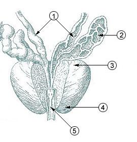
The prostatic urethra, the widest and most dilatable part of the urethra canal, is about 3 cm long.

It runs almost vertically through the prostate from its base to its apex, lying nearer its anterior than its posterior surface; the form of the canal is spindle-shaped, being wider in the middle than at either extremity, and narrowest below, where it joins the membranous portion.

A transverse section of the canal as it lies in the prostate is horse-shoe-shaped, with the convexity directed forward.

The keyhole sign, in ultrasound, is associated with a dilated bladder and prostatic urethra.

Additional images

[edit]

References

[edit]
[edit]
Revisions and contributorsEdit on WikipediaRead on Wikipedia
from Grokipedia
The prostatic urethra is the proximal segment of the male , extending from the neck of the urinary bladder through the central portion of the gland, and serving as a conduit for both and . It measures approximately 3 to 4 cm in length and is the widest part of the male , facilitating the passage of fluids while being enveloped by the prostate's glandular and stromal tissues. Anatomically, the prostatic urethra is positioned inferior to the within the lesser , traversing the from its base to its apex, and is bordered superiorly by the neck and inferiorly by the external urethral . Histologically, it is lined by known as urothelium, which transitions to toward its distal end, and it features prominent internal structures such as the urethral crest (a longitudinal along its posterior ), the (or verumontanum, a mound on the crest where the ejaculatory ducts open), the (a small pouch in the colliculus), and the orifices of the prostatic ducts that drain prostatic secretions. These elements are critical for integrating urinary and reproductive functions, as the here receives contributions from the ejaculatory ducts during emission. The prostatic urethra's location within the makes it clinically significant in conditions like (BPH), where glandular enlargement can compress it and obstruct urine flow, and it is also relevant in due to its proximity to the peripheral and transition zones of the gland. Surgical interventions, such as (TURP), often target this segment to alleviate obstruction while preserving sphincter function.

Anatomy

Location and Course

The prostatic urethra constitutes the initial segment of the male urethra, commencing at the located at the neck and extending inferiorly through the substance of the gland. This segment measures approximately 3 to 4 cm in length, traversing the from its base to the apex before transitioning into the shorter at the inferior border of the . The pathway of the prostatic urethra follows a predominantly vertical trajectory with a subtle anterior concavity, resulting in a characteristic horseshoe-shaped configuration. This curvature arises from the posterior elevation of the urethral wall by the urethral crest, a midline ridge that influences the overall geometry of the lumen. The segment enters the prostate at its superior base, adjacent to the bladder, and exits at the inferior apex, positioned slightly anteriorly within the gland. As the widest and most dilatable portion of the male , the prostatic urethra attains a of approximately 8 , facilitating the passage of urine and while accommodating glandular expansions. It is enveloped by the transition zone of the , which surrounds the proximal urethra and contributes to its positional stability within the pelvic architecture.

Structure and Features

The prostatic urethra exhibits a distinctive internal characterized by a horseshoe-shaped lumen in cross-section, resulting from the prominent posterior urethral crest and relatively flattened anterior and lateral walls. This configuration facilitates the passage of and while accommodating glandular openings. A key feature is the urethral crest, a median longitudinal ridge that extends along the entire posterior wall of the prostatic urethra. At the midpoint of this crest lies the , also known as the verumontanum, which forms a small rounded elevation. The verumontanum serves as the site where the paired ejaculatory ducts open into the urethra, conveying spermatozoa from the and fluid from the . Embedded within the summit of the verumontanum is the , a small blind-ending pouch measuring about 6 mm in length that represents a remnant of the Müllerian duct. Laterally flanking the urethral crest are the prostatic sinuses, paired recesses that receive the openings of approximately 20-30 prostatic ducts draining glandular secretions. These ducts drain the glandular tissue of the , particularly from the transition zone. In terms of zonal anatomy, the transitional zone encircles the proximal portion of the prostatic urethra, while the peripheral zone surrounds its distal segment.

Relations and Blood Supply

The prostatic urethra is situated within the prostate gland, where its anterior relations include the , from which it is separated by retropubic fat and the . Posteriorly, it relates to the , separated by Denonvilliers (also known as rectovesical ), with the on its posterior wall facing the rectal ampulla. Laterally, the prostatic urethra is bordered by the muscles inferiorly, the endopelvic , and the neurovascular bundles that course along the posterolateral aspects of the prostate. The blood supply to the prostatic urethra is primarily derived from the inferior vesical artery, a branch of the , which penetrates the to nourish the urethral wall and surrounding glandular tissue. Additional contributions come from the middle rectal and in some cases. Venous drainage occurs via the , which communicates with the internal iliac veins and connects to the vertebral venous plexus of Batson. Innervation of the prostatic urethra involves a combination of autonomic and somatic components delivered primarily through the prostatic plexus. Sympathetic fibers originate from the (via the hypogastric nerves) and innervate the of the urethral wall and , facilitating contraction during . Parasympathetic innervation arises from the (S2-S4), contributing to glandular secretion within the and relaxation of the . Somatic innervation to the external urethral sphincter at the membranous transition is provided by the (S2-S4), enabling voluntary control of micturition.

Histology

The prostatic urethra is lined by , also known as urothelium, which typically consists of 5-7 layers of cells in its relaxed state. This multilayered structure includes a basal layer of cuboidal or columnar cells attached to the , intermediate layers of polyhedral cells, and a superficial layer of large, binucleate or multinucleate umbrella cells that form a tight barrier. The umbrella cells, characterized by their dome-shaped appearance and asymmetric unit membrane plaques rich in uroplakins, provide essential protection against the osmotic stress and chemical irritants of and passing through the lumen. Beneath the epithelium lies the submucosa, composed of rich in and elastic fibers, which supports the urethral wall and allows for distensibility. This layer contains mucus-secreting glands, often referred to as Littre's glands in the broader context of the male urethra, that lubricate the mucosal surface; it also incorporates vascular and lymphatic elements essential for supply and drainage. In the prostatic segment, these submucosal structures are interspersed with the openings of prostatic ducts, contributing to the secretory environment. The muscularis layer of the prostatic urethra features an inner longitudinal layer and an outer circular layer, which together provide structural support and contribute to urethral tone. These bundles are continuous superiorly with the of the , facilitating coordinated contraction during voiding while maintaining closure at rest. Unlike the , the prostatic segment lacks within its walls, with the external urethral composed of striated muscle located at the transition to the membranous portion. Surrounding the prostatic urethra is the prostate gland tissue, particularly in the periurethral zone, which consists of glandular acini lined by a that secretes prostatic fluid. This glandular component is embedded within a fibromuscular stroma of and fibers, interspersed with elastic fibers that enhance the urethra's dilataility during and . The absence of in this segment underscores its reliance on the surrounding prostatic stroma for stability.

Development

Embryological Origin

The prostatic urethra originates from the endodermal lining of the , specifically its cranial pelvic portion distal to the developing , which forms during the division of the by the between the 4th and 7th weeks of . This endodermal structure gives rise to the pre-prostatic, prostatic, and membranous segments of the male urethra as the differentiates under male hormonal influences. Around the 10th week of gestation, solid epithelial buds emerge from the epithelium along the urethral ridge, invading the surrounding urogenital to initiate formation of the prostatic primordium. These endodermal buds, approximately 15–20 and arising from multiple quadrants of the sinus wall, elongate and branch within the , progressively enclosing and shaping the developing prostatic urethra as the stroma condenses around it. The process continues into the second trimester, with mesenchymal signaling factors such as fibroblast growth factors (FGF7 and ) promoting bud outgrowth and patterning. Remnants of the Müllerian (paramesonephric) ducts contribute to the formation of the , a small in the posterior wall of the prostatic urethra at the verumontanum. In contrast, the Wolffian (mesonephric) ducts, preserved by androgens, migrate caudally and incorporate into the prostatic urethra as precursors to the ejaculatory ducts, which open at the same site. Androgen signaling, driven by testosterone produced by fetal Leydig cells in the testes starting around week 8 and peaking by week 12, is essential for prostatic development. Testosterone diffuses to the urogenital , where it is converted to (DHT) by , activating receptors to induce mesenchymal differentiation into prostatic stroma and stabilize epithelial bud formation. This hormonal influence ensures male-specific urethral and prostatic morphogenesis, with disruptions leading to developmental anomalies.

Anatomical Development

The prostatic urethra displays , being absent in females where the develops into the vestibule and lower , while in males it forms following differentiation around the 7th week of when the indifferent gonads differentiate into testes under SRY influence. In male embryos, testosterone production by Leydig cells begins around week 9, promoting differentiation of the into the prostatic urethra precursor through dihydrotestosterone-mediated signaling in the . By week 16 of fetal development, the and emerge as distinct structures, with prostatic buds elongating, branching, and canalizing from the epithelium around the verumontanum, establishing the foundational anatomical separation. The prostatic urethra derives from the endodermal , integrating with mesodermal stroma to form its conduit role. Pubertal development, driven by an androgen surge from testicular Leydig cells, induces significant enlargement via signaling in stromal cells, which in turn widens the prostatic urethra to adult dimensions of 3-4 cm in length and 0.5-1 cm in diameter. This growth involves increased ductal branching and epithelial proliferation, transforming the prepubertal narrow lumen into a more robust passage accommodating and .

Function

Role in Urination

The prostatic urethra serves as the initial segment of the male , acting as a conduit that transports from the neck to the during micturition. In healthy individuals, its relatively wide diameter—typically around 8 mm—facilitates high urinary flow rates, often exceeding 15 ml/s at peak, without significant obstruction, enabling efficient emptying of the . The layers surrounding the prostatic urethra are primarily under control via the hypogastric nerves (T11-L2), which promote contraction during the bladder storage phase to maintain continence by closing the at the neck and preventing reflux into the upper urinary tract. During the voiding phase of , these muscles relax—facilitated by reduced sympathetic activity and parasympathetic input via release—coordinating with contraction (parasympathetic, S2-S4) to reduce resistance and propel forward. The prostatic urethra's elastic compliance further buffers intraluminal pressure fluctuations, allowing passive dilation to accommodate the detrusor-generated force without excessive backpressure on the . Mucus secreted by submucosal glands and contributions from the urothelium lubricates the inner surface of the prostatic urethra, minimizing during urine passage and protecting the epithelial lining from mechanical irritation. This is essential for smooth, unobstructed flow, particularly given the transitional epithelium's in maintaining barrier integrity.

Role in Ejaculation

The prostatic urethra functions as the primary conduit for semen emission, serving as the site where prostatic fluid mixes with spermatozoa transported via the ejaculatory ducts, which open into it at the verumontanum. Prostatic fluid accounts for approximately 25-30% of total volume, is alkaline in nature, and is rich in enzymes that contribute to composition. This mixing occurs during the emission phase, depositing the components into the urethral lumen for subsequent transport. Sympathetic innervation from the thoracolumbar (T11-L2) triggers peristaltic contractions in the of the and prostatic urethra, propelling the inferiorly toward the . Concurrently, sympathetic-mediated closure of the at the neck prevents by sealing off the outlet. These coordinated contractions ensure unidirectional flow of through the prostatic urethra without backflow. Prostatic secretions neutralize the acidic environment of the distal , which could otherwise impair viability, and supply key nutrients such as to enhance . (PSA), a prominent enzyme in these secretions, facilitates post-emission, thereby promoting efficient progression. ions, highly concentrated in prostatic fluid, further support and essential for fertilization. Emission through the prostatic transitions seamlessly into the expulsion phase, where somatic innervation via the induces rhythmic contractions of the distal to forcefully propel along the penile . This coordination between proximal and distal striated muscle activity completes the ejaculatory process.

Clinical Significance

Associated Pathologies

(BPH) involves the non-malignant enlargement of the prostate's transitional zone, which compresses the prostatic urethra and leads to bladder outlet obstruction. This compression increases voiding pressure, resulting in such as urinary frequency, urgency, , and weak stream. BPH affects approximately 50% of men by age 60 and up to 90% by age 85 (histologic prevalence). Prostatitis encompasses inflammation of the prostate, which can be bacterial or non-bacterial, often causing irritation and pain in the prostatic urethra. Symptoms include , urethral discomfort, , and urinary frequency or hesitancy. Chronic forms of prostatitis impact 10-15% of men during their lifetime, contributing to recurrent episodes of urethral symptoms. Prostate cancer, primarily accounting for over 95% of cases, typically originates in the peripheral zone but can invade the prostatic urethra in advanced stages, leading to , urinary obstruction, or difficulty voiding. Such invasion disrupts urethral patency and may exacerbate . Prostate-specific antigen (PSA) screening plays a key role in early detection to prevent progression to invasive disease. Urethral strictures in the prostatic segment are uncommon but can occur following infections such as bacterial , resulting in formation that narrows the urethral lumen and impairs flow. This narrowing may cause obstructive symptoms including reduced stream and incomplete emptying. Congenital anomalies like cysts arise from remnants of the Müllerian duct and can protrude into the prostatic urethra, potentially causing obstruction, recurrent infections, or in affected individuals. These cysts are typically midline and may lead to if significantly enlarged.

Diagnostic and Therapeutic Procedures

Diagnostic procedures for evaluating issues involving the prostatic urethra primarily include , which allows direct visualization of the urethral lumen and structures such as the verumontanum to identify obstructions or abnormalities. Uroflowmetry assesses urinary flow rates, with reduced rates indicating potential obstruction due to prostatic enlargement. Transrectal ultrasound (TRUS) measures volume and guides biopsies, providing insights into how prostatic growth impacts the urethra. The (PSA) blood test screens for conditions like cancer that may affect the prostatic urethra, with elevated levels prompting further evaluation. Therapeutic interventions target relief from obstructions or infections in the prostatic urethra. (TURP) removes excess tissue endoscopically to alleviate (BPH)-related obstruction, often requiring temporary catheterization post-procedure. Alpha-blockers, such as tamsulosin, provide symptomatic relief by relaxing smooth muscles in the and neck, improving urine flow through the prostatic urethra. For bacterial involving the prostatic urethra, antibiotics are administered for 4-6 weeks or longer to eradicate infection. In prostate cancer cases, brachytherapy implants radioactive seeds into the prostate, delivering targeted radiation that may affect the prostatic urethra and lead to urinary complications. Radical prostatectomy removes the and surrounding tissues, with risks including due to proximity to the urethral sphincter. Catheterization is preferred in the prostatic urethra owing to its dilatability, facilitating management of acute retention or post-procedural drainage. (MRI) aids in staging cancer by assessing urethral involvement and tumor extent.

References

Add your contribution
Related Hubs
User Avatar
No comments yet.