Hubbry Logo
Thompson testThompson testMain
Open search
Thompson test
Community hub
Thompson test
logo
8 pages, 0 posts
0 subscribers
Be the first to start a discussion here.
Thompson test
Thompson test
from Wikipedia

Thompson test
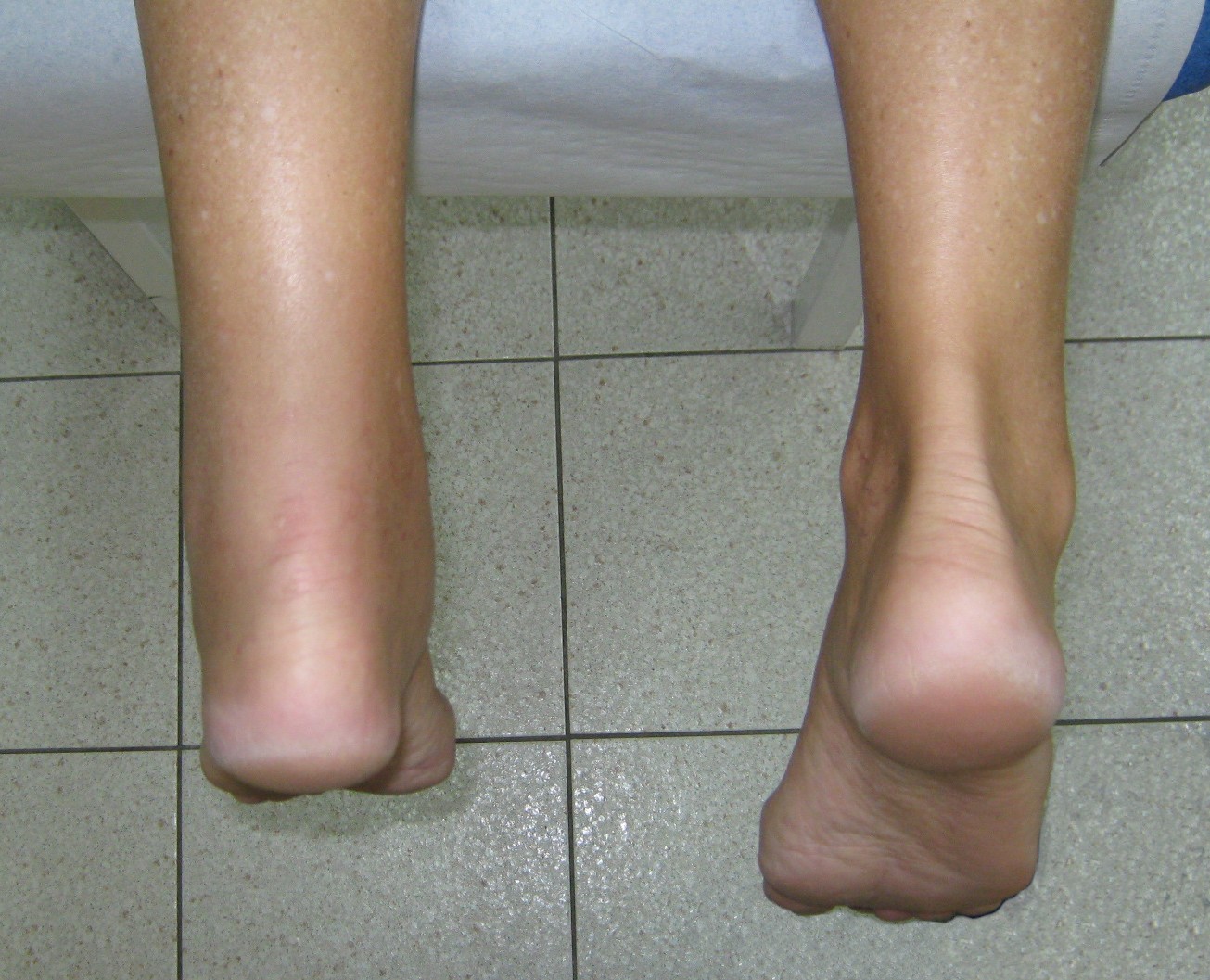
Left Achilles tendon rupture
SynonymsSimmonds' test
Simmonds-Thompson test

The Thompson test (also called Simmonds' test or Simmonds-Thompson test) is used in lower limb examination to test for the rupture of the Achilles tendon.[1][2] The patient lies face down with feet hanging off the edge of the bed. If the test is positive, there is no movement of the foot (normally plantarflexion) on squeezing the corresponding calf, signifying likely rupture of the Achilles tendon.[3]

Interpretation of results

[edit]

Recent research has indicated that while the test is an accurate detector of achilles rupture, it is unable to distinguish between partial tear (tear of the gastrocnemius or soleal portion only) and a complete tear of both portions. [4]

Complete tear of achilles tendon in ultrasound with Simmonds' test

History

[edit]

The test is named after Franklin Adin Simmonds (1910-1983), an English orthopaedic surgeon at the Rowley Bristow Hospital, Surrey.[5]

References

[edit]
Revisions and contributorsEdit on WikipediaRead on Wikipedia
from Grokipedia
The Thompson test, also known as the Simmonds-Thompson test or calf squeeze test, is a straightforward clinical examination used to evaluate the integrity of the , primarily to diagnose complete ruptures by assessing the function of the gastrocnemius-soleus muscle complex. Performed with the patient lying prone and the feet dangling off the examination table, the test involves the examiner squeezing the midpoint of the calf muscle; in an intact tendon, this contraction causes passive plantarflexion of the foot, whereas a lack of movement indicates a positive result suggestive of rupture. Originally described by British orthopedic surgeon Franklin A. Simmonds in 1957 as a method to confirm through calf compression, the test was further elaborated by American surgeon Theodore C. Thompson in , who emphasized its reliance on the soleus muscle's attachment to the for plantarflexion. Despite its simplicity and widespread use in emergency and orthopedic settings—particularly for acute injuries in athletes such as those involving sudden dorsiflexion forces—the test is not infallible, as a positive result can occasionally occur with isolated gastrocnemius tears or partial ruptures without complete discontinuity. The Thompson test demonstrates high diagnostic accuracy, with reported sensitivity ranging from 96% to 100% and specificity from 93% to 100%, making it a reliable initial screening tool, though confirmatory imaging such as or MRI is typically recommended to differentiate partial from complete and guide treatment decisions like surgical repair or conservative management. It carries no significant risks when performed gently and is often combined with other exams, such as the Matles test, to enhance overall diagnostic confidence in cases of suspected pathology.

Background

Achilles Tendon Anatomy

The , also known as the calcaneal tendon, is the largest and strongest in the , primarily composed of fibers arranged in a hierarchical structure that provides tensile strength and elasticity. It forms through the confluence of tendinous fibers from the gastrocnemius and soleus muscles, constituting the distal portion of the group, with the gastrocnemius contributing lateral and medial heads and the soleus adding its fibers approximately 4 cm proximal to the ankle joint. This composition results in a tendon that exhibits a characteristic spiral twist, with soleus fibers inserting medially and gastrocnemius fibers laterally upon reaching the . The tendon originates from the distal ends of the triceps surae muscles in the posterior lower leg and inserts onto the posterosuperior aspect of the , the largest tarsal bone, in a broad, roughened area on its posterior surface. This attachment enables efficient force transmission from the calf muscles to the foot, with the tendon's flattened, ribbon-like shape in its midportion optimizing load distribution. Functionally, the Achilles tendon plays a critical role in plantarflexion of the foot at the ankle joint, facilitating essential movements such as the push-off phase during gait, running, jumping, and climbing stairs. It can withstand tensile forces up to 10 times body weight during intense activities, underscoring its biomechanical importance in locomotion and stability. The blood supply to the Achilles tendon is derived from branches of the posterior tibial artery in the proximal and distal thirds and the fibular (peroneal) artery in the middle third, entering via the musculo-tendinous junction, osseous insertion, and surrounding paratenon. However, the tendon exhibits relatively poor vascularity overall, with a hypovascular watershed area located 2-6 cm proximal to the calcaneal insertion, rendering this midportion particularly vulnerable to ischemic injury and degeneration. Innervation includes motor supply from the tibial nerve (S1-S2 for gastrocnemius and L4-S2 for soleus) to the originating muscles, while sensory innervation is provided by the sural nerve, which courses along the tendon's lateral border and contributes to proprioception and pain signaling.

Achilles Tendon Rupture

Achilles tendon rupture is a significant injury involving the complete or partial tear of the , the strongest tendon in the , which connects the calf muscles to the bone and is essential for plantarflexion of the foot. This condition disrupts the transmission of force from the gastrocnemius and soleus muscles, leading to impaired during and activities requiring push-off. Ruptures most commonly occur in the mid-substance of the tendon, typically 2 to 6 cm proximal to its calcaneal insertion, in a region of relative hypovascularity that predisposes it to degenerative changes and failure under stress. The injury results in a sudden loss of plantarflexion power, often manifesting as an inability to perform single-leg raises or tiptoe walking effectively. Epidemiologically, Achilles tendon ruptures are more prevalent in men, with a male-to-female ratio of approximately 3:1, and the highest incidence occurs in individuals aged 30 to 50 years, particularly among recreational athletes. The annual incidence varies geographically but has been reported to range from 2.5 to 40 cases per 100,000 person-years, with a noted increasing trend over recent decades, largely attributed to greater participation in sports. Risk factors include prior , which weakens the tendon structure; use of fluoroquinolone antibiotics, which can induce tendinopathy and increase rupture risk by up to fourfold, especially when combined with ; and systemic administration, which impairs synthesis and tendon healing. Other contributors encompass older age, , and participation in sports involving explosive movements, such as or . The primary mechanism of injury is acute, resulting from eccentric loading of the during forceful contraction of the calf muscles, often in a plantarflexed foot suddenly subjected to dorsiflexion, as occurs during push-off phases in sports like , , or sprinting. Pathophysiologically, this overload exceeds the tendon's tensile strength, causing either a mid-substance tear—accounting for about 75% of cases—or avulsion from the calcaneal insertion in the remainder, exacerbated by underlying degenerative changes such as mucoid degeneration or collagen disorganization in the hypovascular zone. These tears lead to formation, inflammatory response, and functional deficits in ankle plantarflexion. Clinically, patients typically present with a sudden "pop" or snapping sensation in the posterior ankle or calf, accompanied by acute pain that may subside to a dull ache, followed by swelling, ecchymosis, and localized tenderness. In complete ruptures, a palpable defect or gap may be evident in the , particularly in non-obese individuals, along with visible bruising tracking distally. Functional signs include weakness in plantarflexion, an with reduced push-off, and difficulty bearing weight on the affected side, often prompting patients to describe the injury as feeling like they were struck in the calf.

Procedure

Patient Positioning

The patient is positioned prone (face down) on an examination table, with the feet and ankles extending over the edge to permit unrestricted movement and dangling in a neutral, unsupported alignment. This setup allows for clear visualization and access to the lower legs while minimizing interference from gravitational or supportive forces on the ankles. The knees are maintained in extension to evaluate the integrated function of the triceps surae (gastrocnemius and soleus muscles). If selective involvement of the gastrocnemius is suspected, the knee on the affected side may be flexed to 90 degrees, which slackens the gastrocnemius and isolates the soleus for more targeted assessment. Patient comfort is prioritized by instructing relaxation during positioning, as muscle tension can alter test outcomes; in cases of acute with severe , the may be poorly tolerated, potentially warranting alternatives like kneeling on a stool or bench with the flexed, provided the feet remain freely suspended. No absolute contraindications exist beyond inability to assume the position safely, but providers should monitor for exacerbated .

Examination Technique

The Thompson test examination technique centers on the calf squeeze maneuver, performed after the patient is positioned prone with the feet extending beyond the edge of the examination table. The examiner stabilizes the with one hand if needed and places both hands on the mid-belly of the gastrocnemius and soleus muscles of the triceps surae. A firm, sudden squeeze is then applied using both hands to contract the triceps surae, simulating physiological muscle activation and transmission of force through the . The examiner closely observes the ankle for movement, noting any visible plantarflexion as the expected normal response to the contraction. To enhance accuracy, the test is conducted bilaterally for direct comparison between limbs, repeated as necessary to confirm findings, and accompanied by to identify any asymmetry or defect in the region during the maneuver. No specialized equipment is required, allowing this to function as a straightforward bedside clinical assessment.

Interpretation

Positive Result

A positive result in the Thompson test is characterized by the absence or minimal plantarflexion of the foot when the calf muscles are squeezed, indicating a disruption in the transmission of force from the gastrocnemius-soleus complex to the foot. This lack of movement occurs despite visible or palpable contraction of the calf muscles, distinguishing it from normal responses where the foot passively plantarflexes. Associated physical signs often accompany this observation, including a flattened or atrophic appearance of the calf contour due to the loss of integrity and a palpable defect or gap at the rupture site along the posterior ankle. In some cases, the foot may exhibit slight dorsiflexion or remain completely stationary during the squeeze. The underlying mechanism involves the complete disconnection of the , which prevents the contractile force generated by the calf muscles from being effectively relayed to the triceps surae insertion on the , resulting in no mechanical linkage to produce plantarflexion. Clinically, a positive Thompson test strongly suggests a complete and typically warrants confirmatory imaging, such as or MRI, to delineate the extent of the injury and guide management decisions. This finding is particularly reliable in acute presentations and helps differentiate full ruptures from partial tears or other lower extremity pathologies.

Negative Result

A negative result in the Thompson test is characterized by observable plantarflexion of the foot, where the toes point downward, upon squeezing the calf muscle. This movement occurs due to the intact continuity of the , allowing the gastrocnemius and soleus muscles to transmit force to the foot. Bilateral symmetry is typically assessed by comparing the degree of plantarflexion on the affected side to the contralateral leg, with comparable movement indicating preserved function on both sides. This comparison helps confirm the integrity of the , as asymmetry may suggest underlying pathology even if movement is present. The implications of a negative result generally suggest the absence of a complete , providing reassurance in the context of acute injury evaluation. However, persistent symptoms such as , swelling, or may still necessitate further assessment for partial tears or other injuries, often via imaging modalities like or MRI. Proper technique, including firm compression and relaxation of the patient, is essential to minimize errors.

Diagnostic Performance

Sensitivity and Specificity

The Thompson test demonstrates high diagnostic accuracy for detecting complete Achilles tendon ruptures, with reported sensitivity ranging from 96% to 100% across clinical studies. This high sensitivity indicates that the test effectively identifies most cases of complete rupture, making it a reliable initial screening tool in acute settings. A seminal prospective study by Maffulli involving 174 patients confirmed a sensitivity of 96% (95% CI: 0.91-0.99), using surgical exploration or imaging as the reference standard. Specificity for the Thompson test is similarly robust, ranging from 93% to 100%, reflecting a low rate of false positives particularly in acute presentations where swelling and pain may complicate assessment. In the same Maffulli study, specificity was 93% (95% CI: 0.75-0.99), supporting its utility in confirming rupture without overdiagnosing intact tendons. A systematic review by Reiman et al. corroborated these values, reporting sensitivity of 96% (95% CI: 0.93-0.99) and specificity of 93% (95% CI: 0.75-0.99) from a primary study based on available evidence. The test's positive likelihood ratio (LR+) approximates 13-14, substantially increasing the post-test probability of rupture when positive and underscoring its strong diagnostic support in clinical . Conversely, the negative likelihood ratio (LR-) is 0.04-0.05, providing excellent ability to rule out complete rupture when the test is negative. These metrics highlight the Thompson test's superior performance for complete tears compared to partial ones, where sensitivity may decrease due to preserved partial continuity, as noted in evaluations of clinical diagnostics.

Limitations and Variations

The Thompson test exhibits lower sensitivity for partial or chronic Achilles tendon ruptures compared to complete acute ruptures, with reported ranges of 38% to 84% for acute cases and even lower (12% to 46%) for chronic ones, potentially leading to missed diagnoses in these scenarios. False-negative results can occur in the early post-injury phase due to significant swelling or formation, which may obscure the tendon's discontinuity and allow residual movement during the calf squeeze. Additionally, compensation by accessory muscles, such as the flexor digitorum longus, flexor hallucis longus, tibialis posterior, or peroneus muscles, can produce plantar flexion despite a rupture, further reducing diagnostic reliability. The test is operator-dependent, requiring skilled application of to the calf to adequately assess integrity; inadequate squeeze technique can yield false negatives, particularly in patients lacking examiner . Reliability diminishes in obese individuals, where excess complicates and calf compression, or in those with comorbidities like ankle from or prior trauma, which may alter baseline plantar flexion mechanics. Variations of the Thompson test include a knee-flexed modification, performed with the at 90 degrees to specifically isolate and evaluate the soleus component of the Achilles tendon complex, as the gastrocnemius is relaxed in this position. It is often combined with the Matles test for enhanced accuracy, where prone knee flexion at 90 degrees assesses relative dorsiflexion asymmetry. As a non-standalone diagnostic tool, the Thompson test should be used adjunctively; in equivocal cases, confirmatory imaging such as (sensitivity >95%, specificity ~99%) or MRI is recommended to visualize integrity and guide management.

History

Development by Simmonds

Franklin Adin "Sam" Simmonds (1910–1983) was an English orthopaedic surgeon renowned for his surgical precision and contributions to trauma care. Born on October 31, 1910, he trained at , and St Thomas's Hospital, qualifying in 1935 with MRCS and LRCP, followed by FRCS in 1939. During World War II, he served as a Lieutenant-Colonel in the Royal Army Medical Corps, gaining extensive experience in across , , , and the . After the war, Simmonds became a orthopaedic surgeon at Rowley Bristow Orthopaedic Hospital in , , in 1946, and later at the Royal Surrey County Hospital in from 1951 until his retirement in 1975. In 1957, Simmonds published the initial description of a clinical test for diagnosing Achilles tendon ruptures in The Practitioner, titled "The diagnosis of the ruptured ." The test involved positioning the patient with the feet projecting over the edge of the examination couch and squeezing the calf muscles to assess tendon integrity. He emphasized two key signs of rupture: the relative lack of plantarflexion (equinus) of the injured foot compared to the uninjured side in the , and the absence of passive plantar flexion of the foot upon calf compression. Achilles tendon ruptures often presented diagnostic challenges without advanced imaging. Simmonds' test addressed these gaps by providing a simple, bedside method to evaluate tendon continuity. Subsequent refinements to the test were introduced in the early 1960s.

Adoption by Thompson

Theodore Campbell Thompson (1902–1986), an esteemed American orthopedic surgeon and former surgeon-in-chief at the Hospital for Special Surgery in New York, advanced the clinical evaluation of Achilles tendon ruptures through his work in the field of orthopedics. In 1962, Thompson co-authored a seminal paper in the Journal of Trauma titled "Spontaneous rupture of tendon of Achilles: a new clinical diagnostic test," which built on prior observations to formalize and promote the calf squeeze maneuver as a reliable diagnostic method. Thompson's contributions emphasized the test's inherent simplicity and high efficacy, positioning it as an accessible bedside procedure that required no specialized equipment, making it ideal for busy clinical environments. He specifically detailed its execution with the patient in the , knees extended, and the foot dangling freely off the edge of the examination table to facilitate observation of plantarflexion upon calf compression. Additionally, Thompson highlighted its utility in emergency settings, where swift of ruptures could guide immediate decisions, such as surgical intervention, thereby reducing diagnostic delays. Following Thompson's publication, the maneuver gained dual attribution and became widely known as the Simmonds-Thompson test, reflecting its refined standardization. This recognition propelled its integration into standard orthopedic protocols, achieving widespread adoption in clinical practice by the 1970s as the primary tool for integrity.

References

Add your contribution
Related Hubs
User Avatar
No comments yet.