Hubbry Logo
AnoscopyAnoscopyMain
Open search
Anoscopy
Community hub
Anoscopy
logo
8 pages, 0 posts
0 subscribers
Be the first to start a discussion here.
Anoscopy
Anoscopy
from Wikipedia
Anoscopy
An anoscope, a proctoscope and a rectoscope, and their approximate lengths.
ICD-9-CM49.21
MedlinePlus003890

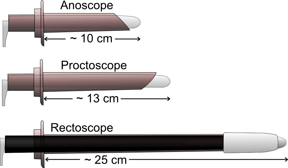
An anoscopy is a medical examination using a small, rigid, tubular instrument called an anoscope (also called a rectal speculum). This is inserted a few centimeters into the anus in order to evaluate problems of the anal canal. Anoscopy is used to diagnose hemorrhoids, anal fissures (tears in the lining of the anus), and some cancers.[1][2]

Process

[edit]

This test is usually done in a doctor's office. The patient is required to remove their underwear, and must either lie on their side on top of an examining table, with their knees bent up towards the chest, or bend forward over the table. The anoscope is 50 to 100 mm long and 8 to 25 mm in diameter. The doctor will coat the anoscope with a lubricant and then gently push it into the anus and rectum. The doctor may ask the patient to "bear down" or push as if they were going to have a bowel movement, and then relax. This helps the doctor insert the anoscope more easily and identify any bulges along the lining of the rectum.

By shining a light into this tube, the doctor will have a clear view of the lining of the lower rectum and anus. The anoscope is pulled out slowly once the test is finished.

The patient will feel pressure during the examination, and the anoscope will make one feel as if they were about to have a bowel movement. This is normal, however, and many patients do not feel pain from anoscopy.

Conditions visible to anoscopy

[edit]

Anoscopy will permit biopsies to be taken, and is used when ligating prolapsed hemorrhoids. It is used in the treatment of warts produced by HPV.

The procedure is done on an outpatient basis.

References

[edit]
Revisions and contributorsEdit on WikipediaRead on Wikipedia
from Grokipedia
Anoscopy is a diagnostic in which a short, rigid, lighted instrument called an anoscope is inserted into the to visualize the , anal sphincter, and distal for signs of injury, disease, or abnormalities such as , fissures, polyps, , infections, or cancer. This examination is particularly useful for evaluating symptoms like , anal pain, itching, discharge, or , and it is often recommended when a digital yields inconclusive results or for screening high-risk individuals, such as HIV-positive men who have sex with men and women aged 35 or older, other HIV-positive individuals aged 45 or older, or patients with a history of anal or persistent anorectal symptoms. High-resolution anoscopy, an advanced variant, enhances detection by applying acetic acid to highlight precancerous lesions and using a colposcope for magnified , making it valuable for early identification of anal intraepithelial neoplasia in at-risk populations.

Overview

Definition

Anoscopy is a diagnostic used to examine the interior of the , , and distal by inserting a lubricated anoscope, a short, rigid, hollow tube typically measuring 3 to 5 inches in length. The anoscope features a light source for illumination and an obturator, a removable plug that aids in smooth insertion while protecting the viewing channel. This procedure allows direct visualization of the and surrounding tissues, often performed as a quick, office-based evaluation without the need for or extensive preparation. Distinguished from related endoscopic techniques, anoscopy is more limited in scope compared to , which employs a longer instrument to inspect deeper portions of the , or , which extends visualization to the for broader colorectal assessment. It is particularly suited for evaluating superficial anorectal structures, such as in cases of . The term "anoscopy" originates from the combining form "ano-," derived from the Latin ānus meaning "ring" (referring to the anus), and the Greek suffix "-scopy," from skopein meaning "to examine" or "to view."

Indications

Anoscopy is primarily indicated for the initial evaluation of , which may stem from conditions such as , , or neoplasms. It is also recommended for assessing anal pain, often associated with issues like thrombosed or fissures. Other common symptoms prompting the procedure include anal itching, anal discharge or , and changes in bowel habits, which can signal underlying anorectal . In high-risk populations, such as individuals with HIV or those infected with high-risk human papillomavirus (HPV) strains, anoscopy—particularly high-resolution anoscopy (HRA)—serves as a key tool for screening anal intraepithelial neoplasia (AIN) and early detection of precancerous changes to prevent anal cancer. As of 2024, updated guidelines based on the ANCHOR study recommend annual assessment of all adults with HIV for anal abnormalities via digital anorectal exam, with HRA offered to men who have sex with men (MSM) and transgender women aged 35 and older, and to other HIV patients aged 45 and older if screening tests (e.g., anal cytology) show abnormalities; frequency is every 1-3 years depending on findings. Additionally, anoscopy may be used to evaluate unexplained or when potentially linked to or occult anorectal sources, particularly after initial digital rectal examination suggests a mass or abnormality. This procedure allows direct visualization of the to identify bleeding sites, such as internal , that could contribute to these systemic symptoms.

Procedure

Preparation

Patients undergoing anoscopy are advised to empty their bowels and prior to the procedure to enhance comfort and visibility during the examination. Additionally, individuals should avoid anal intercourse and the insertion of any objects, medications, creams, or suppositories into the for at least 24 hours beforehand to prevent irritation or interference with the procedure. An may be recommended by the healthcare provider if needed, though it is not routinely required. Healthcare providers prepare for anoscopy by ensuring a private examination room is available, equipped with necessary supplies including lubrication jelly, disposable gloves, an anoscope, a light source, and optional positioning aids such as stirrups to facilitate comfort. Standard anoscopy requires no , bowel preparation, or , as it is a quick, minimally invasive outpatient procedure typically performed without beyond optional topical numbing .

Technique

The technique for performing anoscopy begins with appropriate patient positioning to ensure comfort and access to the anal region. The patient is typically placed in the left lateral decubitus position with the knees flexed toward the chest, though alternatives such as the knee-chest, , or prone positions may be used based on patient needs and clinician preference; draping is applied to maintain throughout the procedure. An optional digital rectal examination may precede the procedure to assess for obstructions or abnormalities, using a lubricated gloved finger. The anoscope, a short tubular instrument (typically 7 cm long and 19 mm in for adults, with a removable obturator), is coated with water-soluble or topical anesthetic jelly such as 2% lidocaine. The separates the buttocks for external , then gently inserts the lubricated anoscope with its obturator in place, advancing it slowly past the anal verge to a depth of approximately 2 to 4 cm or until the dentate line is reached, directing it toward the umbilicus to avoid the anterior vaginal wall in females. Once inserted, the obturator is carefully removed to allow visualization of the and distal through the anoscope, which is equipped with a light source. For a comprehensive circumferential view, the anoscope is gently rotated or withdrawn slightly while observing the mucosa; a slotted anoscope may require multiple insertions for full assessment, while a non-slotted type provides a 360-degree view in one pass. Any obstructing fecal material can be cleared with a during examination. The procedure concludes with slow withdrawal of the anoscope while continuing to inspect the tissues, ensuring no reinsertion of the obturator occurs while inside the instrument. Anoscopy is generally brief, lasting 5 to 10 minutes, and is performed without in an outpatient office setting, though topical anesthetics may be used for patient comfort if indicated.

Diagnostic Findings

Visible Conditions

Anoscopy provides direct visualization of the and distal , allowing identification of various pathologies through the anoscope's illuminated field. This examination reveals abnormalities in the mucosal lining, vascular structures, and surrounding tissues, aiding in the of conditions that may present with symptoms such as bleeding, pain, or discharge. Common findings during anoscopy include internal and external , which appear as swollen, engorged vascular cushions above or below the dentate line, respectively; thrombosed may present as firm, bluish clots, while prolapsed ones extend outward from the . Anal fissures are visualized as linear tears or splits in the anoderm, often posterior and associated with sentinel tags or exposed sphincter muscle, causing sharp pain during passage. Perianal abscesses manifest as tender, fluctuant swellings with and possible drainage, while fistulas appear as abnormal tracts or openings connecting the to the skin, frequently with purulent discharge. Polyps are seen as pedunculated or sessile protrusions from the mucosa, varying in size and color. Inflammatory conditions like are identifiable by diffuse redness, friability, and ulceration of the rectal mucosa, often with mucopurulent exudate indicating infection or involvement. Neoplastic conditions visible on anoscopy encompass benign tumors such as polyps, which may resemble fleshy outgrowths, and anal warts (condyloma acuminata) caused by human papillomavirus (HPV), appearing as multiple, cauliflower-like verrucous lesions on the anoderm. Suspicious lesions for present as irregular, ulcerated, or indurated masses, particularly in high-risk populations, with high-resolution anoscopy enhancing detection of precancerous changes. In normal examinations, anoscopy reveals a smooth, pink mucosal lining transitioning at the dentate line, with regular vascular patterns and no disruptions; tone is indirectly assessed through the ease of instrument passage and mucosal integrity.

Biopsy and Further Evaluation

During anoscopy, sampling of abnormal tissue is performed using specialized inserted through the anoscope to obtain small specimens from suspicious lesions, such as those identified in the or distal . Common instruments include endoscopic cold , which allow precise collection without requiring general in most outpatient settings. Following tissue removal, immediate is achieved by applying direct pressure with a , Monsel's solution, or sticks to minimize bleeding and promote clotting. This process is particularly valuable in high-risk populations, such as individuals with or a history of human papillomavirus (HPV) , where targeted biopsies aid in early detection of precancerous changes. The obtained biopsy samples are subjected to histopathological examination to evaluate for cellular abnormalities, including , , or infectious etiologies. Pathologists classify findings using systems like anal intraepithelial neoplasia (AIN), where low-grade squamous intraepithelial lesions (LSIL; AIN 1) indicate mild often linked to transient HPV infection, while high-grade lesions (HSIL; AIN 2/3) suggest severe with potential progression to . In high-risk cases, results are integrated with HPV testing, such as assays for high-risk HPV types (e.g., HPV-16 and HPV-18), to assess oncogenic potential and guide risk stratification, as these viruses are implicated in over 90% of anal cancers. Infectious analyses may also identify pathogens like or other sexually transmitted infections contributing to the lesion. Based on biopsy and histopathological outcomes, patients are referred for further evaluation to confirm extent of disease and plan management. Abnormal findings, such as confirmed HSIL or , often escalate to high-resolution anoscopy (HRA) for enhanced visualization and additional mapping of lesions using acetic acid application and colposcopy-like magnification. If the pathology suggests involvement beyond the , such as proximal rectal extension or unexplained , referral to is recommended to exclude synchronous colorectal neoplasms. For definitive of invasive cancer or high-grade requiring intervention, surgical consultation with a colorectal specialist is standard, potentially leading to excision, , or oncologic staging.

Risks and Aftercare

Potential Complications

Anoscopy is generally considered a procedure with a low overall risk of complications due to its brief duration and minimally invasive nature. Common minor issues include discomfort during or after the examination, often described as a sensation of pressure or the urge to defecate, which typically resolves quickly. Minor bleeding may occur, particularly in patients with existing . These minor adverse effects are self-limiting and do not usually require intervention. Rare serious complications include perforation of the rectal wall, with an incidence estimated at less than 0.1%, comparable to risks in similar lower endoscopic procedures like flexible . is very uncommon, occurring infrequently post-procedure, though it may be slightly more likely if a is performed. Exacerbation of pre-existing anal fissures can also happen, potentially leading to increased pain or minor tearing of the perianal mucosa or hemorrhoidal tissue. Tearing or abrasion of the perianal skin or mucosa represents another infrequent but possible issue. Certain factors can elevate the risk of complications, including acute of the anal or rectal area, a history of anal surgery or fissures, and non-cooperation, which may hinder smooth insertion of the anoscope. Overall, the procedure's quick execution further contributes to its favorable safety profile.

Post-Procedure Care

Following an anoscopy, are typically advised to rest briefly for a short period, often allowing discharge shortly after the procedure if performed under . To minimize discomfort and promote healing, individuals should avoid straining during bowel movements, heavy lifting, or strenuous activities for at least 24 hours post-procedure. Monitoring for signs such as excessive bleeding or fever is essential during this initial recovery phase, with minor spotting considered normal if a was taken. Due to the minimally invasive nature of anoscopy, most patients can resume normal daily activities within a day or two, though driving should be avoided if was used until fully recovered. Follow-up appointments are scheduled to discuss results, particularly if biopsies were performed, and may include recommendations for ongoing monitoring. If biopsies were taken, a high-fiber diet supplemented with adequate fluid intake is recommended to prevent and straining, facilitating smoother recovery. Patients should contact their healthcare provider promptly if they experience severe unrelieved by over-the-counter medications, persistent or heavy that soaks through a pad hourly, or signs of such as fever exceeding 101°F (38.3°C). Additional soothing measures, such as warm sitz baths several times a day, can help alleviate any mild soreness or discomfort during the first few days.
Add your contribution
Related Hubs
User Avatar
No comments yet.