Hubbry Logo
CholescintigraphyCholescintigraphyMain
Open search
Cholescintigraphy
Community hub
Cholescintigraphy
logo
8 pages, 0 posts
0 subscribers
Be the first to start a discussion here.
Cholescintigraphy
Cholescintigraphy
from Wikipedia
Cholescintigraphy
Normal hepatobiliary scan (HIDA scan). The nuclear medicine hepatobiliary scan is clinically useful in the detection of the gallbladder disease.
ICD-9-CM92.02
OPS-301 code3-707.6

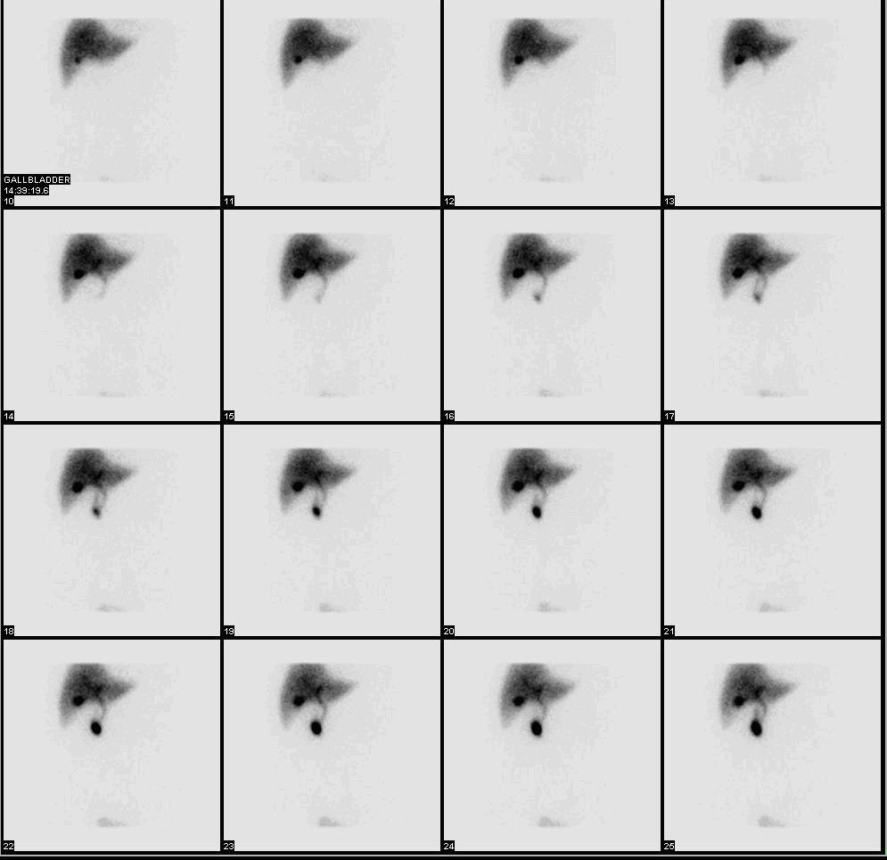
Cholescintigraphy or hepatobiliary scintigraphy is scintigraphy of the hepatobiliary tract, including the gallbladder and bile ducts. The image produced by this type of medical imaging, called a cholescintigram, is also known by other names depending on which radiotracer is used, such as HIDA scan, PIPIDA scan, DISIDA scan, or BrIDA scan.[1][2] Cholescintigraphic scanning is a nuclear medicine procedure to evaluate the health and function of the gallbladder and biliary system. A radioactive tracer is injected through any accessible vein and then allowed to circulate to the liver, where it is excreted into the bile ducts and stored by the gallbladder[3] until released into the duodenum.

Use of cholescintigraphic scans as a first-line form of imaging varies depending on indication. For example for cholecystitis, cheaper and less invasive ultrasound imaging may be preferred,[4] while for bile reflux cholescintigraphy may be the first choice.[5]

Etymology and pronunciation

[edit]

The word cholescintigraphy (/ˌkliˌsɪnˈtɪɡrəfi/) uses combining forms of chole- + scinti(llation) + -graphy, most literally "bile + flash + recording".

Medical use

[edit]

In the absence of gallbladder disease, the gallbladder is visualized within 1 hour of the injection of the radioactive tracer.[citation needed]

If the gallbladder is not visualized within 4 hours after the injection, this indicates either cholecystitis or cystic duct obstruction, such as by cholelithiasis (gallstone formation).[6]

Cholecystitis

[edit]

The investigation is usually conducted after an ultrasonographic examination of the abdominal right upper quadrant for a patient presenting with abdominal pain. If the noninvasive ultrasound examination fails to demonstrate gallstones, or other obstruction to the gallbladder or biliary tree, in an attempt to establish a cause of right upper quadrant pain, a cholescintigraphic scan can be performed as a more sensitive and specific test.[citation needed]

Cholescintigraphy for acute cholecystitis has sensitivity of 97%, specificity of 94%.[7] Several investigators have found the sensitivity being consistently higher than 90% though specificity has varied from 73–99%, yet compared to ultrasonography, cholescintigraphy has proven to be superior.[8] The scan is also important to differentiate between neonatal hepatitis and biliary atresia, because an early surgical intervention in form of Kasai portoenterostomy or hepatoportoenterostomy can save the life of the baby as the chance of a successful operation after 3 months seriously decreases.[9]

Biliary dyskinesia

[edit]

Cholescintigraphy is also used in diagnosis of the biliary dyskinesia.

Radiotracers

[edit]

Most radiotracers for cholescintigraphy are metal complexes of iminodiacetic acid (IDA) with a radionuclide, usually technetium-99m. This metastable isotope has a half-life of 6 hours, so batches of radiotracer must be prepared as needed using a moly cow. A widely recognized trade name for the preparation kits is TechneScan. These radiopharmaceuticals include the following:[10][11]

Nonproprietary drug name (USP format) Common chemical name Acronym Comments
technetium Tc 99m lidofenin hepatobiliary iminodiacetic acid;[12] dimethyl-iminodiacetic acid[10] HIDA An early and widely used tracer; not used as much anymore, as others have progressively replaced it,[13][6] but the term "HIDA scan" is sometimes used even when another tracer was involved, being treated as a catch-all synonym.
technetium Tc 99m iprofenin paraisopropyl-iminodiacetic acid[10] PIPIDA
technetium Tc 99m disofenin diisopropyl-iminodiacetic acid[10] DISIDA
technetium Tc 99m mebrofenin trimethylbromo-iminodiaceticacid[12] BrIDA
  diethyl-iminodiacetic acid[10] EIDA Seems to have been a laboratory tracer but never widely used clinically
  parabutyl-iminodiacetic acid[10] PBIDA Seems to have been a laboratory tracer but never widely used clinically
    BIDA[14] Seems to have been a laboratory tracer but never widely used clinically
    DIDA[14] Seems to have been a laboratory tracer but never widely used clinically

References

[edit]
Revisions and contributorsEdit on WikipediaRead on Wikipedia
from Grokipedia
Cholescintigraphy, also known as hepatobiliary (HIDA) scan or hepatobiliary , is a imaging procedure that employs a to assess the function and patency of the liver, , ducts, and occasionally the by tracking the tracer's pathway through the biliary system. The technique follows the metabolic pathway of , allowing visualization of uptake, biliary excretion, and gallbladder contraction. This procedure is primarily indicated for diagnosing conditions such as acute and chronic , bile duct obstructions, biliary leaks, in infants, and postoperative complications following biliary surgery or . It also evaluates gallbladder to identify , where impaired gallbladder emptying may warrant surgical intervention like . Contraindications include severe to the radiotracer, and relative precautions apply to patients on medications like opioids that can alter biliary . During the procedure, a technetium-99m-labeled derivative (such as mebrofenin) is injected intravenously, followed by serial imaging starting immediately and continuing for up to 60 minutes, with additional views or pharmacologic aids like or cholecystokinin if needed to enhance diagnostic accuracy. Preparation typically involves for at least four hours to promote filling, and the test carries minimal risks, including low comparable to a and rare allergic reactions. Cholescintigraphy offers advantages over by detecting obstruction earlier and providing functional data on biliary dynamics, with high for pathologies.

Background

Definition and purpose

Cholescintigraphy, also known as hepatobiliary scintigraphy or hepatobiliary iminodiacetic acid (HIDA) scan, is a imaging procedure that employs radiotracers to evaluate the and of the liver, , ducts, and . This technique tracks the movement of the radiotracer through the hepatobiliary system using a , providing dynamic images of production, storage, and excretion. The primary purposes of cholescintigraphy include assessing biliary patency, measuring , evaluating bile flow dynamics, and detecting leaks or obstructions within the . It enables quantification of functional parameters, such as the rate of transit from the liver to the , which is essential for diagnosing disorders affecting hepatobiliary motility and integrity. In contrast to anatomical imaging methods like or computed tomography (CT), which focus on structural details such as gallstones or ductal dilation, cholescintigraphy emphasizes physiological function, revealing abnormalities in handling that may elude morphological assessments. Furthermore, it indirectly appraises liver function through the observation of radiotracer uptake by hepatocytes and subsequent biliary , serving as a marker of hepatic synthetic and excretory capacity.

Etymology and history

The term cholescintigraphy derives from the Greek root "chole-," meaning bile, combined with "scintigraphy," a nuclear medicine imaging method based on the Latin scintilla for spark, referring to the detection of gamma ray emissions through scintillation. It is pronounced /ˌkoʊlɪsɪnˈtɪɡrəfi/. A common synonym is the "HIDA scan," an acronym for hepatic iminodiacetic acid, the initial radiotracer employed in this procedure. The origins of hepatobiliary imaging trace back to 1923, when , a halogenated fluorescein , was first utilized for assessing liver function through its hepatic uptake and biliary . This non-radioactive method laid the groundwork for later diagnostic approaches but was limited by the need for invasive sampling and lack of real-time visualization. In the 1950s and 1960s, was labeled with to enable scintigraphic imaging, marking an early shift toward nuclear techniques for evaluating biliary patency and liver function. A major advancement occurred in the 1970s with the development of technetium-99m-labeled (IDA) tracers, originally synthesized as agents due to structural similarities with lidocaine but repurposed after demonstrating hepatobiliary clearance. The first such agent, Tc-99m lidofenin (HIDA), was approved for clinical use by the U.S. in 1982, enabling safer, higher-resolution dynamic imaging of flow. This introduction of dynamic represented a key milestone, allowing real-time functional assessment of the hepatobiliary system beyond static anatomy. In the 1980s, IDA analogs such as disofenin (DISIDA) and later mebrofenin were developed to address limitations of earlier agents, particularly their reduced efficacy in patients with due to lower hepatic extraction fractions. These improved tracers offered higher liver uptake and faster biliary excretion even in elevated levels, leading to the replacement of original HIDA formulations and enhancing diagnostic reliability in complex cases.

Procedure

Patient preparation

Patients undergoing cholescintigraphy are typically required to fast for 4 to 6 hours prior to the procedure to allow for adequate filling, though clear liquids may be permitted in some cases. Exceptions to apply for neonates, who may require specific pretreatments such as , and for critically ill patients, where the procedure may proceed without to expedite . Certain medications that can affect biliary motility, such as opioids (e.g., or ), should be withheld for at least 6 hours before the scan if clinically feasible, and patients are instructed to inform their healthcare provider about all current medications, vitamins, and supplements. Additionally, recent contrast studies involving should be avoided for 48 hours prior, as they may interfere with visualization. Patient instructions emphasize hydration unless contraindicated, removal of jewelry and metal objects, and screening for allergies to potential adjunct agents like or cholecystokinin (CCK). Patients are advised to wear comfortable clothing and may need to change into a . For special populations, pregnant individuals should inform their provider, as the procedure is generally avoided due to risks to the unless benefits outweigh potential harm. Breastfeeding patients must notify their team; milk should be pumped and discarded for 24 hours post-procedure, with alternative feeding arranged. In cases of severe , preparation may include assessment for delayed clearance, but adjustments are individualized.

Imaging protocol

The imaging protocol for cholescintigraphy begins with the intravenous administration of a (Tc-99m)-labeled hepatobiliary agent, such as disofenin or mebrofenin, at a dose of 3-5 mCi (111-185 MBq) for adults, with pediatric dosing scaled by weight (e.g., 0.05 mCi/kg minimum 0.5 mCi). The injection is performed as a rapid bolus via an indwelling , followed immediately by dynamic to capture initial liver uptake. Image acquisition proceeds in phases using a large-field-of-view gamma camera equipped with a low-energy all-purpose or high-resolution collimator. The initial hepatic phase involves serial anterior dynamic images from 0-60 minutes post-injection, typically as 60 one-minute frames in a 128x128 or 256x256 matrix, to assess liver uptake, biliary excretion, and gallbladder visualization, which should occur by 30-60 minutes in normal cases. Additional views, such as left anterior oblique and right lateral, are obtained at 60 minutes to confirm intestinal activity and evaluate the biliary tree. In complex cases, SPECT/CT integration may be employed for anatomical correlation, using a dual-head camera with low-dose non-contrast CT and 140 keV energy window. Adjunct maneuvers are incorporated as needed to enhance diagnostic yield. Delayed imaging is performed up to 4 hours (or 18-24 hours for suspected leaks) if filling or intestinal activity is absent. sulfate (0.04 mg/kg IV over 2-3 minutes, maximum 2-3 mg) may be administered if the is not visualized by 60 minutes, to increase biliary pressure and promote filling within 30 minutes. For assessment in functional studies, cholecystokinin (CCK, sincalide) is infused at 0.02-0.04 mcg/kg IV over 30-60 minutes, with imaging continued for 30-60 minutes post-infusion to measure contraction. The procedure typically lasts 1-2 hours under standard conditions, though it may extend to 4 hours with adjuncts or up to 24 hours for delayed evaluations in cases like bile leaks. Patients are instructed to remain and still during acquisition, with normal breathing to minimize motion artifacts.

Radiotracers

Common agents

The primary radiotracers used in cholescintigraphy are (Tc-99m)-labeled (IDA) analogs, which are hepatobiliary agents designed for efficient hepatic uptake and biliary excretion.01856-6/fulltext) The most commonly employed agents today include Tc-99m-mebrofenin (also known as Choletec) and Tc-99m-disofenin (commonly referred to as DISIDA or Hepatolite), both of which are FDA-approved for hepatobiliary imaging. Tc-99m-mebrofenin exhibits high hepatic extraction efficiency of approximately 98%, making it particularly suitable for patients with compromised liver function, while Tc-99m-disofenin has an extraction efficiency of about 88% and is favored for routine cases due to its established performance and lower cost. An older agent, Tc-99m-lidofenin (the original HIDA compound), is now less commonly used owing to the superior properties of newer analogs. Dosimetry for these agents is standardized to minimize while ensuring adequate quality. For adults, the typical intravenous dose ranges from 111 to 185 MBq (3 to 5 mCi), with adjustments based on patient weight and serum levels; higher doses up to 370 MBq (10 mCi) may be used in severe hyperbilirubinemia to compensate for increased renal clearance. In pediatric patients, dosing is scaled by body weight, typically at 7.4 MBq/kg (200 μCi/kg) with a minimum of 37 MBq (1 mCi), to account for smaller organ sizes and faster clearance. These radiotracers are prepared on-site from commercial containing the lyophilized , which is reconstituted by adding sterile Tc-99m-pertechnetate and allowing labeling to occur at . The have a of up to 12 months when unopened and stored properly, and the labeled product remains stable for 18 hours post-reconstitution for Tc-99m-mebrofenin, with similar stability for Tc-99m-disofenin, after which radiochemical purity may decline due to Tc-99m decay and potential dissociation. Selection of the agent depends on clinical context, particularly liver function and levels. Tc-99m-mebrofenin is preferred when serum exceeds 20 mg/dL or in cases of moderate to severe hepatic dysfunction, as its higher extraction fraction ensures better hepatobiliary visualization despite by . In contrast, Tc-99m-disofenin is selected for standard gallbladder ejection fraction studies or uncomplicated acute evaluations, where its adequate extraction and proven efficacy suffice without the need for higher dosing.

Mechanism and pharmacokinetics

In hepatobiliary scintigraphy, radiotracers such as technetium-99m-labeled (IDA) derivatives are primarily taken up by hepatocytes via organic anion-transporting polypeptides (OATPs), particularly OATP1B1 and OATP1B3, which facilitate the carrier-mediated across the sinusoidal in a manner analogous to bilirubin uptake. This process exhibits high efficiency, with hepatic extraction fractions reaching 90-95% for ideal agents like Tc-99m mebrofenin in healthy individuals, reflecting near-complete first-pass clearance from the bloodstream. Following uptake, the radiotracers undergo minimal intracellular processing within hepatocytes, as they are not conjugated like but are directly transported to the canalicular membrane and excreted into canaliculi via efflux transporters such as (MRP2). This excretion propels the tracer through the biliary tree, with visualization of the and typically occurring within 60 minutes in normal patients, indicating efficient hepatobiliary transit. Renal clearance remains low in healthy individuals, accounting for less than 10% of the injected dose, primarily due to tight binding to plasma proteins that limits glomerular filtration. Pharmacokinetic variations arise in pathological states; hepatocellular dysfunction impairs uptake, reducing the extraction fraction and prolonging plasma , while elevates competing levels, further delaying clearance and . In contexts, the hepatic extraction fraction (HEF) can be quantified to assess liver function, using the formula: HEF=(1activity in blood poolactivity in liver)×100\text{HEF} = \left(1 - \frac{\text{activity in blood pool}}{\text{activity in liver}}\right) \times 100 This simplified metric, derived from region-of-interest analysis of early imaging data, helps differentiate parenchymal from obstructive disease but requires techniques for precision.

Clinical applications

Cholescintigraphy has a broad range of clinical applications beyond disorders, including evaluation of patency for obstructions, detection of biliary leaks (e.g., post-cholecystectomy or trauma), of in infants, and assessment of postoperative complications after biliary surgery or . These uses leverage the tracer's pathway to identify functional or structural abnormalities in the biliary system.

Acute cholecystitis

Cholescintigraphy is indicated for the diagnosis of suspected acute calculous or acalculous , particularly when findings are equivocal or nondiagnostic. In critically ill patients, such as those in intensive care units with suspected acalculous , it serves as a valuable adjunct to due to the latter's limitations in this population, where clinical signs like fever and are often nonspecific. The procedure is especially useful in confirming obstruction, the underlying of acute . The primary diagnostic criterion is non-visualization of the gallbladder within 60 minutes after radiotracer injection, provided hepatic hilar activity is visualized to confirm adequate tracer uptake and biliary excretion. This finding demonstrates a sensitivity of 96% (95% CI: 94%-97%) and specificity of 90% (95% CI: 86%-93%), making cholescintigraphy the most accurate imaging modality for acute cholecystitis compared to ultrasound (sensitivity 81%, specificity 83%) or MRI (sensitivity 85%, specificity 81%). Supporting findings include the rim sign, characterized by increased pericholecystic hepatic activity, which has a 94% positive predictive value for acute cholecystitis and is more frequent in gangrenous cases (57%). Visualization of the bile ducts without gallbladder filling further supports the diagnosis by indicating cystic duct obstruction. To enhance diagnostic performance, augmentation is employed if the is not visualized by 60 minutes; a low-dose intravenous infusion (0.04 mg/kg) promotes contraction, reducing the need for prolonged delayed imaging. This approach improves specificity to 100% and overall accuracy to 98%, compared to 83% specificity and 88% accuracy with conventional protocols, while maintaining sensitivity at 96%. augmentation is superior to delayed imaging alone, with higher positive predictive value and fewer false positives. Positive cholescintigraphy results guide timely surgical interventions, such as , in patients with confirmed acute . False negatives are uncommon (approximately 5%), but can occur in cases of acute superimposed on severe chronic disease, where gallbladder visualization may still happen despite inflammation. In acalculous among critically ill patients, sensitivity may be lower (67%-100%), emphasizing the need for clinical correlation.

Biliary dyskinesia and functional disorders

Cholescintigraphy plays a key role in evaluating patients with chronic biliary pain in the absence of gallstones, serving as the primary diagnostic tool for and dysfunction. These conditions are characterized by impaired motility of the or biliary sphincter, leading to recurrent right upper quadrant pain without structural abnormalities on or other imaging. The test is particularly indicated when symptoms align with functional biliary disorders, such as those meeting Rome criteria for biliary pain, to assess dynamic function rather than . Quantitative assessment of is performed using cholecystokinin (CCK) stimulation during cholescintigraphy, with the (EF) as the central metric. The protocol begins with baseline dynamic following radiotracer injection to visualize tracer uptake in the , followed by intravenous CCK infusion to stimulate contraction. Serial images are acquired over 30-60 minutes post-infusion, with regions of interest drawn over the to obtain counts for EF calculation. The EF is computed using the : EF=(max countspost-CCK counts)max counts×100EF = \frac{(max\ counts - post\text{-}CCK\ counts)}{max\ counts} \times 100 where max counts represent peak gallbladder activity before stimulation, and post-CCK counts reflect the nadir after contraction. A normal EF exceeds 35-40% at 30 minutes post-CCK, indicating adequate gallbladder emptying. Diagnostic thresholds focus on impaired function: an EF below 35% signifies biliary dyskinesia due to poor gallbladder contractility, while prolonged transit time from the hepatic hilum to the duodenum (typically exceeding 9-10 minutes) suggests sphincter of Oddi dysfunction by indicating delayed biliary flow without obstruction. These measures help differentiate functional from mechanical issues, with low EF correlating to histologic findings of chronic cholecystitis in acalculous gallbladders. For sphincter evaluation, quantitative time-activity curves from regions over the biliary tree enhance specificity. The clinical utility of cholescintigraphy lies in its ability to predict symptom relief following in cases, with studies reporting 70-90% success rates for pain resolution in patients with low EF. This predictive value guides surgical decision-making, as abnormal results are associated with favorable outcomes post-, particularly when symptoms are typical. For dysfunction, the test serves as a non-invasive screening tool prior to more invasive manometry, aiding in patient selection for sphincterotomy. Overall, these functional insights improve management of syndromes unresponsive to conservative therapy.

Image interpretation

Normal patterns

In a standard cholescintigraphy procedure, the radiotracer exhibits rapid hepatic uptake, beginning immediately after intravenous injection and achieving uniform distribution throughout the liver within 1 to 5 minutes, with prompt clearance of any initial cardiac blood pool activity. This phase confirms intact hepatocellular function and even tracer handling across the liver lobes. Excretion into the biliary tree follows swiftly, with visualization of the intrahepatic and extrahepatic bile ducts typically occurring between 10 and 30 minutes post-injection, reflecting efficient hepatobiliary transport. The then fills promptly, often becoming apparent by 20 to 60 minutes and usually well visualized by 30 to 40 minutes, demonstrating smooth contour and no evidence of distortion or incomplete filling. Intestinal activity appears in the and proximal small bowel typically within 60 minutes (though delays beyond this can occur in up to 20% of normal individuals), signifying unobstructed transit from the . Key visual criteria for normal patterns include homogeneous radiotracer distribution in the liver without photopenic or hyperintense focal defects, sequential progression of activity from liver to bile ducts, , and bowel, and absence of persistent hepatic retention beyond the early phases. These features establish a baseline for expected biodistribution in individuals with preserved liver and biliary function. Quantitative norms reinforce these observations: visualization occurs in virtually all normal studies by 1 hour, while bile duct transit to the small bowel typically completes within 60 minutes, with the typically peaking in activity before 20 to 30 minutes. Such metrics, derived from standardized protocols, provide thresholds for distinguishing physiologic from pathologic delays. Normal patterns can vary slightly in certain physiologic states; for instance, elderly patients may show modestly slower hepatic uptake and biliary excretion due to age-related reductions in liver , while recent food intake (postprandial state) can inhibit filling unless a period of at least 4 hours precedes the study. Nonetheless, these variations remain within acceptable limits when patient preparation guidelines are followed, ensuring reliable interpretation.

Abnormal findings

Abnormal findings in cholescintigraphy manifest as deviations in radiotracer distribution, transit, and clearance, signaling disruptions in hepatobiliary function. These patterns are identified through dynamic sequences, often requiring delayed acquisitions up to 24 hours for confirmation. Persistent hepatic uptake without biliary represents a critical abnormality, where the liver demonstrates normal initial radiotracer accumulation but fails to release it into the biliary tree, indicating severe parenchymal dysfunction or downstream blockage that impairs clearance. Absent bowel activity further underscores complete obstruction at the level, as no tracer reaches the even on extended , distinguishing it from partial delays. Extrahepatic abnormalities involve ectopic or stagnant tracer accumulation outside the expected biliary pathways. Tracer pooling within the , characterized by prolonged retention without prompt emptying, suggests impaired contractility or outflow resistance, often prompting further functional assessment. Free peritoneal activity appears as diffuse or localized extrabiliary collections, typically in the gallbladder fossa or perihepatic regions, signifying a breach in biliary such as a leak, which may necessitate SPECT/CT for precise localization and quantification. Delayed or variant patterns reflect temporal disruptions in tracer progression. spasm presents as postponed entry of tracer into the , with biliary activity visible but intestinal visualization lagging beyond the typical 60-minute timeframe, potentially mimicking obstruction if not correlated with clinical factors like recent use. Hepatic dysfunction, conversely, is evident from poor initial parenchymal uptake, where blood pool clearance is sluggish (>5-10 minutes) due to reduced extraction efficiency, often requiring alternative imaging agents or prolonged protocols. Quantitative abnormalities provide measurable insights into functional deficits. High residual gallbladder activity following cholecystokinin (CCK) administration, yielding an below 38%, indicates defective emptying and supports evaluation of motility disorders. Compartmental , applied to leak scenarios, quantifies the extent of extravasation by modeling tracer distribution across regions, aiding in assessing leak severity and guiding intervention without relying solely on qualitative visuals.

Safety considerations

Radiation risks

Cholescintigraphy involves the administration of technetium-99m-labeled hepatobiliary agents, typically at 111-296 MBq (3-8 mCi) for adults, resulting in an effective radiation dose of approximately 3-5 mSv for a standard planar imaging study. This dose is comparable to that from an abdominal CT scan, which averages 5-10 mSv, and represents about one year's worth of natural background radiation exposure (around 3 mSv annually). When single-photon emission computed tomography (SPECT) is incorporated, the effective dose increases to up to 7 mSv due to the additional imaging acquisition and potential hybrid SPECT/CT components. Organ-specific absorbed doses are highest to the gallbladder wall, estimated at 20-30 mGy for a typical adult dose, reflecting its role as the primary site of radiotracer accumulation in normal studies. The liver receives 5-10 mGy, as it serves as the initial uptake and excretion site for the agent, while other organs such as the intestines and receive lower doses (e.g., 10-20 mGy to the upper wall). These absorbed doses contribute to a low stochastic risk profile, with the probability of estimated at approximately 1 in 5,000 for a standard procedure, based on linear no-threshold models that extrapolate from higher-dose epidemiological data. In pediatric patients, administered activities are reduced according to the ALARA (as low as reasonably achievable) principle, typically to 1-2 mCi scaled by body weight (e.g., 1.8 MBq/kg with a minimum of 18.5 MBq), yielding an effective dose of less than 2 mSv even in older children. This dose adjustment minimizes exposure in more radiosensitive populations, prioritizing image quality sufficient for diagnosis while adhering to pediatric guidelines from organizations like the Society of Nuclear Medicine and Molecular Imaging. Long-term risks from cholescintigraphy are minimal, with negligible carcinogenic potential at diagnostic levels below 100 mSv, as supported by extensive reviews of procedures showing no observable increase in cancer incidence from single low-dose exposures. Deterministic effects, such as tissue damage, do not occur at these doses, which are far below thresholds (typically >1 Gy) for such outcomes.

Contraindications and complications

Cholescintigraphy has few absolute contraindications. The primary absolute is a history of severe anaphylactic reaction to the radiotracer, such as technetium-99m-labeled derivatives (e.g., disofenin or mebrofenin). to hepatobiliary compounds is also contraindicated, as it may lead to allergic responses. Relative contraindications include recent use of opiates, which should be withheld for at least 6 hours prior to the procedure due to their potential to increase tone and mimic biliary obstruction. Other medications, such as atropine or benzodiazepines, may interfere with contractility and should be avoided if possible. For procedures involving cholecystokinin (CCK or sincalide) infusion to assess ejection fraction, additional contraindications apply, including known allergy to sincalide, intestinal obstruction, and , as sincalide may stimulate preterm labor. In pregnant patients, the procedure should be performed only if benefits outweigh fetal risks, and non-radiation alternatives like are preferred when possible; the radiotracer crosses the with an estimated fetal dose of 0.017 mGy/MBq in early . No interruption of is required after administration of technetium-99m- derivatives. Complications from cholescintigraphy are rare and generally mild. The most common adverse effects include bruising or discomfort at the intravenous injection site of the radiotracer. Allergic reactions to the radiotracer occur infrequently but can range from mild urticaria to severe in sensitized individuals. When is used to augment biliary visualization in cases of suspected , it carries risks such as respiratory depression in non-ventilated patients, contraindication in those with increased (particularly children), allergy, or relative in . Sincalide administration for functional assessment can cause transient abdominal cramping or , especially with rapid infusions (e.g., over 3 minutes, affecting 48-53% of patients), though these effects are minimized with slower infusion protocols (e.g., over 60 minutes). The procedure does not precipitate or choledocholithiasis.

References

Add your contribution
Related Hubs
User Avatar
No comments yet.