Hubbry Logo
search
logo
1740420

Capitulum of the humerus

logo
Community Hub0 Subscribers
Read side by side
from Wikipedia
Capitulum of the humerus
Left humerus seen from front (capitulum visible at bottom right)
Left humerus seen from front (part of the appendicular skeleton)
Details
Identifiers
Latincapitulum humeri
TA98A02.4.04.022
TA21202
FMA23373
Anatomical terms of bone

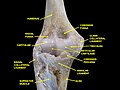
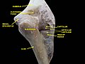
In human anatomy of the arm, the capitulum of the humerus is a smooth, rounded eminence on the lateral portion of the distal articular surface of the humerus. It articulates with the cup-shaped depression on the head of the radius, and is limited to the front and lower part of the bone.

In non-human tetrapods, the name capitellum is generally used, with "capitulum" limited to the anteroventral articular facet of the rib (in archosauromorphs).

Lepidosauromorpha

[edit]

Lepidosaurs show a distinct capitellum and trochlea on the centre of the ventral (anterior in upright taxa) surface of the humerus at the distal end.

Archosauromorpha

[edit]

In non-avian archosaurs, including crocodiles, the capitellum and the trochlea are no longer bordered by distinct etc.- and entepicondyles respectively, and the distal humerus consists two gently expanded condyles, one lateral and one medial, separated by a shallow groove and a supinator process. Romer (1976) homologizes the capitellum in Archosauromorphs with the groove separating the medial and lateral condyles.

In birds, where forelimb anatomy has an adaptation for flight, its functional if not[1] ontogenetic equivalent is the dorsal condyle of the humerus.

Additional images

[edit]

References

[edit]
[edit]
Revisions and contributorsEdit on WikipediaRead on Wikipedia
from Grokipedia
The capitulum of the humerus, also known as the capitellum, is a smooth, rounded eminence forming the lateral portion of the distal articular surface of the humerus, which articulates with the head of the radius to create the humeroradial joint as part of the elbow complex.[1][2] Located on the inferolateral aspect of the humeral condyle, the capitulum presents a convex, knob-like projection that covers the anterior and inferior surfaces of the bone but spares the posterior aspect, and it is separated from the adjacent medial trochlea by a shallow groove.[3][4] This structure facilitates forearm flexion and extension by enabling the radius to pivot smoothly against the humerus during elbow motion, contributing to overall joint stability and the transmission of forces in the upper limb.[1] Above the capitulum lies the radial fossa, a shallow depression that accommodates the radial head during full flexion of the forearm.[1] Clinically, the capitulum is significant due to its vulnerability to intra-articular fractures, which often result from high-energy trauma such as falls on an outstretched hand or direct impacts, particularly affecting adolescents and young adults whose ossification centers are developing.[5] These fractures, classified into types based on fragment size and displacement (e.g., Hahn-Steinthal or Kocher-Lorenz patterns), can lead to complications like avascular necrosis, nonunion, or elbow stiffness if not managed with timely surgical intervention such as open reduction and internal fixation.[5][6] As the first ossification center in the distal humerus—typically appearing between ages 1 and 2—the capitulum serves as a key radiographic landmark for assessing skeletal maturity and diagnosing pediatric elbow injuries.[7]

Overview and Definition

Structure and Location

The capitulum of the humerus is a smooth, rounded eminence situated on the lateral portion of the distal articular surface of the humerus, forming a convex projection that contributes to the overall condylar structure.[8] In tetrapods, this feature is consistently positioned on the anterior aspect of the distal humerus, providing a key component of the elbow region's bony architecture.[9] In humans, the capitulum forms part of the humeral condyle and is oriented anteriorly and inferiorly at approximately 30 degrees relative to the humeral shaft, with a diameter of about 1-2 cm in adults.[10] Medially, it is adjacent to the trochlea, from which it is separated by a shallow groove that delineates the two articular surfaces. Proximally, the capitulum is continuous with the radial fossa, a depression on the anterior humerus.[3] Anatomically, the capitulum overlies the radial fossa anteriorly, accommodating the proximal radius during joint flexion, and lies medial to the lateral epicondyle, which serves as an attachment site for forearm muscles.[8]

Etymology and Terminology

The term capitulum originates from the Latin capitulum, a diminutive form of caput meaning "head," thereby denoting a "small head." This etymology directly reflects the anatomical feature's characteristic rounded, knob-like prominence on the distal humerus, evoking the image of a diminutive cranial structure.[11][12][13] In anatomical nomenclature, capitulum humeri is the preferred Latin term, with the synonymous capitellum employed interchangeably in various texts to describe the same structure.[11][10] The structure is codified in modern standardized systems, including the Terminologia Anatomica (TA98 identifier: A02.4.04.022; TA2 identifier: 1202) and the Foundational Model of Anatomy (FMA identifier: 23373), ensuring consistent usage across international anatomical references.[14] Historically, the capitulum was detailed as a component of the distal humerus in Gray's Anatomy (20th edition, 1918), where it is portrayed as the lateral, rounded eminence articulating with the radius.[15] Earlier allusions trace to 16th-century Renaissance anatomy, notably in Andreas Vesalius' De humani corporis fabrica (1543), which advanced descriptive precision for humeral features within the classical Latin tradition.[16] To avoid confusion, the humeral capitulum must be differentiated from homonymous terms in other contexts, as well as extraneous applications like the botanical capitulum (a compact flower head) or liturgical capitulum (a short scriptural reading).[13][11]

Anatomy in Mammals

Human Anatomy

In human anatomy, the capitulum of the humerus, also known as the capitellum, is a smooth, rounded eminence forming the lateral portion of the distal humerus's articular surface. It measures approximately 15-20 mm in anteroposterior diameter in adults, with studies reporting average widths of about 15.55 mm and depths around 9 mm, contributing to its role in precise articulation without delving into functional aspects. Ossification begins from a secondary center that appears between 1 and 2 years of age, typically around 1 year, and fuses with the humeral shaft by 12-14 years, though fusion can occur as early as 10-12 years in some cases.[17][18][10] The vascular supply to the capitulum primarily arises from perforating vessels of the recurrent radial artery, with contributions from the radial collateral and interosseous recurrent arteries entering posteriorly, ensuring nutrient delivery to the epiphyseal region. Innervation is provided by branches of the radial nerve, which supply the surrounding lateral epicondylar region and periarticular tissues. Gender variations show the capitulum to be slightly larger in males, consistent with overall distal humeral dimensions being more prominent in males across multiple morphometric parameters. With age, the structure exhibits minimal atrophy, maintaining relative stability into adulthood, though it remains susceptible to osteochondritis dissecans in adolescents, particularly affecting the dominant arm in active individuals.[12][19][9][20] On imaging, the capitulum appears as a rounded osseous density on lateral radiographs of the elbow, best visualized in true lateral projections where it projects anteriorly relative to the humeral shaft. Magnetic resonance imaging (MRI) reveals its coverage by hyaline articular cartilage, typically 1-2 mm thick, with T2-weighted sequences highlighting the cartilage's high signal intensity against the low-signal bone, aiding in assessment of surface integrity.[12][21][22]

Comparative Features in Other Mammals

In quadrupedal mammals such as dogs, the capitulum of the humerus is typically more elongated and cylindrical compared to humans, facilitating greater weight-bearing stability during terrestrial locomotion. This morphology supports a stable hinge joint at the elbow, with the capitulum articulating less prominently with the radius to emphasize load distribution across the forelimb.[23][24][25] Among non-human primates like chimpanzees, the capitulum exhibits a more globular shape akin to that in humans, but with enhanced sphericity and smoother contours that permit greater radial mobility and pronation-supination for arboreal and manipulative activities. This adaptation reflects the demands of suspensory and climbing behaviors, where the capitulum's rounded form allows for increased forearm rotation without compromising joint integrity. Three-dimensional analyses confirm that the capitulum in great apes, including chimpanzees, has a larger articular area and more acute angles relative to cercopithecoids, correlating with orthograde postures.[25][26][27] In aquatic mammals such as whales, the capitulum is markedly reduced and often flattened or rudimentary, reflecting the evolution of flipper-like forelimbs with minimal elbow mobility. The humerus in cetaceans is short and robust, with the capitulum and associated structures fused to the radius and ulna, eliminating distinct articulations and prioritizing hydrodynamic stability over terrestrial function. This vestigial form aligns with the loss of medullar cavity and overall forelimb simplification in fully aquatic lifestyles.[28][29][25] Arboreal species like squirrels display a prominent capitulum that is small yet rounded and well-developed for enhanced rotation and flexibility in climbing. The humerus in these rodents is long and slender, with the capitulum supporting agile pronation-supination to navigate branching substrates, distinguishing it from more rigid forms in ground-dwelling relatives. Morphometric studies highlight how this capitular prominence scales with locomotor ecology, promoting rotational freedom in tree-dwelling habits.[25][30][31] Across mammals, capitulum size generally scales positively with body mass, as seen in comparative analyses of humeral geometry where larger species exhibit proportionally broader articular surfaces for load-bearing efficiency.[32][33][34]

Function and Biomechanics

Articulation with Radius

The humeroradial joint is formed by the articulation between the rounded capitulum on the lateral aspect of the distal humerus and the fovea of the head of the radius, creating a synovial hinge joint with ball-and-socket-like characteristics that facilitates forearm flexion-extension and rotation. This joint is part of the broader elbow complex and contributes to the overall stability and mobility of the proximal forearm. The convex curvature of the capitulum precisely matches the shallow concavity of the radial head's fovea, ensuring congruent contact during motion.[35][36][37] The articulating surfaces are both covered by a layer of hyaline articular cartilage, which varies in thickness from approximately 0.4 to 1.8 mm across the distal humerus, providing a smooth, low-friction interface. This cartilage layer on the capitulum and radial head helps distribute compressive forces and absorb shock during joint loading. The synovial membrane lining the joint cavity secretes synovial fluid, a viscous lubricant rich in hyaluronic acid, which further reduces friction and nourishes the avascular cartilage.[38][37] Stability of the humeroradial articulation is primarily provided by surrounding ligaments, including the annular ligament, which forms a strong fibrous band encircling about 80% of the radial head and attaching to the anterior and posterior margins of the ulnar radial notch, thereby holding the radial head securely against the capitulum. The radial collateral ligament, originating from the lateral epicondyle of the humerus, blends with the annular ligament and reinforces the lateral aspect of the joint against varus stresses. Additionally, the joint capsule envelops the articulation, being notably thin anteriorly (to allow hinge motion) and thicker laterally where it integrates with the collateral structures for enhanced reinforcement.[39][40][41][42]

Role in Elbow Joint Movements

The capitulum of the humerus plays a pivotal role in facilitating pronation and supination of the forearm by providing a spherical articulation surface for the radial head, allowing rotation around a longitudinal axis. During these movements, the superior surface of the radial head rotates directly against the capitulum, while the ridge of the radial head glides within the groove separating the capitulum from the trochlea, enabling a total range of motion of approximately 160–180 degrees in humans (typically 80 degrees of pronation and 90 degrees of supination from neutral). This pivot-like interaction at the humeroradial joint ensures smooth forearm rotation without significant translation, contributing to the precision required for hand positioning in daily activities.[43][44] In elbow flexion and extension, the capitulum supports the rolling and gliding arthrokinematics of the radial head, which translates along its surface to maintain joint congruence. Flexion, ranging from 0 to approximately 145 degrees, involves an anterior (volar) glide and roll of the radial head on the capitulum, allowing the forearm to approach the humerus while distributing shear forces. Conversely, extension features a posterior (dorsal) glide of the radial head, with rolling prominent at the extremes of motion to prevent excessive tension on surrounding soft tissues. These kinematics complement the primary hinge action at the ulnohumeral joint, enhancing overall elbow stability during sagittal plane movements.[45][46] The capitulum also contributes to load distribution and joint stability in the elbow complex, bearing a significant portion of axial compressive forces through the radiocapitellar articulation. In elbow extension under axial loading, the radiocapitellar joint transmits approximately 60% of the total load, with the remainder borne by the ulnotrochlear joint, which helps prevent overload on the ulna during weight-bearing activities. Additionally, the capitulum aids in resisting varus stress by forming part of the lateral joint articulation, where up to 75% of varus stability at 90 degrees of flexion derives from bony congruence and compressive forces across the radiocapitellar interface, supplemented by the lateral collateral ligament complex.[47][48] Biomechanically, the capitulum's rounded geometry influences torque dynamics around the elbow's lateral axis, where simplified torque calculations (τ=F×d\tau = F \times d)—with τ\tau as torque, FF as applied force, and dd as perpendicular distance from the capitulum's rotation axis—determine stress thresholds for joint integrity. Excessive varus torque, often exceeding 50 Nm in high-impact scenarios, can overwhelm these mechanics, leading to radiocapitellar subluxation by disrupting the radial head's centered position on the capitulum. This underscores the capitulum's role in balancing rotational and compressive loads to safeguard against instability.[47]

Comparative Anatomy in Reptiles

Lepidosauromorpha

In lepidosaurs, which include squamates (lizards and snakes) and rhynchocephalians (tuatara), the capitulum of the humerus forms part of the distal articular surface, characterized by a distinct, rounded capitellum for radial articulation positioned ventrally and preaxially, adjacent to a shallower trochlea for the ulna. These structures are separated by a shallow sulcus, with the capitellum appearing relatively smaller in proportion to the overall humerus compared to more erect-limbed vertebrates, reflecting adaptations to a sprawling posture.[49] In lizards such as the iguana (Iguana iguana), the capitulum supports the sprawling gait typical of lepidosaurs, facilitating lateral undulation and weight-bearing during terrestrial locomotion while permitting only limited humeral rotation at the elbow joint. This configuration allows for protraction and retraction in a primarily horizontal plane, with kinematic studies in species like the central bearded dragon (Pogona vitticeps) showing restricted long-axis rotation to maintain stability during slow walking and faster gaits.[49][50][51] Among snakes, which represent the limbless extreme within Squamata, the humerus and its capitulum are vestigial or entirely absent in adults due to secondary limb reduction, though embryonic remnants may persist briefly before resorption. This loss aligns with the evolution of serpentine locomotion, eliminating the need for forelimb articulations.[52] The capitulum is notably present in basal lepidosaurs like the tuatara (Sphenodon punctatus), where it exhibits a moderately expanded form aiding in lateral flexion of the forelimb, a primitive trait conserved from early rhynchocephalians and contributing to subtle rotational movements during foraging and climbing.[53][49] In these taxa, the articular cartilage at the capitulum enhances durability for terrestrial stresses as described in foundational osteological analyses.[49]

Archosauromorpha

In archosauromorphs, the capitulum of the humerus exhibits modifications adapted to diverse locomotor strategies, particularly in crocodilians and birds as extant representatives, with fossil evidence illuminating variations in extinct forms like dinosaurs. In crocodilians, the distal humerus features distinct capitellar (lateral) and trochlear (medial) condyles separated by a shallow intercondylar groove, which accommodates the radius and ulna during elbow flexion and extension.[54] This configuration supports a semi-erect forelimb posture during terrestrial "high walks" and galloping, enabling greater limb elevation and stability compared to sprawling lepidosaurs, while maintaining amphibious versatility.[55] The groove's shallow depth reflects the elbow's limited rotation, prioritizing robust load-bearing for quadrupedal support in predatory ambushes.[54] In birds, the capitulum is reconfigured into a prominent dorsal condyle equivalent, which articulates with both the radius and ulna to facilitate wing folding essential for perching and powered flight, while the ventral capitulum is notably reduced to minimize mass and drag.[56] This dorsal emphasis allows the antebrachium to fold tightly against the body during rest, enhancing aerodynamic efficiency during soaring and flapping. In raptorial species like eagles, the dorsal condyle is elongated to provide a pivotal articulation that supports precise wing adjustments for hunting dives and maneuvers.[57] The intercondylar incisura between dorsal and ventral portions further aids in constraining motion to protraction and retraction, integrating seamlessly with the carpus for coordinated wing extension in flight—contrasting the more hinge-like mammalian elbow by permitting greater rotational freedom at the wrist.[56] Variations among extinct archosauromorphs, particularly dinosaurs, highlight evolutionary trends toward enhanced forelimb functionality. These adaptations underscore archosauromorph shifts from sprawling to erect postures, optimizing the humerus for aerial and terrestrial innovations absent in mammalian lineages.

Development and Evolution

Embryological Development

The capitulum of the humerus arises from the lateral aspect of the distal humeral chondrification center, which forms during weeks 6-7 of gestation as part of the initial mesenchymal condensations in the developing limb bud.[58] By week 8, this center differentiates further, with the capitellar anlage emerging as a distinct precartilaginous structure within the lateral condyle, coinciding with the recognition of the elbow joint interzone.[58] This early separation allows for the specific shaping of the capitulum as a rounded eminence for radial articulation. Chondrogenesis of the capitulum involves the condensation of mesenchymal cells into a cartilaginous template, regulated by homeobox (Hox) genes that pattern the proximodistal axis of the limb.[59] Specifically, Hoxa11 plays a key role in influencing distal limb patterning and promoting chondrocyte differentiation by acting upstream of transcription factors such as Runx2, ensuring proper cartilage formation in the distal humerus.[60] These genetic controls integrate with signaling pathways like BMP and Ihh to drive the maturation of the cartilaginous anlage.[58] During prenatal growth, the capitellar cartilage undergoes phased expansion, with the hypertrophic zone of chondrocytes enlarging and mineralizing the surrounding matrix to prepare for ossification.[9] This process transitions via endochondral ossification, where the cartilage model is gradually replaced by bone, beginning at birth with the appearance of the capitellar ossification center around 1-12 months postnatally.[9] The hypertrophic zone's expansion thus bridges embryonic cartilage development to postnatal bony maturation. Congenital anomalies such as capitellar aplasia are rare and typically manifest as isolated absences of the structure, potentially leading to elbow instability.[61] These defects have been linked to disruptions in limb patterning pathways during embryogenesis.[61]

Evolutionary Origins and Variations

The capitulum of the humerus first appeared as a small eminence on the distal end of the humerus in early tetrapods during the Late Devonian to Carboniferous fin-to-limb transition, facilitating the evolution of propped postures and limited terrestrial support from aquatic fin-like structures.[62] In Carboniferous and early Permian tetrapods, such as the temnospondyl Eryops megacephalus, the capitulum enlarged significantly, dominating the lateral portion of the distal humerus and enabling greater elbow flexion for weight-bearing on land; for example, in Eryops specimens, the capitulum width measures approximately 33.8 mm relative to a distal humeral width of 93.7 mm.[63] This enlargement represents a key adaptation in the progressive evolution of the elbow joint among temnospondyls, contrasting with the ancestral condition of a short humerus featuring a small capitulum and separate ulnar condyle.[63] By the Permian period, the capitulum was fully present in synapsids, the stem group to mammals, though it remained relatively small and permitted restricted elbow mobility compared to later forms. In basal synapsids such as pelycosaurs (e.g., Dimetrodon), the capitulum is notably reduced, with a width of about 22.5 mm on a humerus of distal width 102 mm, reflecting a transitional morphology adapted to sprawling gaits and partial aquatic habits.[63] Fossil evidence from Permian pelycosaur humeri, including those of eupelycosaurs like Gordodon kraineri, shows variations in distal flattening and capitular prominence, bridging earlier tetrapod designs toward more derived amniote configurations.[64] In therapsids, the mammalian ancestors arising in the late Permian, the capitulum underwent refinement, becoming more rounded and integrated with a twisted humeral shaft to support increasingly parasagittal forelimb postures associated with enhanced metabolic efficiency. This shift paralleled broader postcranial changes in synapsids, where forelimb variability increased dramatically by the late Permian, predating dinosaur dominance and enabling diverse locomotor modes. Within diapsids, the sister clade to synapsids, the capitulum diverged by the Triassic into distinct forms: more pronounced in lepidosauromorphs for flexible, sprawling locomotion, and variably reduced or specialized in archosauromorphs, as seen in early forms like Prolacerta. Across Amniota, the capitulum is a conserved feature, with its morphology tracking ecological adaptations, from the sprawling limbs of basal amniotes to the specialized extremities of derived clades.[65]

Clinical and Pathological Aspects

Fractures and Injuries

Capitellar fractures are uncommon injuries, accounting for less than 1% of all elbow fractures and approximately 6% of distal humeral fractures.[66] They occur in adolescents and adults, typically resulting from high-energy trauma such as falls on an outstretched hand, though low-energy mechanisms can also contribute.[67] In the AO/OTA classification system, these are categorized as partial articular fractures in the coronal plane, specifically type 13-B3.[66] The primary types include coronal shear fractures, often referred to as Hahn-Steinthal fractures, which involve a large osseous fragment with attached subchondral bone; avulsion fractures (Kocher-Lorenz type), characterized by a smaller fragment limited to articular cartilage; and more complex variants such as comminuted (type III) or those extending into the trochlea (type IV McKee).[66] Displacement is defined as greater than 2 mm and typically requires surgical intervention to restore joint congruity.[66] The mechanism of injury generally involves hyperextension of the elbow combined with a varus force, where the radial head shears against the capitellum during axial loading from a fall on the outstretched hand.[66] Approximately 50% of cases are associated with radial head dislocation or subluxation, which complicates stability and necessitates careful assessment.[66] Initial management depends on displacement and fragment size. Nondisplaced fractures with less than 2 mm of separation can be treated conservatively with immobilization in a long-arm cast for 3 to 4 weeks, followed by protected range-of-motion exercises.[66] For displaced fractures, open reduction and internal fixation (ORIF) is the standard approach, using headless compression screws such as Herbert screws to secure capitellar fragments while preserving the subchondral surface and avoiding intra-articular hardware prominence.[66]

Associated Conditions and Imaging

Panner's disease, also known as osteochondrosis of the capitellum, represents avascular necrosis of the capitellar epiphysis primarily affecting children aged 5 to 10 years, often those engaged in throwing sports due to repetitive valgus stress.[68] This self-limiting condition typically resolves spontaneously with conservative management, including rest and activity modification, leading to full recovery without residual deformity in most cases.[69] Osteochondritis dissecans (OCD) of the capitellum is a more common non-traumatic pathology in adolescents, particularly overhead athletes aged 12 to 16 years, resulting from repetitive microtrauma and vascular compromise to the subchondral bone.[70] Lesions most frequently involve the anterolateral aspect of the capitellum, accounting for the majority of cases and causing lateral elbow pain, swelling, and limited extension.[71] Recent advancements as of 2025 include arthroscopic drilling and fixation techniques for stable lesions to promote revascularization.[72] In rheumatoid arthritis, the capitellum is a frequent site of erosive changes due to chronic synovitis, with radiographic erosions commonly observed on the capitellar surface alongside the lateral epicondyle.[73] These erosions contribute to progressive joint destruction and instability, occurring in 20% to 65% of rheumatoid patients with elbow involvement.[73] Tumors of the capitellum are exceedingly rare, comprising less than 1% of primary bone neoplasms, with chondroblastoma serving as a representative example of an epiphyseal benign lesion that can mimic OCD through lytic destruction and pain.[74] Such tumors typically arise in the second decade and require histologic confirmation for diagnosis.[75] Diagnostic imaging plays a crucial role in evaluating capitellar pathologies. Plain radiographs, particularly lateral views, assess alignment and detect joint effusion via the sail sign, where the displaced anterior fat pad appears elevated.[76] Computed tomography (CT) provides detailed evaluation of lesion size and fragment displacement in OCD, with three-dimensional mapping revealing typical surface areas of 50 to 200 mm² in the posterolateral capitellum.[77] Magnetic resonance imaging (MRI) excels in assessing cartilage integrity, using T2-weighted sequences to identify defects greater than 5 mm and subchondral instability through high-signal rims or cysts.[78] Prognosis for capitellar OCD is guided by the Nelson classification, which stages lesions from I (intact cartilage with subchondral changes) to IV (displaced fragment or loose body).[79] Stable stages I and II often heal with nonoperative care, while stage III lesions, indicating partial detachment, typically require drilling to stimulate revascularization and promote union.[80]

References

User Avatar
No comments yet.