Hubbry Logo
Cardinal ligamentCardinal ligamentMain
Open search
Cardinal ligament
Community hub
Cardinal ligament
logo
8 pages, 0 posts
0 subscribers
Be the first to start a discussion here.
Cardinal ligament
Cardinal ligament
from Wikipedia
Cardinal ligament
Vessels of the uterus and its appendages, rear view. (Cardinal ligament not visible, but location can be inferred from position of uterine artery and uterine vein.)
Uterus and right broad ligament, seen from behind. (Cardinal ligament not labeled, but broad ligament visible at center.)
Details
Identifiers
Latinligamentum cardinale, ligamentum transversum cervicis, ligamentum transversalis colli
TA98A09.1.03.031
A09.1.03.022
TA23839
FMA77064
Anatomical terminology

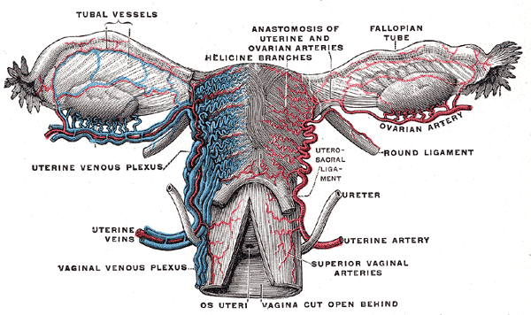
The cardinal ligament (also transverse cervical ligament, lateral cervical ligament,[1] or Mackenrodt's ligament[2][1]) is a major ligament of the uterus formed as a thickening of connective tissue of the base of the broad ligament of the uterus. It extends laterally (on either side) from the cervix and vaginal fornix to attach onto the lateral wall of the pelvis. The female ureter, uterine artery, and inferior hypogastric (nervous) plexus course within the cardinal ligament. The cardinal ligament supports the uterus.[1]

Structure

[edit]

The cardinal ligament is a paired structure on the lateral side of the uterus. It originates from the lateral part of the cervix.[3]

Attachments

[edit]

It attaches the cervix to the lateral pelvic wall by its attachment to the obturator fascia of the obturator internus muscle.[4] It attaches to the uterosacral ligament.[3]

Relations

[edit]

It is continuous externally with the fibrous tissue surrounding the pelvic blood vessels.[4]

Function

[edit]

The cardinal ligament supports the uterus, providing lateral stability to the cervix.[1]

Clinical significance

[edit]

The cardinal ligament may be affected in hysterectomy.[5][6] Due to its proximity to the ureters, it can get damaged during ligation of the ligament. It is routinely cut during some uterine operations, although this can have side effects.[3]

See also

[edit]

References

[edit]
[edit]
Revisions and contributorsEdit on WikipediaRead on Wikipedia
from Grokipedia
The cardinal ligament, also known as the transverse cervical ligament or Mackenrodt's ligament, is a paired structure in the female pelvis composed of connective tissue that provides essential apical support to the uterus, cervix, and upper vagina by attaching these organs to the lateral pelvic walls. It was first described by German gynecologist Alwin Mackenrodt in 1895. Anatomically, the cardinal ligament arises as a thickening of the parametrium and at the base of the broad ligament, extending laterally from the base of the and the lateral to insert on the pelvic sidewall near the ischial spines; it measures approximately 10 cm in length and is subdivided into distal, intermediate, and proximal portions. Unlike true skeletal , it consists primarily of loose areolar containing fibers, fibroblasts, cells, blood vessels (including the ), nerves from the , and lymphatics, with minimal elastic fibers and no bony attachments. Functionally, the cardinal ligaments form a hammock-like network that, in conjunction with the uterosacral ligaments, pubocervical , and musculature, stabilizes the pelvic organs in a vertical orientation and prevents their downward , particularly under intra-abdominal . Clinically, the cardinal ligaments are significant in gynecological procedures such as , where they are divided to access the uterine vessels, though this carries risks of ureteral injury due to the ureters' close proximity as they traverse the ligaments; alterations in their content are also implicated in , and their lymphatic drainage plays a role in the spread of .

Overview

Definition and nomenclature

The cardinal ligament is a paired structure consisting of a thickening of the parametrium and located at the base of the broad ligament. It extends from the base of the , specifically the and upper , to the lateral wall, providing foundational structural support within the female . This ligament is also known by several alternative names, including Mackenrodt's ligament, transverse cervical ligament, and lateral cervical ligament, reflecting variations in anatomical across historical and regional contexts. Its official Latin term is ligamentum cardinale. In its basic role, the cardinal ligament offers primary lateral support to the and , helping to stabilize these structures against descent and maintain pelvic organ positioning. This naming was first introduced by Josef Kocks in 1880.

Historical background

The cardinal was first formally named in 1880 by German surgeon Josef Kocks, who identified it as a key supportive structure extending from the base of the broad ligament to the pelvic sidewall. This initial description highlighted its central position in stabilizing the and upper . Fifteen years later, in 1895, German gynecologist Alwin Mackenrodt provided a detailed anatomical account based on studies of fetal and adult specimens, renaming it the transverse cervical and underscoring its critical role in uterine suspension. Mackenrodt's work established it as a distinct entity, often bearing his thereafter. The nomenclature evolved in the early 20th century, with "cardinal ligament" gaining prominence to emphasize its pivotal, hinge-like function in pelvic mechanics, while "transverse cervical ligament" persisted as a descriptive alternative. This dual usage reflected broader shifts in toward functional significance. Earlier 19th-century references occasionally alluded to similar structures under vague fascial descriptions, but systematic recognition began with Kocks and Mackenrodt. By the 1930s, the structure was firmly integrated into major anatomy texts, including editions of , solidifying its status in . Mid-20th-century anatomical reviews sparked debates over the cardinal ligament's true nature, questioning whether it constituted a discrete ligament or merely a fascial condensation within the parametrium. For example, Moritz in 1913 challenged its distinctiveness, viewing it as an extension of surrounding tissues, a perspective echoed in later works like Range and Woodburne's 1957 description of it as a loose areolar mesenteric fold. These discussions influenced pelvic anatomy classifications through the century. In the 2010s, reappraisals in pelvic floor surgery literature, informed by imaging and histological studies, reaffirmed its composite structure while resolving some historical ambiguities.

Anatomy

Gross structure and attachments

The cardinal ligaments are paired structures located on either side of the , forming a hammock-like base that provides foundational support within the . These ligaments appear as bilateral fan-shaped condensations of the parametrium and , thickening laterally to adopt a triangular configuration in their proximal portions. Overall, each measures approximately 10 cm in total length and is subdivided into three distinct sections: distal, intermediate, and proximal. The distal section, also known as the cervical portion, averages 2.1 cm in length and 2.0 cm in thickness, representing the most medial and robust part of the ligament. The intermediate section extends laterally from the , measuring about 3.4 cm in length and 1.8 cm in width. The proximal section, or pelvic portion, is the longest at approximately 4.6 cm in length and 2.1 cm in maximum width, forming a triangular shape that anchors to the pelvic sidewall. Medially, the cardinal ligaments attach to the lateral aspect of the and the upper , often merging with the distal uterosacral ligaments to create a cardinal-uterosacral . Laterally, the proximal ends connect to the pelvic sidewall at a triangular site, with the apex positioned near the first branching of the and vein. Caudally, they blend with the superior fascia of the muscle, while cranially, they remain continuous with the base of the broad ligament. Posteriorly, the ligaments are confluent with the uterosacral ligaments, contributing to the overall apical support network. This arrangement positions the cardinal ligaments posterior to the and superior to the .

Relations and composition

The cardinal ligament maintains close spatial relationships with several key pelvic structures, influencing surgical and diagnostic approaches in the region. The traverses the intermediate portion of the ligament superiorly, where it is crossed by the and at a point approximately 2 cm above the , forming the classic "" configuration. The and course through the cranial (vascular) part of the ligament, providing a conduit for these vessels en route to the . In the caudal (neural) part, the ligament incorporates branches of the , which supply autonomic innervation to pelvic organs. Laterally, the ligament attaches to the pelvic sidewall in proximity to the and approximately 2 cm superior to the , near the and fascia. At the tissue level, the cardinal ligament consists primarily of loose areolar reinforced by fibers, with only a few elastic fibers contributing to its flexibility. It is functionally divided into a cranial vascular segment, which contains the uterine vessels and associated lymphatics, and a caudal neural segment, which houses autonomic nerves from the . The ligament includes extensions of parametrial tissue, blending with the surrounding and to form a mesentery-like structure that envelops neurovascular elements. The blood supply to the cardinal ligament arises from branches of the , including contributions from the uterine and vaginal arteries, which traverse its vascular segment. Lymphatic drainage follows the vascular pathways, primarily to the internal and , with parametrial lymphatics playing a key role in regional spread of pelvic malignancies.

Development

Embryonic origin

The cardinal ligament originates from mesodermal condensations within the of the urogenital , which emerges around the 6th week of as an elevation along the dorsal of the . These condensations form part of the mesenchymal tissue surrounding the developing paramesonephric (Müllerian) ducts, which arise as invaginations on the lateral aspects of the urogenital under the influence of genes such as Wnt4 and Wnt9b. By the 7th week, the caudal ends of the Müllerian ducts fuse to create the uterovaginal , with the surrounding beginning to differentiate into supportive connective tissues that will contribute to . The forms as a component of the parametrium, the sheath enveloping the , through the progressive fusion and organization of and mesenchymal elements around the elongating and fusing Müllerian ducts. This process integrates with the development of the broad ligament, creating a horizontal shelf from the genital ridges that suspends the uterine between the pelvic sidewalls by the 10th week. The cardinal itself emerges distinctly during the 10th week of intrauterine life as paired thickenings at the base of the broad ligament, providing initial stabilization to the descending and upper amid the expanding . By the third fetal month, it manifests as a fibrous sheet of in the lateral cervical wall, deepening the continuity with the parametrium. Hormonal factors play an indirect role in this early differentiation, as the female —characterized by the absence of and testosterone from the regressing mesonephric (Wolffian) ducts—permits the unhindered growth of Müllerian-derived structures and their mesenchymal supports. Mesenchymal cells in the genital cord condense into and fibrous elements by the 3rd month, influenced by epithelial-mesenchymal interactions, though overt estrogenic effects on supportive differentiation become prominent only in later fetal and postnatal stages. By the fourth month, the parametrium fully differentiates into a peritoneal covering with embedded connective tissues, solidifying the ligament's role in early pelvic architecture.

Anatomical variations

The cardinal ligament displays notable variations in length and among individuals, often influenced by reproductive and status. In nulliparous women, the average length measures approximately 59 mm at rest, increasing to 70 mm in parous women—an 18% elongation—while women with exhibit even greater lengths of about 94 mm at rest and 113 mm during . also varies, with straightening observed primarily in prolapse cases ( ratio of 1.15 during Valsalva, a 18% reduction compared to parous women), whereas parity alone does not significantly alter ligament . Left and right cardinal ligaments typically show similar lengths, suggesting limited bilateral asymmetry in this dimension, though individual differences in overall pelvic anatomy can lead to subtle variations in insertion points. Demographic factors contribute to these structural differences, particularly parity and potential ethnic influences on composition. Multiparous women often present with elongated cardinal ligaments compared to nulliparous counterparts, reflecting cumulative effects of on remodeling, though specific thinning or weakening is more pronounced in those with associated disorders rather than parity alone. Studies indicate histological variations, such as reduced density and altered fiber arrangement in prolapsed cases, which may correlate with weakness in multiparous individuals. Regarding ethnicity, research highlights racial differences in integrity, with East Asian women (e.g., Chinese) demonstrating potentially stronger ligamentous support through higher expression in related structures compared to Caucasian women, though direct measurements of cardinal ligament length or thickness across populations remain limited. Debates persist in anatomical literature regarding the cardinal ligament's discrete nature, with some experts viewing it as a continuum of parametrial rather than a well-defined , leading to inconsistencies in its identification on and during . This perspective emphasizes its role as a thickening within the broader , where proximal extensions may vary, sometimes blending more extensively with the broad ligament in certain individuals. Such definitional variability underscores the need for context-specific assessments in clinical evaluations.

Function

Mechanical support

The cardinal ligament functions primarily as a lateral stabilizer for the and upper , collaborating with the uterosacral ligaments to create a supportive framework that prevents uterine descent and maintains pelvic organ position. This paired structure anchors the reproductive organs against downward displacement, particularly in the upright posture where gravitational forces are prominent. In terms of load-bearing, the cardinal ligament distributes mechanical forces arising from intra-abdominal pressure to the pelvic sidewalls, effectively resisting prolapse during straining or increased abdominal loading. Its attachments to the pelvic sidewall enable this force transmission, allowing the ligament to counter both static gravitational pull and dynamic stresses. Biomechanical analyses demonstrate that the cardinal ligaments assume a substantial share of the supportive tension, bearing approximately 60% of the total load in models simulating normal pelvic support with a 1 Newton force applied to the viscera. The ligament's mechanical efficacy relies on interactions with adjacent structures, where tension is sustained via connections to the pubocervical and rectovaginal fasciae, integrating it into the broader endopelvic fascial network. Collagen fibers within the cardinal ligament provide the requisite tensile strength, oriented primarily in the plane of the tissue to optimize resistance to elongation and rupture under load.

Vascular and neural roles

The cardinal ligament serves as a conduit for key vascular structures in the , primarily transmitting the from its origin at the to the . This artery, located in the superior or vascular portion of the ligament (known as the parametrium), provides essential supply to the and adjacent structures, including branches that form the spiral arteries critical for endometrial . The ligament also houses the uterine vein and associated vaginal and vesical veins in its inferior region, facilitating venous drainage from the pelvic organs into the internal iliac veins via interconnected plexuses that help manage return during varying physiological demands. Additionally, lymphatic vessels within the cardinal ligament drain from the , , and toward the pelvic lymph nodes, supporting immune surveillance and fluid balance in the reproductive tract. In terms of neural contributions, the caudal or deep portion of the cardinal ligament (paracervix) contains branches of the , a mixed autonomic network formed by sympathetic fibers from the and parasympathetic fibers from the . These branches extend to innervate the , , and , providing sympathetic input for and inhibition of smooth muscle contraction, while parasympathetic fibers promote glandular and organ . This innervation enables coordinated autonomic regulation of pelvic organ function, including and vaginal lubrication. Physiologically, the vascular elements of the cardinal ligament play a vital role in modulating blood flow to accommodate cyclical and gestational changes; for instance, increased flow during supports placental development and fetal nutrition, while adjustments in spiral dynamics facilitate endometrial shedding during . The neural components facilitate autonomic control of pelvic organ motility, such as detrusor contraction and uterine activity, ensuring efficient visceral responses to hormonal and neural signals. Ureteral vessels and nerves course adjacent to the cardinal ligament, approximately 2 cm lateral to the , where their proximity may influence ureteral through shared autonomic pathways.

Clinical significance

Surgical considerations

The cardinal ligament plays a in gynecological surgeries, particularly , where precise dissection is essential to maintain pelvic support while minimizing risks to adjacent structures. In transvaginal approaches, the distal portion of the ligament, including the cardinal-uterosacral confluence (CUSC), is typically clamped and ligated as the initial pedicle to secure the base and facilitate uterine removal. The intermediate segment is generally avoided during dissection due to its proximity to the , which courses through the ligament, thereby reducing the potential for iatrogenic injury. The proximal portion, conversely, is often utilized for suspension post- to reinforce apical support and prevent . Advanced techniques emphasize preservation of the ligament's supportive and neural components. The CUSC suspension procedure reattaches the vaginal apex to the cardinal-uterosacral complex, providing durable level I support and lowering the incidence of post-surgical . In nerve-sparing radical hysterectomy, meticulous dissection preserves the caudal neural segment within the , mitigating postoperative and while ensuring oncologic adequacy through complete cardinal removal. Preoperative imaging aids in identifying anatomical variations of the cardinal ligament to optimize surgical planning. (MRI) delineates the ligament's attachments and relations in three dimensions, facilitating assessment of deviations that could impact dissection. , particularly transvaginal or transrectal approaches, detects variations such as fibrotic changes or vascular anomalies preoperatively, enhancing procedural safety. Intraoperatively, the ligament's location is confirmed by palpating the pulsation of the , which traverses its vascular segment, guiding accurate clamping and ligation. The cardinal ligament's close relations to the , which tunnels through it superior to the , underscore the need for vigilant identification to avoid complications. Potential complications from improper handling include ureteral injury, occurring in approximately 1-2% of hysterectomies due to or mechanical trauma during cardinal ligament division. Bleeding may also arise from the ligament's vascular segment, particularly if the branches are inadequately ligated, necessitating prompt to prevent significant hemorrhage.

Pathological involvement

The cardinal ligament plays a critical role in pelvic support, and its weakening or elongation is a key factor in (POP), leading to uterine or vaginal descent, particularly in stages II-IV. This structural compromise results from alterations in ligament length, direction, and distribution, often exacerbated by oxidative to the supportive tissues. Approximately 30% of parous women experience stage II or greater prolapse, with major risk factors including vaginal childbirth, which stretches the ligament, and , which reduces estrogen-mediated tissue integrity. Endometriosis can infiltrate the cardinal ligament, causing secondary and chronic due to ectopic endometrial tissue growth and inflammatory response. This involvement is identifiable on , often presenting as nodules in the transverse view, and contributes to distorted pelvic . Adenomyosis, while primarily uterine, may coexist and amplify ligament-related symptoms through shared inflammatory pathways. affects 10-15% of reproductive-age women, with deep infiltrating forms potentially targeting parametrial structures like the cardinal ligament. In , the serves as a primary route for lymphatic spread, with parametrial lymph nodes within the acting as a common site for early . This spread influences staging, where MRI is used to assess involvement and tumor extension. Advanced cases necessitate resection of the during radical hysterectomy to achieve clear margins and control disease progression. The cardinal ligament is also implicated in pelvic inflammatory disease, where chronic infection can lead to parametritis with thickening and adhesions in the parametrium, compromising ligament function. In Ehlers-Danlos syndrome, inherent defects result in ligament fragility, increasing susceptibility to and hemorrhagic complications, with disorders affecting up to 75% of affected individuals.

References

Add your contribution
Related Hubs
User Avatar
No comments yet.