Hubbry Logo
End-plate potentialEnd-plate potentialMain
Open search
End-plate potential
Community hub
End-plate potential
logo
8 pages, 0 posts
0 subscribers
Be the first to start a discussion here.
End-plate potential
End-plate potential
from Wikipedia
A sample endplate potential (EPP; an average of 10 single EPPs) is shown at the top, and sample miniature endplate potentials (mEPPs) are shown at the bottom. Note the differences in the scales on the X- and Y-axes. Both are taken from recordings at the mouse neuromuscular junction.

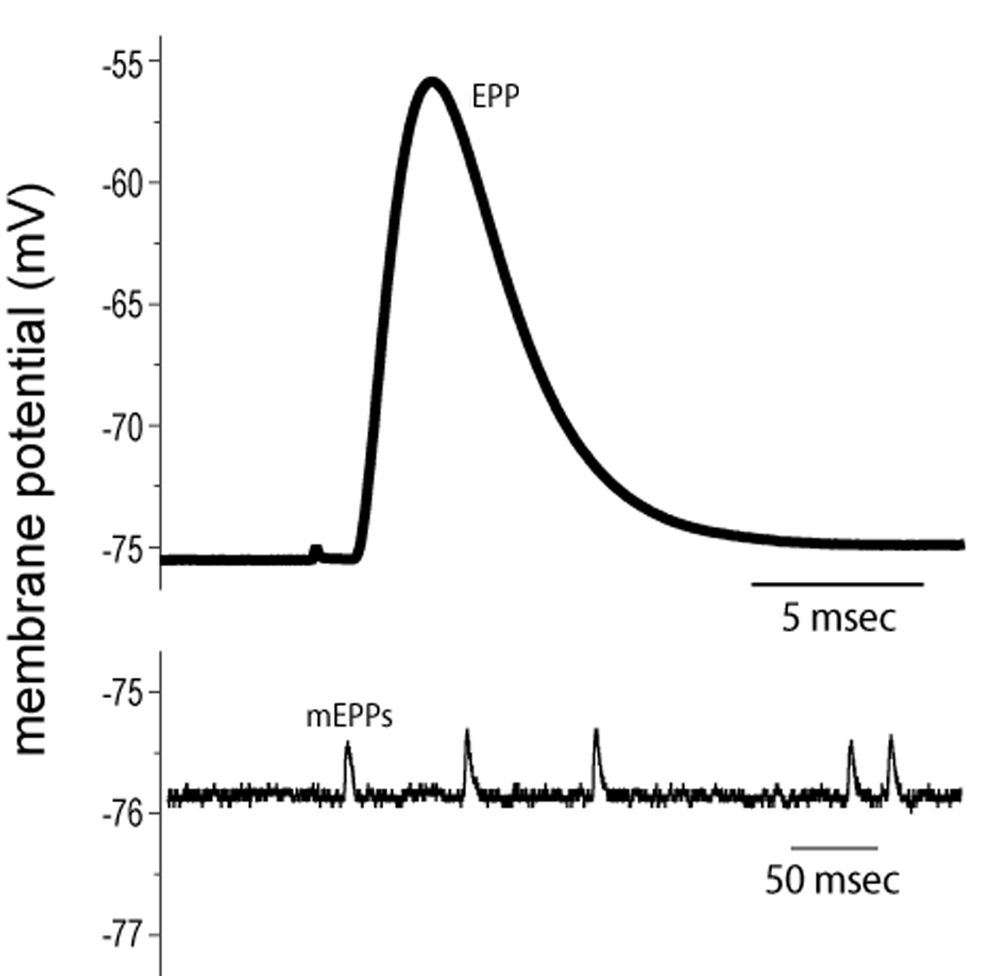
End plate potentials (EPPs) are the voltages which cause depolarization of skeletal muscle fibers caused by neurotransmitters binding to the postsynaptic membrane in the neuromuscular junction. They are called "end plates" because the postsynaptic terminals of muscle fibers have a large, saucer-like appearance. When an action potential reaches the axon terminal of a motor neuron, vesicles carrying neurotransmitters (mostly acetylcholine) are exocytosed and the contents are released into the neuromuscular junction. These neurotransmitters bind to receptors on the postsynaptic membrane and lead to its depolarization. In the absence of an action potential, acetylcholine vesicles spontaneously leak into the neuromuscular junction and cause very small depolarizations in the postsynaptic membrane. This small response (~0.4mV)[1] is called a miniature end plate potential (MEPP) and is generated by one acetylcholine-containing vesicle. It represents the smallest possible depolarization which can be induced in a muscle.

Neuromuscular junction

[edit]
Signal transmission from nerve to muscle at the motor end plate.

The neuromuscular junction is the synapse that is formed between an alpha motor neuron (α-MN) and the skeletal muscle fiber. In order for a muscle to contract, an action potential is first propagated down a nerve until it reaches the axon terminal of the motor neuron. The motor neuron then innervates the muscle fibers to contraction by causing an action potential on the postsynaptic membrane of the neuromuscular junction.

Acetylcholine

[edit]

End plate potentials are produced almost entirely by the neurotransmitter acetylcholine in skeletal muscle. Acetylcholine is the second most important excitatory neurotransmitter in the body following glutamate. It controls the somatosensory system which includes the senses of touch, vision, and hearing. It was the first neurotransmitter to be identified in 1914 by Henry Dale. Acetylcholine is synthesized in the cytoplasm of the neuron from choline and acetyl-CoA. Choline acetyltransferase is the enzyme that synthesizes acetylcholine and is often used as a marker in research relating to acetylcholine production. Neurons that utilize acetylcholine are called cholinergic neurons and they are very important in muscle contraction, memory, and learning.[2]

Ion channels

[edit]

The polarization of membranes is controlled by sodium, potassium, calcium, and chloride ion channels. There are two types of ion channels involved in the neuromuscular junction and end plate potentials: voltage-gated ion channel and ligand-gated ion channel. Voltage gated ion channels are responsive to changes in membrane voltage which cause the voltage gated ion channel to open and allows certain ions to pass through. Ligand gated ion channels are responsive to certain molecules such as neurotransmitters. The binding of a ligand to the receptor on the ion channel protein causes a conformational change which allows the passing of certain ions.

Presynaptic membrane

[edit]

Normally the resting membrane potential of a motor neuron is kept at -70mV to -50 with a higher concentration of sodium outside and a higher concentration of potassium inside. When an action potential propagates down a nerve and reaches the axon terminal of the motor neuron, the change in membrane voltage causes the calcium voltage gated ion channels to open allowing for an influx of calcium ions. These calcium ions cause the acetylcholine vesicles attached to the presynaptic membrane to release acetylcholine via exocytosis into the synaptic cleft.[3]

Postsynaptic membrane

[edit]

EPP are caused mostly by the binding of acetylcholine to receptors in the postsynaptic membrane. There are two different kinds of acetylcholine receptors: nicotinic and muscarinic. Nicotinic receptors are ligand gated ion channels for fast transmission. All acetylcholine receptors in the neuromuscular junction are nicotinic. Muscarinic receptors are G protein-coupled receptors that use a second messenger. These receptors are slow and therefore are unable to measure a miniature end plate potential (MEPP). They are located in the parasympathetic nervous system such as in the vagus nerve and the gastrointestinal tract. During fetal development acetylcholine receptors are concentrated on the postsynaptic membrane and the entire surface of the nerve terminal in the growing embryo is covered even before a signal is fired. Five subunits consisting of four different proteins from four different genes comprise the nicotinic acetylcholine receptors therefore their packaging and assembly is a very complicated process with many different factors. The enzyme muscle-specific kinase (MuSK) initiates signaling processes in the developing postsynaptic muscle cell. It stabilizes the postsynaptic acetylcholine receptor clusters, facilitates the transcription of synaptic genes by muscle fiber nuclei, and triggers differentiation of the axon growth cone to form a differentiated nerve terminal.[4] Substrate laminin induces advanced maturation of the acetylcholine receptor clusters on the surfaces of myotubes.[5]

Initiation

[edit]

Synaptic vesicles

[edit]

All neurotransmitters are released into the synaptic cleft via exocytosis from synaptic vesicles. Two kinds of neurotransmitter vesicles exist: large dense core vesicles and small clear core vesicles. Large dense core vesicles contain neuropeptides and large neurotransmitters that are created in the cell body of the neuron and then transported via fast axonal transport down to the axon terminal. Small clear core vesicles transport small molecule neurotransmitters that are synthesized locally in the presynaptic terminals. Finalized neurotransmitter vesicles are bound to the presynaptic membrane. When an action potential propagates down the motor neuron axon and arrives at the axon terminal, it causes a depolarization of the axon terminal and opens calcium channels. This causes the release of the neurotransmitters via vesicle exocytosis.

After exocytosis, vesicles are recycled during a process known as the synaptic vesicle cycle. The retrieved vesicular membranes are passed through several intracellular compartments where they are modified to make new synaptic vesicles. They are then stored in a reserve pool until they are needed again for transport and release of neurotransmitters.

Unlike the reserve pool, the readily releasable pool of synaptic vesicles is ready to be activated. Vesicle depletion from the readily releasable pool occurs during high frequency stimulation of long duration and the size of the evoked EPP reduces. This neuromuscular depression is due to less neurotransmitter release during stimulation. In order for depletion not to occur, there must be a balance between repletion and depletion which can happen at low stimulation frequencies of less than 30 Hz.[6]

When a vesicle releases its neurotransmitters via exocytosis, it empties its entire contents into the synaptic cleft. Neurotransmitter release from vesicles is therefore stated to be quantal because only whole numbers of vesicles can be released. In 1970, Bernard Katz from the University of London won the Nobel Prize for Physiology or Medicine for statistically determining the quantal size of acetylcholine vesicles based on noise analysis in the neuromuscular junction. Using a book on mechanical statistics[clarification needed], he was able to infer the size of individual events going on at the same time.

The synaptic vesicles of acetylcholine are clear core synaptic vesicles with a diameter of 30 nm. Each acetylcholine vesicle contains approximately 5000 acetylcholine molecules. The vesicles release their entire quantity of acetylcholine and this causes miniature end plate potentials (MEPPs) to occur which are less than 1mV in amplitude and not enough to reach threshold.[7]

Miniature end plate potentials (MEPPs)

[edit]

Miniature end plate potentials are the small (~0.4mV) depolarizations of the postsynaptic terminal caused by the release of a single vesicle into the synaptic cleft. Neurotransmitter vesicles containing acetylcholine collide spontaneously with the nerve terminal and release acetylcholine into the neuromuscular junction even without a signal from the axon. These small depolarizations are not enough to reach threshold and so an action potential in the postsynaptic membrane does not occur.[8] During experimentation with MEPPs, it was noticed that often spontaneous action potentials would occur, called end plate spikes in normal striated muscle without any stimulus. It was believed that these end plate spikes occurred as a result of injury or irritation of the muscles fibers due to the electrodes. Recent experiments have shown that these end plate spikes are actually caused by muscle spindles and have two distinct patterns: small and large. Small end plate spikes have a negative onset without signal propagation and large end plate spikes resemble motor unit potentials (MUPs). Muscle spindles are sensory receptors that measure muscle elongation or stretch and relay the information to the spinal cord or brain for the appropriate response.[9]

Threshold potential ("All or None")

[edit]

When an action potential causes the release of many acetylcholine vesicles, acetylcholine diffuses across the neuromuscular junction and binds to ligand-gated nicotinic receptors (non-selective cation channels) on the muscle fiber. This allows for increased flow of sodium and potassium ions, causing depolarization of the sarcolemma (muscle cell membrane). The small depolarization associated with the release of acetylcholine from an individual synaptic vesicle is called a miniature end-plate potential (MEPP), and has a magnitude of about +0.4mV. MEPPs are additive, eventually increasing the end-plate potential (EPPs) from about -100mV up to the threshold potential of -60mV, at which level the voltage-gated ion channels in the postsynaptic membrane open, allowing a sudden flow of sodium ions from the synapse and a sharp spike in depolarization. This depolarization voltage spike triggers an action potential which propagates down the postsynaptic membrane leading to muscle contraction. It is important to note that EPPs are not action potentials, but that they trigger action potentials. In a normal muscular contraction, approximately 100-200 acetylcholine vesicles are released causing a depolarization that is 100 times greater in magnitude than a MEPP. This causes the membrane potential to depolarize +40mV (100 x 0.4mV = 40mV) from -100mV to -60mV where it reaches threshold.[7]

Action potential phases

[edit]

Once the membrane potential reaches threshold, an action potential occurs and causes a sharp spike in membrane polarity. There are five phases of an action potential: threshold, depolarization, peak, repolarization, and hyperpolarization.

Threshold is when the summation of MEPPs reaches a certain potential and induces the opening of the voltage-gated ion channels. The rapid influx of sodium ions causes the membrane potential to reach a positive charge. The potassium ion channels are slower-acting than the sodium ion channels and so as the membrane potential starts to peak, the potassium ion channels open and causes an outflux of potassium to counteract the influx of sodium. At the peak, the outflux of potassium equals the influx of sodium, and the membrane does not change polarity.

During repolarization, the sodium channels begin to become inactivated, causing a net efflux of potassium ions. This causes the membrane potential to drop down to its resting membrane potential of -100mV. Hyperpolarization occurs because the slow-acting potassium channels take longer to deactivate, so the membrane overshoots the resting potential. It gradually returns to resting potential and is ready for another action potential to occur.

During the action potential before the hyperpolarization phase, the membrane is unresponsive to any stimulation. This inability to induce another action potential is known as the absolute refractory period. During the hyperpolarization period, the membrane is again responsive to stimulations but it requires a much higher input to induce an action potential. This phase is known as the relative refractory period.

Once the action potential has finished in the neuromuscular junction, the used acetylcholine is cleared out of the synaptic cleft by the enzyme acetylcholinesterase. Several diseases and problems can be caused by the inability of enzymes to clear away the neurotransmitters from the synaptic cleft leading to continued action potential propagation.[10]

Clinical applications

[edit]
Patient with myasthenia gravis showing typical symptom of eyelid droop

Current research is attempting to learn more about end plate potentials and their effect on muscle activity. Many current diseases involve disrupted end plate potential activity. In Alzheimer patients, beta amyloid attaches to the acetylcholine receptors and inhibits acetylcholine binding. This causes less signal propagation and small EPPs that do not reach threshold. By analyzing brain processes with acetylcholine, doctors can measure how much beta amyloid is around and use it to judge its effects on Alzheimer's.[11] Myasthenia gravis is an autoimmune disease, where the body produces antibodies targeted against the acetylcholine receptor on the postsynaptic membrane in the neuromuscular junction. Muscle fatigue and weakness, worsened with use and improved by rest, is the hallmark of the disease. Because of the limited amount of acetylcholine receptors that are available for binding, symptomatic treatment consists of using an acetylcholinesterase inhibitor to reduce the breakdown of acetylcholine in the neuromuscular junction, so that enough acetylcholine will be present for the small number of unblocked receptors. A congenital abnormality caused by a deficiency in end-plate acetylcholine esterase (AChE) might be a pathophysiologic mechanism for myasthenic gravis. In a study on a patient with AChE deficiency, doctors noted that he had developed severe proximal and truncal muscle weakness with jittering in other muscles. It was found that a combination of the jitter and blocking rate of the acetylcholine receptors caused a reduced end-plate potential similar to what is seen in cases of myasthenia gravis.[12] Research of motor unit potentials (MUPs) has led to possible clinical applications in the evaluation of the progression of pathological diseases to myogenic or neurogenic origins by measuring the irregularity constant related. Motor unit potentials are the electrical signals produced by motor units that can be characterized by amplitude, duration, phase, and peak, and the irregularity coefficient (IR) is calculated based on the peak numbers and amplitudes.[13] Lambert–Eaton myasthenic syndrome is a disorder where presynaptic calcium channels are subjected to autoimmune destruction which causes fewer neurotransmitter vesicles to be exocytosed. This causes smaller EPPs due to less vesicles being released. Often the smaller EPPs do not reach threshold which causes muscle weakness and fatigue in patients. Many animals use neurotoxins to defend themselves and kill prey. Tetrodotoxin is a poison found in the certain poisonous fishes such as pufferfish and triggerfish which blocks the sodium ion channels and prevents an action potential on the postsynaptic membrane. Tetraethylammonium found in insects blocks potassium channels. Alpha neurotoxin found in snakes binds to acetylcholine receptors and prevents acetylcholine from binding. Alpha-latrotoxin found in black widow spiders causes a massive influx of calcium at the axon terminal and leads to an overflow of neurotransmitter release. Botulinum toxin produced by the bacteria Clostridium botulinum is the most powerful toxic protein. It prevents release of acetylcholine at the neuromuscular junction by inhibiting docking of the neurotransmitter vesicles.

See also

[edit]

References

[edit]
[edit]
Revisions and contributorsEdit on WikipediaRead on Wikipedia
from Grokipedia
The end-plate potential (EPP) is a graded, localized of the postsynaptic membrane at the (NMJ), where a synapses with a fiber, typically shifting the from approximately -90 mV to -40 mV or more. This potential is triggered by the release of () from the presynaptic terminal, which binds to nicotinic acetylcholine receptors (nAChRs) on the motor end plate, a specialized region of the muscle membrane characterized by junctional folds that increase the surface area for receptor density. Unlike propagating s, the EPP is non-propagating but sufficiently large under normal conditions—approximately 40-50 mV in amplitude—to reliably initiate a muscle that propagates along the fiber to trigger contraction. The mechanism of EPP generation begins with an arriving at the terminal, which opens voltage-gated calcium channels and allows Ca²⁺ influx, prompting the of synaptic vesicles containing ACh into the synaptic cleft. Each vesicle releases about 5,000-10,000 ACh molecules, and the simultaneous release of hundreds of vesicles (a multiquantal response) during produces the full EPP, as opposed to the smaller miniature end-plate potentials (mEPPs) caused by spontaneous release of a single vesicle quantum. The bound ACh opens ligand-gated cation channels in nAChRs, permitting a net influx of Na⁺ (and some Ca²⁺) that depolarizes the end plate, with the process rapidly terminated by (AChE) in the cleft to prevent prolonged activation. The reversal potential of the EPP is near 0 mV, reflecting the non-selective cation permeability of these channels. Physiologically, the EPP serves as the critical link in neuromuscular transmission, ensuring 1:1 fidelity between motor nerve impulses and muscle contractions essential for voluntary movement, with a built-in safety factor where even partial blockade of transmission (e.g., by ) may not fully abolish responses due to the EPP's suprathreshold . Disruptions in EPP generation underlie disorders like , where autoantibodies against nAChRs reduce receptor density and weaken the potential, leading to fatigable weakness. Miniature EPPs, occurring spontaneously at rates of 0.5-2 Hz, provide baseline synaptic activity and may contribute to trophic maintenance of the NMJ, though their is only about 0.5-1 mV.

Overview

Definition

The end-plate potential (EPP) is a graded of the postsynaptic at the , the between a and a fiber. It represents the local change in resulting from synaptic transmission that initiates muscle fiber excitation. Under normal physiological conditions, the EPP amplitude is typically around 50 mV, sufficient to reliably trigger an in the muscle fiber. This arises from an influx of cations through ligand-gated channels in the end-plate , shifting the potential from the muscle's resting level of approximately -90 mV toward threshold. EPPs are measured using intracellular microelectrodes inserted into fibers in isolated nerve-muscle preparations, where the potential change is recorded following electrical of the motor nerve. To isolate the EPP without triggering a propagating , low concentrations of or other blockers are often applied, allowing direct observation of the graded response. In contrast to excitatory postsynaptic potentials (EPSPs) at synapses, which are smaller (typically 0.5-5 mV) and often require temporal or spatial to reach threshold, EPPs have a much larger and directly couple to without . This distinction underscores the neuromuscular junction's role in ensuring reliable, one-to-one transmission for control.

Physiological Role

The end-plate potential (EPP) plays a central role in neuromuscular transmission by depolarizing the motor end-plate region of the fiber, which activates voltage-gated sodium channels and initiates an that propagates along the . This arises from the influx of sodium ions through ligand-gated channels, ensuring a rapid and localized change in from the resting state of approximately -90 mV. In the context of excitation-contraction coupling, the EPP guarantees reliable synaptic transmission at the , which is essential for precise control of voluntary movements and reflex responses. Unlike central synapses, the operates with a high safety factor, where a single EPP consistently triggers a full muscle contraction due to its robust amplitude, preventing transmission failures under normal conditions. This one-to-one fidelity supports the coordinated recruitment of motor units necessary for graded force generation in skeletal muscles. Typically, the EPP amplitude measures around 50 mV, far exceeding the muscle threshold of approximately -65 mV (requiring about 25 mV ), which ensures an "all-or-none" response in the muscle fiber without partial activations. This margin, often termed the safety factor of about 25 mV, provides resilience against variations in release or receptor sensitivity, maintaining efficient neuromuscular function across diverse physiological demands.

Neuromuscular Junction

Presynaptic Components

The presynaptic terminal at the is formed by the distal branches of the , which lose their myelin sheath upon reaching the fiber and expand into a series of synaptic boutons covering the motor end-plate region. These terminals contain specialized presynaptic active zones, which are electron-dense regions of the plasma membrane where synaptic vesicles dock and undergo . Active zones are organized by a scaffold of proteins including and , and they feature clusters of P/Q-type voltage-gated calcium channels (VGCCs, Cav2.1) precisely positioned to facilitate rapid release. These VGCCs are anchored via interactions with RIM proteins and muscle-derived β2 in the , ensuring their alignment opposite postsynaptic densities for efficient . Within the presynaptic terminal, synaptic vesicles are stored in clusters near active zones and in a reserve pool, each vesicle containing approximately 5,000–10,000 molecules of (ACh), the responsible for . Upon arrival of an , depolarization opens the clustered VGCCs, allowing calcium influx that triggers the of docked vesicles; in mammalian neuromuscular junctions, approximately 150–200 vesicles are released per , corresponding to the quantal content that ensures reliable muscle activation. This calcium-triggered occurs with high fidelity due to the low release probability per active zone (around 0.22 in mice) balanced by the large number of active zones (about 700 per terminal). Key presynaptic proteins orchestrate vesicle docking and fusion, with synaptotagmin I serving as the primary calcium sensor on the vesicle membrane. Synaptotagmin I binds calcium ions through its C2 domains upon VGCC-mediated influx, undergoing a conformational change that promotes SNARE complex assembly (involving syntaxin, SNAP-25, and synaptobrevin) to drive synchronous vesicle fusion with the presynaptic membrane. This mechanism ensures ultrafast release kinetics, typically within 0.2–0.5 milliseconds, critical for the temporal precision of neuromuscular transmission.

Postsynaptic Components

The postsynaptic components of the neuromuscular junction reside on the muscle fiber membrane and are adapted to detect and transduce the signal released from presynaptic vesicles. The motor end-plate forms a specialized, convoluted region of this membrane, marked by extensive junctional folds that invaginate deeply into the underlying . These folds expand the effective surface area by approximately eight to ten times compared to an unfluted membrane, optimizing the postsynaptic apparatus for rapid and reliable signal reception. The crests of the junctional folds host a dense array of nicotinic acetylcholine receptors (nAChRs), pentameric ligand-gated cation channels composed of α1, β1, δ, ε (in adult muscle), and γ (in fetal) subunits. This arrangement positions the receptors in close apposition to presynaptic active zones, with a receptor of approximately 10,000 per μm², which supports the of excitatory postsynaptic potentials at the end-plate. Spanning the synaptic cleft is the , a thin layer that includes (AChE) to ensure prompt termination of . The predominant AChE isoform at is the asymmetric A12 form, consisting of 12 catalytic subunits organized into three tetramers linked by disulfide bonds to a triple-helical Q (ColQ) tail; this tail anchors the enzyme to the via interactions with proteoglycans such as , preventing diffusion and localizing hydrolysis of . Adjacent to the end-plate, at the mouths of the junctional folds, voltage-gated (primarily Nav1.4 in ) cluster at elevated densities to amplify and propagate the local into a muscle . density in this perijunctional zone yields a 5- to 10-fold higher than in extrajunctional membrane regions, which ensures robust excitation despite safety factors in transmission.

Generation of End-Plate Potential

Neurotransmitter Release

When an arrives at the presynaptic terminal of the at the , it causes of the presynaptic membrane. This opens voltage-gated calcium channels, predominantly of the P/Q-type, allowing calcium ions to enter the terminal. The influx of calcium ions rapidly elevates the intracellular calcium concentration to approximately 100 μM near the release sites. This transient increase binds to calcium sensors, such as synaptotagmin, which trigger the synchronous of approximately 200-300 synaptic vesicles through SNARE complex-mediated fusion with the presynaptic membrane. The release process exhibits a quantal nature, wherein each synaptic vesicle represents one quantum of neurotransmitter, containing roughly 5,000 to 10,000 molecules of acetylcholine. These vesicles fuse at specialized active zones, ensuring efficient and localized discharge of acetylcholine into the synaptic cleft.

Receptor Binding and Ion Flow

Upon neurotransmitter release, acetylcholine (ACh) diffuses across the synaptic cleft and binds to postsynaptic nicotinic acetylcholine receptors (nAChRs) on the motor end plate. These nAChRs are ligand-gated cation channels composed of five subunits in a heteropentameric arrangement: two α1 subunits, one β1 subunit, one δ subunit, and one ε subunit (α₁₂β₁δε) in adult skeletal muscle. ACh binds with high affinity at two orthosteric sites located at the interfaces between the α1-δ and α1-ε subunits, inducing a conformational change that rapidly opens the ion channel within microseconds. The opened channel pore, approximately 7 Å in diameter, is selectively permeable to cations, permitting influx of Na⁺ ions down their and efflux of ions. This results in a net inward depolarizing current, as the driving force for Na⁺ entry (from extracellular ~145 mM to intracellular ~12 mM) exceeds that for exit (from intracellular ~155 mM to extracellular ~4 mM), with the reversal potential for the nAChR current near 0 mV. Single-channel conductance is approximately 50 pS, and the collective activation of thousands of channels generates the end-plate potential (EPP). The EPP exhibits a rapid time course, rising in less than 1 ms to a peak of 40-50 mV (depolarizing the from a of ~ -90 mV toward -40 mV) before decaying over 5-10 ms. This decay is primarily driven by the of ACh by (AChE), which exhibits a high catalytic turnover rate of approximately 10⁴ s⁻¹, rapidly clearing the from the cleft and terminating receptor activation.

Miniature End-Plate Potentials

Characteristics

Miniature end-plate potentials (MEPPs) are small, spontaneous depolarizations observed at the , arising from the random fusion of single synaptic vesicles containing with the presynaptic membrane, independent of action potentials. These events occur stochastically, with a typical of approximately 1 Hz in mammalian preparations, though this can vary widely from 0.1 to 10 Hz depending on , , and experimental conditions. The amplitude of MEPPs generally ranges from 0.4 to 1 mV, reflecting the postsynaptic response to the release of one quantum of . The time course of an MEPP is brief, with a rise time of 1-2 ms and a half-decay time of about 3-5 ms, resulting in an overall duration of 5-15 ms, as recorded intracellularly in muscle fibers. According to the quantal hypothesis, each MEPP represents the elementary unit—or quantum—of synaptic transmission, while an evoked end-plate potential (EPP) is the near-synchronous summation of 50-200 such quanta during normal nerve stimulation. Although MEPP amplitudes exhibit variability across end-plates due to differences in receptor and the location of the recording relative to the release site, the mean quantal size remains remarkably stable under physiological conditions, underscoring the consistency of single-vesicle release efficacy. This stability supports the vesicle-based quantal model proposed in foundational studies.

Relation to Synaptic Transmission

The quantal model of synaptic transmission at the , developed by del Castillo and Katz in the , posits that the end-plate potential (EPP) arises from the synchronous release of multiple discrete packets, or quanta, of from the presynaptic terminal. This model is expressed mathematically as EPP=npqEPP = n \cdot p \cdot q, where nn represents the number of available release sites, pp is the probability of quantal release at each site (typically ranging from 0.2 to 0.5 under physiological conditions), and qq is the quantal size, equivalent to the amplitude of a miniature end-plate potential (MEPP). Miniature end-plate potentials, being spontaneous single-quantal events, thus provide a direct measure of qq, allowing researchers to quantify the postsynaptic response to one quantum of transmitter. A key feature of this model is the safety factor, which describes the excess amplitude of the EPP over the threshold required to trigger a muscle action potential, ensuring reliable transmission even under suboptimal conditions such as partial receptor blockade or reduced release. In normal mammalian neuromuscular junctions, the EPP amplitude is typically 3 to 5 times the threshold value (around 15-20 mV), providing a buffer against fluctuations in quantal release or postsynaptic sensitivity. This redundancy is evident in the high quantal content (m=npm = n \cdot p, often 50-200 quanta per impulse), which amplifies the postsynaptic depolarization far beyond what a single quantum could achieve. Experimental manipulations have validated the quantal model by selectively altering its components. Application of , a competitive at receptors, reduces qq by diminishing the postsynaptic response to each quantum without affecting presynaptic release, leading to smaller, more variable EPPs that reveal underlying quantal fluctuations matching a . Conversely, elevating extracellular calcium concentration increases npn \cdot p by enhancing the release probability, thereby boosting EPP and quantal content, as demonstrated in neuromuscular preparations where transmission failures decrease under high-calcium conditions. These findings underscore how MEPPs serve as a foundational unit for understanding evoked synaptic transmission reliability.

Propagation to Action Potential

Threshold Mechanism

The end-plate potential (EPP) generated at the must depolarize the muscle fiber membrane sufficiently to reach the threshold for initiation. In fibers, the resting is approximately -90 mV, and an EPP amplitude of about 15-20 mV is typically required to depolarize the membrane to a threshold of around -70 mV, thereby activating voltage-gated Na⁺ channels situated at the perimeter of the end-plate region. This threshold mechanism operates according to the all-or-none principle, where depolarization exceeding the threshold triggers a full regenerative that propagates along the muscle fiber, while EPPs below threshold fail to activate sufficient Na⁺ influx and dissipate locally without further propagation. The safety margin provided by normal EPP amplitudes—often exceeding 40-50 mV—ensures reliable threshold crossing under physiological conditions, preventing transmission failure. The geometry of the end-plate plays a in facilitating this process, as the postsynaptic junctional folds increase the surface area and direct the flow of depolarizing current from the synaptic cleft toward adjacent excitable , thereby amplifying the effectiveness of the EPP in reaching threshold at voltage-gated Na⁺ channels. This structural adaptation enhances the spatial efficiency of current spread without altering the intrinsic threshold properties of the muscle .

Muscle Fiber Depolarization

Upon reaching the from the end-plate potential, voltage-gated sodium channels located in the peri-junctional region of the muscle membrane activate, initiating an . This spreads bidirectionally along the from the initiation site at the and into the transverse tubules () through the regenerative activation of voltage-gated sodium channels, ensuring rapid and uniform excitation across the . The high density of sodium channels near the end-plate region facilitates this efficient initiation, preventing decrement of the signal as it propagates. The in exhibits distinct phases that reflect the sequential activation of channels. The rising phase occurs due to rapid influx of Na⁺ through voltage-gated sodium channels, depolarizing the membrane from its of approximately -90 mV to a peak of about +30 mV. This is followed by the falling phase, driven by Na⁺ channel inactivation and efflux of K⁺ through voltage-gated potassium channels, repolarizing the membrane toward its resting level. An after-hyperpolarization phase then briefly brings the potential below resting due to lingering K⁺ conductance, with the entire lasting 2-5 ms in fibers. This depolarization couples to through excitation-contraction coupling mechanisms in the . Voltage-sensitive dihydropyridine receptors (DHPRs), acting as voltage sensors rather than significant Ca²⁺ channels in , undergo a conformational change that mechanically activates ryanodine receptors (RyRs) on the . This interaction triggers Ca²⁺ release from the into the , where Ca²⁺ binds to , enabling actin-myosin cross-bridge formation and force generation. Seminal studies have identified specific domains in the DHPR α1 subunit as critical for this orthograde signaling to RyRs.

Clinical Relevance

Pathological Conditions

In , an autoimmune disorder, autoantibodies target postsynaptic nicotinic acetylcholine receptors (nAChRs) at the , leading to their internalization, degradation, and complement-mediated destruction, which reduces the total number of functional receptors. This receptor loss directly diminishes the amplitude of the end-plate potential (EPP) by decreasing the postsynaptic response to (ACh) release, thereby reducing the quantal size (the EPP contribution from each vesicle) and compromising the safety factor that ensures reliable muscle activation. The condition affects approximately 20 per 100,000 individuals (1 in 5,000) and is more prevalent in women, particularly those under 50 years of age. Botulism arises from botulinum neurotoxin, produced by , which enters motor nerve terminals and cleaves SNARE proteins such as SNAP-25, syntaxin, and synaptobrevin, thereby preventing the fusion of ACh-containing vesicles with the presynaptic membrane. This enzymatic action blocks evoked ACh release, effectively reducing the release probability (p), thereby decreasing the quantal content (m) in the quantal model of synaptic transmission and resulting in EPPs too small to trigger action potentials, which manifests as and . Lambert-Eaton myasthenic syndrome involves autoantibodies directed against presynaptic P/Q-type voltage-gated calcium channels, causing their internalization and functional loss, which impairs calcium influx necessary for ACh vesicle . Consequently, the probability of quantal release (p) is lowered, yielding reduced EPP amplitudes and initial ; however, repetitive stimulation can facilitate transmission as residual calcium accumulates in the terminal, broadening the action potential and enhancing subsequent calcium entry to partially compensate for the deficit.

Pharmacological Applications

Non-depolarizing neuromuscular blockers, such as vecuronium, function as competitive antagonists at postsynaptic nicotinic receptors (nAChRs) on the motor end-plate, thereby preventing (ACh) from binding and generating a sufficient end-plate potential (EPP) to trigger . This antagonism reduces EPP amplitude in a dose-dependent manner, facilitating muscle relaxation during without initial . In contrast, depolarizing blockers like succinylcholine act as ACh agonists, binding to nAChRs and inducing an initial robust EPP that causes transient muscle fasciculations, followed by persistent receptor activation leading to desensitization and blockade of subsequent EPPs. This mechanism results in prolonged of the end-plate membrane, rendering it unresponsive to further ACh release and producing suitable for short-term procedures like endotracheal . Cholinesterase inhibitors, exemplified by neostigmine, enhance EPP amplitude and duration by inhibiting , the enzyme responsible for ACh hydrolysis in the synaptic cleft, thus prolonging availability at the . Low concentrations of neostigmine (e.g., 10⁻⁶ M) increase miniature end-plate current amplitude and extend the decay time constant without altering conductance, effectively amplifying synaptic transmission. Therapeutically, this potentiation counters the reduced EPPs in conditions like , where neostigmine administration improves muscle strength by sustaining ACh effects. Recent advancements for include FcRn inhibitors such as nipocalimab (FDA-approved April 2025), which decrease circulating autoantibodies to enhance nAChR function and EPP amplitude. Botulinum neurotoxins are employed therapeutically in low doses to inhibit presynaptic ACh release, attenuating EPPs and muscle contractions in conditions such as , , and . In experimental contexts, α-bungarotoxin serves as a high-affinity, irreversible of nAChRs, binding specifically to the receptor's ACh site and abolishing EPPs, which enables precise labeling and quantification of end-plate receptors using radiolabeled or fluorescent conjugates. With a in the picomolar to nanomolar range, it has been instrumental in mapping receptor distribution and density at the . Additionally, quantal analysis of EPPs employs calcium modulators to dissect presynaptic release mechanisms; elevating extracellular Ca²⁺ increases the quantal content (m) of the EPP by enhancing vesicle release probability (p), as shown in foundational studies where Ca²⁺ influx directly scales the number of quanta released per nerve impulse. Conversely, Ca²⁺ chelators like EGTA reduce p, allowing researchers to isolate the binomial parameters of quantal transmission without altering postsynaptic sensitivity.

References

Add your contribution
Related Hubs
User Avatar
No comments yet.