Hubbry Logo
Threshold potentialThreshold potentialMain
Open search
Threshold potential
Community hub
Threshold potential
logo
8 pages, 0 posts
0 subscribers
Be the first to start a discussion here.
Contribute something
Threshold potential
Threshold potential
from Wikipedia
A. A schematic view of an idealized action potential illustrates its various phases as the action potential passes a point on a cell membrane. B. Actual recordings of action potentials are often distorted compared to the schematic view because of variations in electrophysiological techniques used to make the recording.

In electrophysiology, the threshold potential is the critical level to which a membrane potential must be depolarized to initiate an action potential. In neuroscience, threshold potentials are necessary to regulate and propagate signaling in both the central nervous system (CNS) and the peripheral nervous system (PNS).

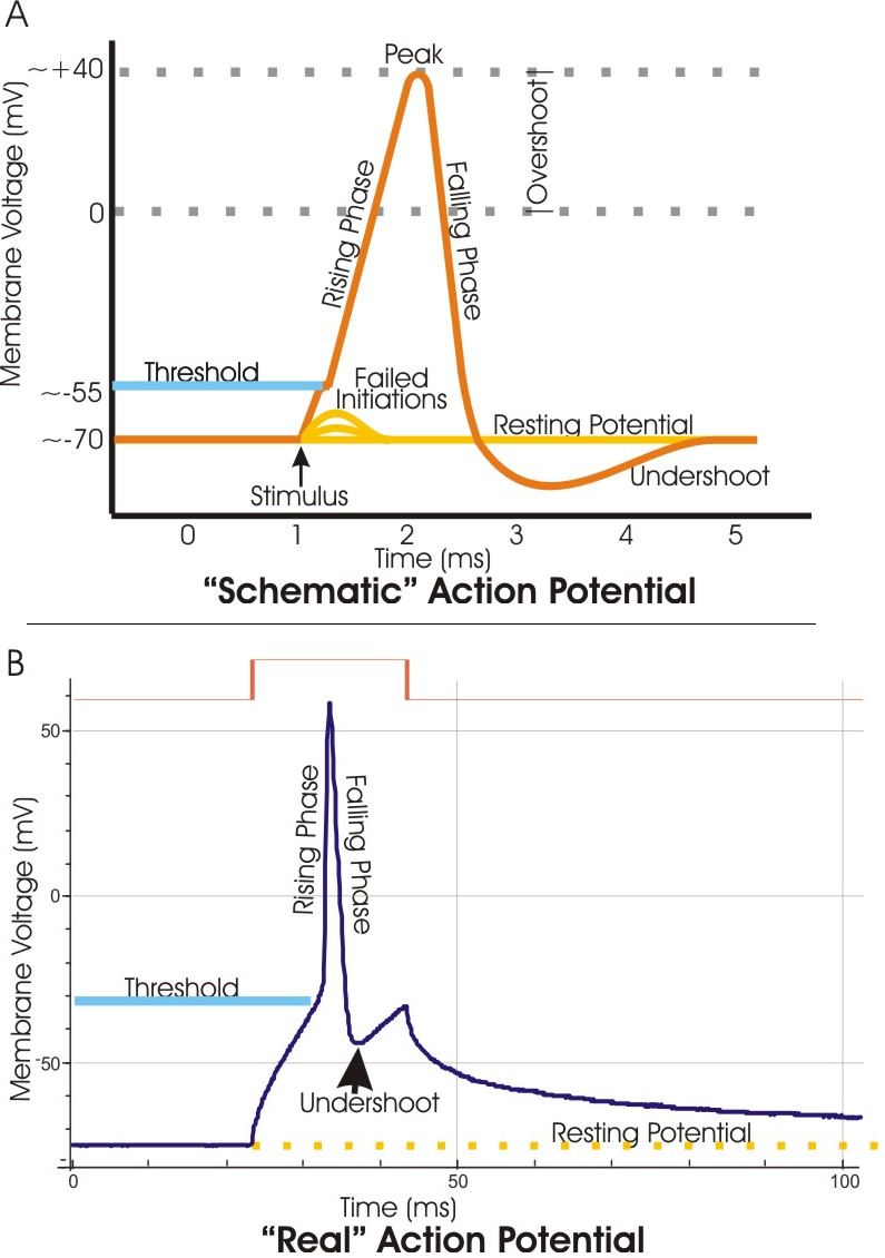
Most often, the threshold potential is a membrane potential value between –50 and –55 mV,[1] but can vary based upon several factors. A neuron's resting membrane potential (–70 mV) can be altered to either increase or decrease likelihood of reaching threshold via sodium and potassium ions. An influx of sodium into the cell through open, voltage-gated sodium channels can depolarize the membrane past threshold and thus excite it while an efflux of potassium or influx of chloride can hyperpolarize the cell and thus inhibit threshold from being reached.

Discovery

[edit]

Initial experiments revolved around the concept that any electrical change that is brought about in neurons must occur through the action of ions. The German physical chemist Walther Nernst applied this concept in experiments to discover nervous excitability, and concluded that the local excitatory process through a semi-permeable membrane depends upon the ionic concentration. Also, ion concentration was shown to be the limiting factor in excitation. If the proper concentration of ions was attained, excitation would certainly occur.[2] This was the basis for discovering the threshold value.

Along with reconstructing the action potential in the 1950s, Alan Lloyd Hodgkin and Andrew Huxley were also able to experimentally determine the mechanism behind the threshold for excitation. It is known as the Hodgkin–Huxley model. Through use of voltage clamp techniques on a squid giant axon, they discovered that excitable tissues generally exhibit the phenomenon that a certain membrane potential must be reached in order to fire an action potential. Since the experiment yielded results through the observation of ionic conductance changes, Hodgkin and Huxley used these terms to discuss the threshold potential. They initially suggested that there must be a discontinuity in the conductance of either sodium or potassium, but in reality both conductances tended to vary smoothly along with the membrane potential.[3]

They soon discovered that at threshold potential, the inward and outward currents, of sodium and potassium ions respectively, were exactly equal and opposite. As opposed to the resting membrane potential, the threshold potential's conditions exhibited a balance of currents that were unstable. Instability refers to the fact that any further depolarization activates even more voltage-gated sodium channels, and the incoming sodium depolarizing current overcomes the delayed outward current of potassium.[4] At resting level, on the other hand, the potassium and sodium currents are equal and opposite in a stable manner, where a sudden, continuous flow of ions should not result. The basis is that at a certain level of depolarization, when the currents are equal and opposite in an unstable manner, any further entry of positive charge generates an action potential. This specific value of depolarization (in mV) is otherwise known as the threshold potential.

Physiological function and characteristics

[edit]

The threshold value controls whether or not the incoming stimuli are sufficient to generate an action potential. It relies on a balance of incoming inhibitory and excitatory stimuli. The potentials generated by the stimuli are additive, and they may reach threshold depending on their frequency and amplitude. Normal functioning of the central nervous system entails a summation of synaptic inputs made largely onto a neuron's dendritic tree. These local graded potentials, which are primarily associated with external stimuli, reach the axonal initial segment and build until they manage to reach the threshold value.[5] The larger the stimulus, the greater the depolarization, or attempt to reach threshold. The task of depolarization requires several key steps that rely on anatomical factors of the cell. The ion conductances involved depend on the membrane potential and also the time after the membrane potential changes.[6]

Resting membrane potential

[edit]

The phospholipid bilayer of the cell membrane is, in itself, highly impermeable to ions. The complete structure of the cell membrane includes many proteins that are embedded in or completely cross the lipid bilayer. Some of those proteins allow for the highly specific passage of ions, ion channels. Leak potassium channels allow potassium to flow through the membrane in response to the disparity in concentrations of potassium inside (high concentration) and outside the cell (low). The loss of positive(+) charges of the potassium(K+) ions from the inside of the cell results in a negative potential there compared to the extracellular surface of the membrane.[7] A much smaller "leak" of sodium(Na+) into the cell results in the actual resting potential, about –70 mV, being less negative than the calculated potential for K+ alone, the equilibrium potential, about –90 mV.[7] The sodium-potassium ATPase is an active transporter within the membrane that pumps potassium (2 ions) back into the cell and sodium (3 ions) out of the cell, maintaining the concentrations of both ions as well as preserving the voltage polarization.

Depolarization

[edit]

However, once a stimulus activates the voltage-gated sodium channels to open, positive sodium ions flood into the cell and the voltage increases. This process can also be initiated by ligand or neurotransmitter binding to a ligand-gated channel. More sodium is outside the cell relative to the inside, and the positive charge within the cell propels the outflow of potassium ions through delayed-rectifier voltage-gated potassium channels. Since the potassium channels within the cell membrane are delayed, any further entrance of sodium activates more and more voltage-gated sodium channels. Depolarization above threshold results in an increase in the conductance of Na sufficient for inward sodium movement to swamp outward potassium movement immediately.[3] If the influx of sodium ions fails to reach threshold, then sodium conductance does not increase a sufficient amount to override the resting potassium conductance. In that case, subthreshold membrane potential oscillations are observed in some type of neurons. If successful, the sudden influx of positive charge depolarizes the membrane, and potassium is delayed in re-establishing, or hyperpolarizing, the cell. Sodium influx depolarizes the cell in attempt to establish its own equilibrium potential (about +52 mV) to make the inside of the cell more positive relative to the outside.

Variations

[edit]

The value of threshold can vary according to numerous factors. Changes in the ion conductances of sodium or potassium can lead to either a raised or lowered value of threshold. Additionally, the diameter of the axon, density of voltage activated sodium channels, and properties of sodium channels within the axon all affect the threshold value.[8] Typically in the axon or dendrite, there are small depolarizing or hyperpolarizing signals resulting from a prior stimulus. The passive spread of these signals depend on the passive electrical properties of the cell. The signals can only continue along the neuron to cause an action potential further down if they are strong enough to make it past the cell's membrane resistance and capacitance. For example, a neuron with a large diameter has more ionic channels in its membrane than a smaller cell, resulting in a lower resistance to the flow of ionic current. The current spreads quicker in a cell with less resistance, and is more likely to reach the threshold at other portions of the neuron.[3]

The threshold potential has also been shown experimentally to adapt to slow changes in input characteristics by regulating sodium channel density as well as inactivating these sodium channels overall. Hyperpolarization by the delayed-rectifier potassium channels causes a relative refractory period that makes it much more difficult to reach threshold. The delayed-rectifier potassium channels are responsible for the late outward phase of the action potential, where they open at a different voltage stimulus compared to the quickly activated sodium channels. They rectify, or repair, the balance of ions across the membrane by opening and letting potassium flow down its concentration gradient from inside to outside the cell. They close slowly as well, resulting in an outward flow of positive charge that exceeds the balance necessary. It results in excess negativity in the cell, requiring an extremely large stimulus and resulting depolarization to cause a response.

Tracking techniques

[edit]

Threshold tracking techniques test nerve excitability, and depend on the properties of axonal membranes and sites of stimulation. They are extremely sensitive to the membrane potential and changes in this potential. These tests can measure and compare a control threshold (or resting threshold) to a threshold produced by a change in the environment, by a preceding single impulse, an impulse train, or a subthreshold current.[9] Measuring changes in threshold can indicate changes in membrane potential, axonal properties, and/or the integrity of the myelin sheath.

Threshold tracking allows for the strength of a test stimulus to be adjusted by a computer in order to activate a defined fraction of the maximal nerve or muscle potential. A threshold tracking experiment consists of a 1-ms stimulus being applied to a nerve in regular intervals.[10] The action potential is recorded downstream from the triggering impulse. The stimulus is automatically decreased in steps of a set percentage until the response falls below the target (generation of an action potential). Thereafter, the stimulus is stepped up or down depending on whether the previous response was lesser or greater than the target response until a resting (or control) threshold has been established. Nerve excitability can then be changed by altering the nerve environment or applying additional currents. Since the value of a single threshold current provides little valuable information because it varies within and between subjects, pairs of threshold measurements, comparing the control threshold to thresholds produced by refractoriness, supernormality, strength-duration time constant or "threshold electrotonus" are more useful in scientific and clinical study.[11]

Tracking threshold has advantages over other electrophysiological techniques, like the constant stimulus method. This technique can track threshold changes within a dynamic range of 200% and in general give more insight into axonal properties than other tests.[12] Also, this technique allows for changes in threshold to be given a quantitative value, which when mathematically converted into a percentage, can be used to compare single fiber and multifiber preparations, different neuronal sites, and nerve excitability in different species.[12]

"Threshold electrotonus"

[edit]

A specific threshold tracking technique is threshold electrotonus, which uses the threshold tracking set-up to produce long-lasting subthreshold depolarizing or hyperpolarizing currents within a membrane. Changes in cell excitability can be observed and recorded by creating these long-lasting currents. Threshold decrease is evident during extensive depolarization, and threshold increase is evident with extensive hyperpolarization. With hyperpolarization, there is an increase in the resistance of the internodal membrane due to closure of potassium channels, and the resulting plot "fans out". Depolarization produces has the opposite effect, activating potassium channels, producing a plot that "fans in".[13]

The most important factor determining threshold electrotonus is membrane potential, so threshold electrotonus can also be used as an index of membrane potential. Furthermore, it can be used to identify characteristics of significant medical conditions through comparing the effects of those conditions on threshold potential with the effects viewed experimentally. For example, ischemia and depolarization cause the same "fanning in" effect of the electrotonus waveforms. This observation leads to the conclusion that ischemia may result from over-activation of potassium channels.[14]

Clinical significance

[edit]

The role of the threshold potential has been implicated in a clinical context, namely in the functioning of the nervous system itself as well as in the cardiovascular system.

Febrile seizures

[edit]

A febrile seizure, or "fever fit", is a convulsion associated with a significant rise in body temperature, occurring most commonly in early childhood. Repeated episodes of childhood febrile seizures are associated with an increased risk of temporal lobe epilepsy in adulthood.[15]

With patch clamp recording, an analogous state was replicated in vitro in rat cortical neurons after induction of febrile body temperatures; a notable decrease in threshold potential was observed. The mechanism for this decrease possibly involves suppression of inhibition mediated by the GABAB receptor with excessive heat exposure.[15]

ALS and diabetes

[edit]

Abnormalities in neuronal excitability have been noted in amyotrophic lateral sclerosis and diabetes patients. While the mechanism ultimately responsible for the variance differs between the two conditions, tests through a response to ischemia indicate a similar resistance, ironically, to ischemia and resulting paresthesias. As ischemia occurs through inhibition of the sodium-potassium pump, abnormalities in the threshold potential are hence implicated.[12]

Arrhythmia

[edit]

Since the 1940s, the concept of diastolic depolarization, or "pacemaker potential", has become established; this mechanism is a characteristic distinctive of cardiac tissue.[16] When the threshold is reached and the resulting action potential fires, a heartbeat results from the interactions; however, when this heartbeat occurs at an irregular time, a potentially serious condition known as arrhythmia may result.

Use of medications

[edit]

A variety of drugs can present prolongation of the QT interval as a side effect. Prolongation of this interval is a result of a delay in sodium and calcium channel inactivation; without proper channel inactivation, the threshold potential is reached prematurely and thus arrhythmia tends to result.[17] These drugs, known as pro-arrhythmic agents, include antimicrobials, antipsychotics, methadone, and, ironically, antiarrhythmic agents.[18] The use of such agents is particularly frequent in intensive care units, and special care must be exercised when QT intervals are prolonged in such patients: arrhythmias as a result of prolonged QT intervals include the potentially fatal torsades de pointes, or TdP.[17]

Role of diet

[edit]

Diet may be a variable in the risk of arrhythmia. Polyunsaturated fatty acids, found in fish oils and several plant oils,[19] serve a role in the prevention of arrhythmias.[20] By inhibiting the voltage-dependent sodium current, these oils shift the threshold potential to a more positive value; therefore, an action potential requires increased depolarization.[20] Clinically therapeutic use of these extracts remains a subject of research, but a strong correlation is established between regular consumption of fish oil and lower frequency of hospitalization for atrial fibrillation, a severe and increasingly common arrhythmia.[21]

Notes

[edit]

References

[edit]
[edit]
Revisions and contributorsEdit on WikipediaRead on Wikipedia
from Grokipedia
The threshold potential is the critical membrane voltage in excitable cells, such as neurons and cells, at which voltage-gated sodium channels sufficiently activate to trigger the rapid influx of sodium ions, initiating the phase of an . In typical neurons, this threshold is reached at approximately -55 mV, a level that exceeds the resting of about -70 mV and follows graded depolarizations from synaptic inputs or other stimuli. Once attained, the threshold potential sets off an all-or-none regenerative process, where the propagates along the without decrement, enabling efficient signal transmission over long distances in the . This mechanism is fundamental to neuronal communication, , and , with the precise threshold value influenced by factors like density and local environmental conditions at sites such as the . Variations in threshold potential can occur across cell types and physiological states, underscoring its role in modulating excitability and preventing spurious firing in response to subthreshold stimuli.

Definition and Fundamentals

Core Definition

The threshold potential is the critical membrane voltage level, approximately -55 mV (ranging from -60 to -50 mV) in neurons, at which voltage-gated sodium channels activate sufficiently to produce a net inward current that initiates the regenerative phase of an . This level represents the point of instability in membrane excitability, where depolarizing stimuli overcome the stabilizing influences of potassium efflux and the sodium-potassium pump, leading to rapid self-amplifying . It is distinct from related concepts such as rheobase, defined as the minimal current amplitude of infinite duration required to depolarize the to threshold, and chronaxie, the pulse duration needed to reach threshold using a current twice the rheobase value. These parameters quantify neuronal excitability in response to electrical stimuli and are foundational to understanding strength-duration relationships in excitable tissues. The threshold is characterized in strength-duration curves by the Lapicque-Weiss equation: I=Irh(1+τt)I = I_{\mathrm{rh}} \left(1 + \frac{\tau}{t}\right) where II is the stimulus current amplitude, IrhI_{\mathrm{rh}} is the rheobase current, tt is the stimulus duration, and τ\tau is the (typically around 0.1–1 ms in neurons). This hyperbolic relationship describes how shorter stimuli require higher currents to achieve threshold, reflecting the membrane's capacitive and resistive properties. From a typical resting of -70 mV, the membrane must depolarize by approximately 10–20 mV to attain threshold, marking the transition from subthreshold graded potentials to the all-or-none .

Role in Excitable Cells

The threshold potential functions as the critical "all-or-nothing" trigger for initiating s in excitable cells, including neurons, cardiac myocytes, and cells, where membrane depolarization must reach this specific voltage level to evoke a full response or none at all. In neurons, this threshold ensures that only stimuli strong enough to depolarize the membrane to approximately -55 mV will generate a propagating , preventing weak signals from eliciting partial responses. Similarly, in cardiac myocytes, crossing the threshold around -70 mV triggers a complete , while in cells it is around -60 mV, linking electrical excitation directly to mechanical contraction. Reaching the threshold initiates rapid, self-sustaining depolarization that propagates signals reliably over long distances without decrement, enabling efficient communication in these cell types. In neurons, this propagation occurs along axons, often via saltatory conduction in myelinated fibers, ensuring swift transmission of nerve impulses from the central nervous system to effectors. For cardiac myocytes, threshold crossing in interconnected cells allows the action potential to spread across the myocardium, coordinating synchronized contractions essential for heart function. In skeletal muscle cells, the propagated action potential travels along the sarcolemma and into T-tubules, activating contraction throughout the fiber for precise motor control. This threshold mechanism is pivotal in neuronal synaptic transmission, where summed excitatory postsynaptic potentials at the must exceed the threshold to fire an , thereby relaying information across synapses to influence downstream neurons or muscles. In cardiac rhythm generation, pacemaker cells in the spontaneously approach and cross their threshold during phase 4 , initiating each heartbeat and setting the pace for the entire conduction system. Overall, the threshold potential governs excitability, determining whether environmental stimuli translate into functional outputs like nerve signaling or muscle contractions.

Historical Development

Early Observations

In the , conducted extensive experiments on the electrical excitability of , employing highly sensitive galvanometers to measure bioelectric currents and identify the minimal electrical stimulus required to produce a response. His work demonstrated that excitation occurred only when the stimulus intensity exceeded a certain threshold, a finding detailed in publications such as his preliminary report on frog currents and his 1850 study on the laws of electrical nerve irritation. These observations established the electrical basis of nerve signaling and introduced quantitative aspects of stimulus thresholds in excitable tissues. Building on these foundations, early 19th-century experiments utilized galvanic stimulation—constant direct currents—to probe "" thresholds in isolated muscle preparations, typically from frogs. Researchers like Carlo Matteucci applied varying current strengths to muscle-nerve setups, revealing that contractions ensued only above a minimal intensity, which varied with factors such as placement and tissue condition. Matteucci's investigations in the and quantified these thresholds, linking electrical to inherent tissue properties and advancing the understanding of excitation limits without invoking vitalistic forces. By 1902, Julius Bernstein integrated these insights into his seminal membrane theory, positing that cell membranes maintain a through selective permeability to ions, with the threshold for excitation arising from a transient increase in permeability to other ions during stimulation. This model explained the all-or-nothing nature of nerve responses and preceded modern concepts by attributing threshold dynamics to permeability shifts rather than simple . Bernstein's framework provided a biophysical rationale for the minimal stimuli observed earlier, influencing subsequent quantitative models of action potentials.

Key Experimental Discoveries

In the late 1920s and early 1930s, Joseph Erlanger and Herbert Spencer Gasser conducted pioneering electrophysiological studies on compound action potentials in mammalian trunks, demonstrating that threshold potentials vary systematically with fiber diameter. Using the cathode-ray oscillograph to record extracellular potentials, they classified fibers into groups (A, B, and C) based on conduction velocity and size, revealing that larger-diameter A-fibers exhibit lower excitation thresholds compared to smaller B- and C-fibers, which require stronger stimuli to initiate action potentials. This work highlighted threshold heterogeneity within bundles, attributing variations to differences in fiber excitability and influencing subsequent models of conduction. Building on these foundations, and advanced the understanding of threshold potential through their voltage-clamp experiments on the in the early 1950s. By clamping the to specific voltages and measuring ionic currents, they isolated the rapid inward sodium current responsible for , showing that threshold occurs at a membrane potential of approximately -57 mV (about 6-8 mV above the of -65 mV), where sodium influx begins to dominate and trigger regenerative excitation. These studies quantified the voltage- and time-dependent activation of sodium channels, establishing that threshold marks the instability point where from sodium entry overcomes outward and leak currents. Hodgkin and Huxley formalized these findings in a that describes dynamics at threshold, emphasizing the dominance of sodium conductance. The core equation governing the rate of change in VV is: dVdt=IgNam3h(VENa)gKn4(VEK)gL(VEL)Cm\frac{dV}{dt} = \frac{I - g_\mathrm{Na} m^3 h (V - E_\mathrm{Na}) - g_\mathrm{K} n^4 (V - E_\mathrm{K}) - g_\mathrm{L} (V - E_\mathrm{L})}{C_m} where II is the applied current, gNag_\mathrm{Na}, gKg_\mathrm{K}, and gLg_\mathrm{L} are maximum conductances for sodium, , and leak channels, mm, hh, and nn are gating variables, ENaE_\mathrm{Na}, EKE_\mathrm{K}, and ELE_\mathrm{L} are reversal potentials, and CmC_m is . At threshold, the inward sodium term gNam3h(VENa)g_\mathrm{Na} m^3 h (V - E_\mathrm{Na}) rapidly increases due to voltage-dependent of mm gates, driving dV/dt>0dV/dt > 0 and initiating the upstroke. This model accurately predicted propagation and excitability, validated against experimental data from axons.

Biophysical Mechanisms

Resting Membrane Potential

The resting membrane potential represents the baseline electrical state of the in excitable cells, such as neurons, typically maintained at approximately -70 mV, with the interior of the cell negative relative to the exterior. This potential arises primarily from the unequal distribution of ions across the membrane and the membrane's selective permeability to those ions, particularly (K⁺). The sodium-potassium pump plays a crucial role in sustaining this potential by actively transporting three sodium ions (Na⁺) out of the cell and two ions into the cell per cycle, counteracting passive ion leaks and establishing concentration gradients essential for the negative interior charge. The quantitative description of the resting membrane potential is provided by the Goldman-Hodgkin-Katz (GHK) voltage equation, which accounts for the contributions of multiple ions based on their permeabilities and concentration gradients: Vm=RTFln(PK[K+]o+PNa[Na+]o+PCl[Cl]iPK[K+]i+PNa[Na+]i+PCl[Cl]o)V_m = \frac{RT}{F} \ln \left( \frac{P_K [K^+]_o + P_{Na} [Na^+]_o + P_{Cl} [Cl^-]_i}{P_K [K^+]_i + P_{Na} [Na^+]_i + P_{Cl} [Cl^-]_o} \right) where VmV_m is the membrane potential, RR is the gas constant, TT is the absolute temperature, FF is Faraday's constant, PP denotes permeability coefficients, and subscripts oo and ii indicate extracellular and intracellular concentrations, respectively; note that chloride (Cl⁻) terms are inverted due to its negative charge. In typical neurons, the membrane's high permeability to K⁺ relative to Na⁺ and Cl⁻ (with PKPNaP_K \gg P_{Na}) results in a resting potential close to the K⁺ equilibrium potential, around -90 mV, but shifted toward less negative values by minor Na⁺ influx. Leak channels, particularly potassium leak channels, are integral to stabilizing the by allowing passive K⁺ efflux, which generates the dominant negative charge inside the cell, while the Na⁺/K⁺ ATPase continuously restores ion gradients to prevent dissipation. This steady-state balance ensures the remains polarized, serving as the essential starting point from which depolarizing stimuli can drive the potential toward threshold.

Depolarization Process

The depolarization process in excitable cells originates from the resting , a stable state where the intracellular environment is negatively charged relative to the , typically around -70 mV in neurons. This process is initiated by an external stimulus, such as a synaptic input or sensory signal, which triggers the opening of ligand-gated or mechanically sensitive ion channels, leading to an initial influx of positively charged ions—primarily sodium (Na⁺) in neurons or calcium (Ca²⁺) in certain muscle cells—into the cell. This selective permeability shift causes a gradual, partial , shifting the membrane potential from its resting value toward less negative levels, often by 5-15 mV depending on stimulus strength. As this initial depolarization progresses, the membrane voltage enters the activation range of voltage-gated channels embedded in the plasma membrane, prompting a small number of these channels—particularly voltage-gated Na⁺ channels—to transition from closed to open states. The resulting additional influx of Na⁺ s further reduces the membrane's negative charge, creating a loop where the rising voltage activates even more channels, exponentially increasing permeability and accelerating the rate. In cells relying on Ca²⁺, a similar mechanism operates through voltage-gated calcium channels, amplifying the inward current and driving the potential upward. The threshold potential, generally around -55 mV in neurons, marks the critical instability point in this sequence, where the becomes self-sustaining and regenerative, ensuring the propagates as an all-or-nothing without reverting to rest. At this juncture, the process transitions from stimulus-dependent to autonomous, as the cumulative influx overwhelms opposing forces like efflux, committing the to rapid overshoot. This regenerative nature distinguishes threshold from subthreshold s, which dissipate without full activation.

Ionic Currents at Threshold

At the threshold potential, typically around -50 to -55 mV in neuronal membranes, voltage-gated sodium channels undergo rapid , permitting a substantial influx of Na⁺ ions that drives regenerative depolarization and propels the membrane potential toward the overshoot phase of the action potential. This Na⁺ current, denoted as INaI_{Na}, dominates the ionic flux at this critical juncture, as the channels' activation gates open in response to the voltage shift, while their inactivation gates remain largely inactive initially. Contemporaneously, delayed rectifier potassium channels, characterized by their slower kinetics, begin to open but contribute minimally to the net current at threshold due to the opposing Na⁺ influx; these channels, modeled with activation variable nn in the Hodgkin-Huxley framework, facilitate outward K⁺ flow that eventually counters . The inactivation gates of sodium channels, governed by the hh variable, progressively close during this phase, curtailing further Na⁺ entry and thereby delineating the threshold boundary by preventing sustained excitation from subthreshold perturbations. The electrochemical gradients underlying these currents are quantified by the , which computes the equilibrium potential for each ion: Eion=RTzFln([ion]o[ion]i)E_{\text{ion}} = \frac{RT}{zF} \ln \left( \frac{[\text{ion}]_o}{[\text{ion}]_i} \right) where RR is the , TT is in , zz is the ion's valence, and FF is Faraday's constant. For Na⁺ in typical mammalian neurons, with extracellular concentration [Na+]o145[\text{Na}^+]_o \approx 145 mM and intracellular [Na+]i12[\text{Na}^+]_i \approx 12 mM at 37°C, ENa+60E_{Na} \approx +60 mV, creating a strong inward driving force at threshold. In contrast, for K⁺ with [K+]o4[\text{K}^+]_o \approx 4 mM and [K+]i140[\text{K}^+]_i \approx 140 mM, EK90E_K \approx -90 mV, resulting in negligible outward K⁺ current near threshold but poised for activation during peak .

Factors Influencing Threshold

Cellular Variations

The threshold potential exhibits notable variations across excitable cell types, largely attributable to differences in voltage-gated ion channel densities and subcellular architectures. In neurons, this value typically ranges from -50 to -40 mV, enabling rapid action potential initiation due to high densities of sodium channels, particularly at the axon initial segment. In contrast, cardiac myocytes display a lower threshold of -65 to -50 mV, resulting from comparatively reduced sodium channel densities that contribute to the distinct, slower depolarization kinetics observed in cardiac action potentials. Skeletal muscle fibers have a threshold potential around -55 mV, modulated by the intricate geometry of , which influences membrane capacitance and the spatial distribution of voltage-sensitive channels to ensure synchronized excitation across the fiber.

Environmental and Pathophysiological Factors

Environmental factors such as hypoxia can modulate the threshold potential in neurons by altering function, thereby increasing cellular excitability. During hypoxic conditions, the voltage threshold for initiation decreases, allowing neurons to fire with less synaptic input due to enhanced sodium influx through voltage-gated sodium channels. This shift is mediated by hypoxia-induced upregulation of sodium currents, which facilitates and lowers the required to reach threshold. Similarly, influences neuronal threshold potential by promoting greater excitability through intrinsic membrane property changes. Elevated temperatures reduce the voltage threshold for generation in both excitatory and inhibitory neurons, leading to faster rates and increased firing propensity via modulation of temperature-sensitive channels like TRPV4. This results in heightened neuronal responsiveness, as the membrane reaches firing threshold more readily under . Pathophysiological alterations in extracellular ion concentrations, particularly hyperkalemia, affect threshold potential by depolarizing the resting membrane potential. Elevated extracellular potassium levels shift the resting potential toward less negative values (e.g., from -90 mV to -80 mV), partially inactivating voltage-gated sodium channels and effectively raising the threshold potential required for action potential initiation. This mechanism reduces neuronal excitability despite the initial depolarization, as the voltage difference between resting and threshold potentials narrows while sodium channel availability decreases. Genetic mutations, such as those in the SCN1A gene encoding the NaV1.1 , can shift the threshold potential in affected neurons. Loss-of-function mutations in SCN1A, common in certain epilepsies, lead to higher thresholds for firing by impairing excitability, particularly in . Computational models of these mutants demonstrate elevated voltage thresholds for single s and repetitive firing, altering overall network dynamics without altering baseline cellular variations.

Measurement Techniques

Direct Recording Methods

Direct recording methods for threshold potential involve invasive electrophysiological techniques that enable precise of the membrane voltage at which a regenerative is initiated in isolated cellular preparations. These approaches typically employ current-clamp configurations to simulate natural while monitoring voltage responses, allowing researchers to identify the threshold as the point where activation leads to an all-or-nothing spike rather than passive decay. Such methods are fundamental for studying excitability in neurons and other excitable cells, providing direct insight into the biophysical determinants of firing. Intracellular microelectrode recordings represent a cornerstone technique, pioneered in the study of axons, where fine glass micropipettes filled with solution are inserted into the cell interior to both record and inject current. Depolarizing current pulses of incrementally increasing amplitude are applied, and the threshold is determined as the membrane voltage at which the response transitions from a subthreshold to a rapid upstroke driven by voltage-gated sodium influx, typically around -55 to -40 mV in mammalian neurons. This method allows for high-fidelity capture of the regenerative phase, revealing how threshold varies with stimulus ramp speed or holding potential, and has been instrumental in validating ionic models of excitability. For instance, in classic experiments, threshold was observed to shift with changes in external sodium concentration, confirming its dependence on inward currents. Brief applications of this approach also inform indirect methods like threshold electrotonus by establishing baseline excitability metrics in single fibers. Patch-clamp techniques, particularly whole-cell configurations in current-clamp mode, extend these measurements to smaller mammalian neurons and isolated cells where microelectrode impalement is challenging. A glass forms a high-resistance seal on the , enabling intracellular access without excessive damage; current steps are then injected to depolarize the , and threshold is quantified as the voltage initiating the action potential overshoot. This variant offers superior space-clamp control, minimizing voltage escape in dendrites, and has quantified threshold values in diverse cell types, such as cortical pyramidal neurons at approximately -50 mV under physiological conditions. Advantages include low noise and the ability to dialyze the intracellular milieu for pharmacological studies, though series resistance artifacts must be compensated to ensure accurate threshold detection. Strength-duration curve plotting further refines threshold assessment by varying stimulus pulse width while measuring the minimal current required to elicit an , often using the same intracellular or patch-clamp setups. The curve, typically hyperbolic, plots threshold current against duration on a semi-log scale, with rheobase defined as the minimal current for an infinitely long , representing the steady-state threshold under constant depolarization. Seminal formulations describe this relationship as Ith=Irh(1+[τ](/page/Tau)t)I_{th} = I_{rh} \left(1 + \frac{[\tau](/page/Tau)}{t}\right), where IthI_{th} is threshold current, IrhI_{rh} is rheobase, τ\tau is the , and tt is duration; in neuronal preparations, rheobase currents range from 0.1 to 1 nA for intracellular . This analysis distinguishes passive charging from active threshold crossing and is used to characterize excitability changes, such as hyperpolarizing shifts in threshold during repetitive firing.

Threshold Electrotonus Approach

The threshold electrotonus approach is a specialized electrophysiological technique designed to evaluate the threshold potential in intact peripheral nerve bundles without invasive intracellular recordings. Developed by Bostock and Baker, it employs automated threshold tracking to monitor changes in axonal excitability induced by prolonged subthreshold polarizing currents, typically lasting 100 ms, applied via surface electrodes. These currents, set at ±40% of threshold intensity, alter the membrane potential electrotonically, and the method continuously adjusts the test stimulus intensity to maintain a constant compound action potential amplitude, thereby tracking threshold variations over time. This non-invasive procedure allows assessment of multi-fiber nerve responses in vivo, providing insights into nodal and internodal membrane properties. Threshold electrotonus curves, derived from these measurements, plot the percentage change in threshold against time during the polarizing current. For depolarizing currents, the curves often display an S-shaped profile, characterized by an initial rapid rise (fast phase, peaking within 10-20 ms) followed by a transient decline and a slower secondary rise (slow phase, developing over 50-100 ms). These phases reflect the differential activation of fast and slow gating mechanisms, primarily involving voltage-dependent potassium channels that modulate sodium channel availability and membrane accommodation. Hyperpolarizing currents produce opposing shifts, with an initial decrease in threshold followed by an overshoot, highlighting inward rectifier properties. In clinical applications, threshold electrotonus is particularly useful for detecting subtle axonal dysfunction in conditions affecting nerve excitability, such as demyelination or channelopathies, by quantifying deviations in curve morphology. For instance, analysis of TE peaks can reveal abnormal threshold changes, with typical normal depolarizing shifts of 10-20% in the late phase indicating intact slow gating; reductions or exaggerations in these values signal impaired function or membrane polarization. This quantitative profiling, often implemented using systems like QTRAC, enables early and monitoring of peripheral neuropathies through standardized protocols.

Clinical Relevance

Neurological Disorders

Altered threshold potential plays a significant role in the of several neurological disorders, where disruptions in neuronal excitability contribute to disease progression. In febrile seizures, which predominantly affect children aged 6 months to 5 years, elevated body temperature during fever lowers the activation threshold for neuronal firing, promoting hyperexcitability and synchronized activity that can precipitate . This temperature-dependent reduction in threshold is evidenced by studies showing that increases the excitability of hippocampal pyramidal cells, dentate granule cells, and inhibitory , thereby enhancing the likelihood of seizure induction. Furthermore, lower fever temperatures are associated with a higher risk of seizure recurrence, indicating a dynamically reduced threshold in susceptible individuals. Mechanisms involve fever-induced changes in function, such as modulation of hyperpolarization-activated currents (I_h) via HCN channels, which amplify rebound and network excitability during prolonged episodes. In (ALS), a progressive targeting motor neurons, reduced threshold potential in affected cells arises from hyperactivity, leading to early hyperexcitability that exacerbates neuronal loss. Persistent sodium currents (I_NaP) are elevated in ALS motor neurons, particularly through upregulation of NaV1.6 at initial segments and hyperpolarizing shifts in NaV1.3 , which lower the voltage threshold for initiation. This hyperactivity, observed in both SOD1 mutant mouse models and patient-derived neurons, contributes to repetitive firing and cortical hyperexcitability detectable even presymptomatically. The imbalance is compounded by reduced expression, such as KCNQ2, failing to counteract sodium influx and further promoting . Diabetes-related neuropathy, a common complication of chronic , involves shifts in threshold potential driven by on voltage-gated sodium channels, resulting in hyperexcitability and . upregulates NaV1.3 and NaV1.7 in neurons, causing a negative shift in voltage-dependent activation and delayed inactivation of tetrodotoxin-sensitive currents, which lowers the threshold for generation. from further modifies channel function, such as reducing NaV1.8 peak currents while enhancing overall excitability through post-translational changes. Byproducts like , generated under hyperglycemic conditions, directly activate NaV1.8, amplifying nociceptive signaling and contributing to and .

Cardiovascular Conditions

In (LQTS), delayed repolarization due to genetic mutations in ion channels, such as those affecting or sodium currents, prolongs the action potential duration in ventricular cardiomyocytes, creating a substrate for early afterdepolarizations (EADs) that can reach the threshold potential and trigger . This prolongation extends the vulnerable window during which premature stimuli are more likely to evoke re-excitation, as demonstrated in murine models where LQTS variants lower the re-excitation threshold at longer coupling intervals compared to controls. Consequently, the altered excitability dynamics in LQTS heighten the risk of polymorphic , with clinical manifestations often linked to adrenergic triggers that exacerbate repolarization instability. Myocardial ischemia lowers the stimulation threshold in ventricular myocytes primarily through extracellular accumulation and , which depolarize the resting and enhance excitability early in the ischemic process. This reduction in threshold facilitates the initiation of ectopic beats and reentrant circuits, promoting the transition to , particularly when combined with conduction slowing in the ischemic border zone. Studies in isolated perfused hearts show that this threshold decrease occurs transiently at potassium levels below 6 mmol/L, underscoring ischemia's role in acute arrhythmogenesis during . In (), heterogeneous threshold changes across atrial tissue arise from regional variations in expression and remodeling, leading to nonuniform refractoriness that sustains reentrant wavefronts. Such heterogeneity, often exacerbated by persistent Na+ current increases, creates areas of differential excitability where duration dispersion drives formation and AF maintenance, as evidenced in genetic models. This spatial variability in threshold recovery contributes to the self-perpetuating nature of AF, with pharmacological interventions targeting APD uniformity showing potential to reduce inducibility.

Therapeutic Interventions

Therapeutic interventions targeting threshold potential primarily involve pharmacological agents that modulate function to alter neuronal and cardiac excitability, as well as dietary strategies to influence electrolyte balance. Voltage-gated , such as , increase the threshold potential for initiation by stabilizing inactivated sodium channels and reducing repetitive firing in hyperexcitable tissues. This mechanism prevents seizures in by elevating the stimulus intensity required to elicit motor responses, as demonstrated in studies where raised motor thresholds by approximately 5% compared to . Similarly, in cardiac contexts, class I antiarrhythmics like these blockers raise the threshold potential to suppress re-entrant arrhythmias, particularly in conditions such as . Lidocaine, a class Ib , specifically stabilizes threshold potential in ischemic myocardial tissue by preferentially binding to inactivated sodium channels under acidic and depolarized conditions prevalent during ischemia. This action prolongs the and elevates the ventricular fibrillatory threshold, reducing the incidence of life-threatening ventricular arrhythmias at therapeutic plasma levels of 1.5–5 μg/mL. Clinical evidence supports its use in acute ischemic settings to interrupt re-entrant circuits, though its role has diminished with newer agents due to limited efficacy in non-ischemic arrhythmias. Dietary interventions play a supportive role in modulating threshold potential through electrolyte homeostasis, particularly in conditions involving potassium dysregulation. In hyperkalemia-related cardiac conditions, such as those complicating arrhythmias, restricting potassium intake via low-potassium diets helps normalize extracellular potassium levels, thereby restoring resting membrane potential and threshold excitability to prevent conduction abnormalities. Moderate hyperkalemia initially lowers the difference between resting and threshold potentials, heightening excitability, while severe levels inactivate sodium channels and elevate the effective threshold; dietary restriction aids in reversing these shifts to maintain stable cardiac rhythm. Additionally, omega-3 polyunsaturated fatty acids from dietary sources like fish oil modulate cardiac ion channel function, including sodium channels, to increase the threshold for membrane depolarization and reduce arrhythmogenic potential in ischemic heart disease. Supplementation with eicosapentaenoic and docosahexaenoic acids has been shown to inhibit sodium influx, thereby stabilizing action potentials and conferring cardioprotective effects against ventricular arrhythmias.

References

Add your contribution
Related Hubs
Contribute something
User Avatar
No comments yet.