Hubbry Logo
Muscle spindleMuscle spindleMain
Open search
Muscle spindle
Community hub
Muscle spindle
logo
8 pages, 0 posts
0 subscribers
Be the first to start a discussion here.
Muscle spindle
Muscle spindle
from Wikipedia
Muscle spindle
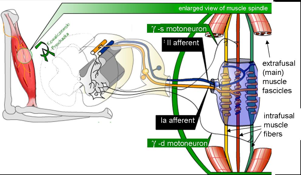
Mammalian muscle spindle showing typical position in a muscle (left), neuronal connections in spinal cord (middle) and expanded schematic (right). The spindle is a stretch receptor with its own motor supply consisting of several intrafusal muscle fibres. The sensory endings of a primary (group Ia) afferent and a secondary (group II) afferent coil around the non-contractile central portions of the intrafusal fibres. Gamma motor neurons activate the intrafusal muscle fibres, changing the resting firing rate and stretch-sensitivity of the afferents. [a]
Details
Part ofMuscle
SystemMusculoskeletal
Identifiers
Latinfusus neuromuscularis
MeSHD009470
THH3.11.06.0.00018
FMA83607
Anatomical terminology

Muscle spindles are stretch receptors within the body of a skeletal muscle that primarily detect changes in the length of the muscle. They convey length information to the central nervous system via afferent nerve fibers. This information can be processed by the brain as proprioception. The responses of muscle spindles to changes in length also play an important role in regulating the contraction of muscles, for example, by activating motor neurons via the stretch reflex to resist muscle stretch.

The muscle spindle has both sensory and motor components.

Structure

[edit]

Muscle spindles are found within the belly of a skeletal muscle. Muscle spindles are fusiform (spindle-shaped), and the specialized fibers that make up the muscle spindle are called intrafusal muscle fibers. The regular muscle fibers outside of the spindle are called extrafusal muscle fibers. Muscle spindles have a capsule of connective tissue, and run parallel to the extrafusal muscle fibers unlike Golgi tendon organs which are oriented in series.[citation needed]

Composition

[edit]

Muscle spindles are composed of 5–14 muscle fibers, of which there are three types: dynamic nuclear bag fibers (bag1 fibers), static nuclear bag fibers (bag2 fibers), and nuclear chain fibers.[2][3]

A
Light microscope photograph of a muscle spindle with H&E stain

Primary type Ia sensory fibers (large diameter) spiral around all intrafusal muscle fibres, ending near the middle of each fibre. Secondary type II sensory fibers (medium diameter) end adjacent to the central regions of the static bag and chain fibres.[3] These fibres send information by stretch-sensitive mechanically gated ion-channels of the axons.[4]

The motor part of the spindle is provided by motor neurons: up to a dozen gamma motor neurons also known as fusimotor neurons.[5] These activate the muscle fibres within the spindle. Gamma motor neurons supply only muscle fibres within the spindle, whereas beta motor neurons supply muscle fibres both within and outside of the spindle. Activation of the neurons causes a contraction and stiffening of the end parts of the muscle spindle muscle fibers.[citation needed]

Fusimotor neurons are classified as static or dynamic according to the type of muscle fibers they innervate and their effects on the responses of the Ia and II sensory neurons innervating the central, non-contractile part of the muscle spindle.

  • The static axons innervate the chain or static bag2 fibers. They increase the firing rate of Ia and II afferents at a given muscle length (see schematic of fusimotor action below).
  • The dynamic axons innervate the bag1 intrafusal muscle fibers. They increase the stretch-sensitivity of the Ia afferents by stiffening the bag1 intrafusal fibers.

Efferent nerve fibers of gamma motor neurons also terminate in muscle spindles; they make synapses at either or both of the ends of the intrafusal muscle fibers and regulate the sensitivity of the sensory afferents, which are located in the non-contractile central (equatorial) region.[6]

Function

[edit]

Stretch reflex

[edit]

When a muscle is stretched, primary type Ia sensory fibers of the muscle spindle respond to both changes in muscle length and velocity and transmit this activity to the spinal cord in the form of changes in the rate of action potentials. Likewise, secondary type II sensory fibers respond to muscle length changes (but with a smaller velocity-sensitive component) and transmit this signal to the spinal cord. The Ia afferent signals are transmitted monosynaptically to many alpha motor neurons of the receptor-bearing muscle. The reflexly evoked activity in the alpha motor neurons is then transmitted via their efferent axons to the extrafusal fibers of the muscle, which generate force and thereby resist the stretch. The Ia afferent signal is also transmitted polysynaptically through interneurons (Ia inhibitory interneurons), which inhibit alpha motorneurons of antagonist muscles, causing them to relax.[7]

Sensitivity modification

[edit]

The function of the gamma motor neurons is not to supplement the force of muscle contraction provided by the extrafusal fibers, but to modify the sensitivity of the muscle spindle sensory afferents to stretch. Upon release of acetylcholine by the active gamma motor neuron, the end portions of the intrafusal muscle fibers contract, thus elongating the non-contractile central portions (see "fusimotor action" schematic below). This opens stretch-sensitive ion channels of the sensory endings, leading to an influx of sodium ions. This raises the resting potential of the endings, thereby increasing the probability of action potential firing, thus increasing the stretch-sensitivity of the muscle spindle afferents.

Recent transcriptomic and proteomic studies have identified unique gene expression profiles specific to muscle spindle regions. Distinct macrophage populations, known as muscle spindle macrophages (MSMPs), have been observed, suggesting an immunological component in muscle spindle maintenance and function.[8] Immunostaining and sequencing have enabled tissue-level identification of novel markers, contributing to an advanced cellular atlas of the muscle spindle. Regarding the structural-functional correlation; muscle spindle density is not uniform across the musculoskeletal system. Recent biomechanical modeling suggests that spindle abundance correlates with muscle fascicle length and fiber velocity during dynamic movement, emphasizing the relationship between muscle structure and proprioceptive requirements.[9]

How does the central nervous system control gamma fusimotor neurons? It has been difficult to record from gamma motor neurons during normal movement because they have very small axons. Several theories have been proposed, based on recordings from spindle afferents.

  • 1) Alpha-gamma coactivation. Here it is posited that gamma motor neurons are activated in parallel with alpha motor neurons to maintain the firing of spindle afferents when the extrafusal muscles shorten.[10]
  • 2) Fusimotor set: Gamma motor neurons are activated according to the novelty or difficulty of a task. Whereas static gamma motor neurons are continuously active during routine movements such as locomotion, dynamic gamma motorneurons tend to be activated more during difficult tasks, increasing Ia stretch-sensitivity.[11]
  • 3) Fusimotor template of intended movement. Static gamma activity is a "temporal template" of the expected shortening and lengthening of the receptor-bearing muscle. Dynamic gamma activity turns on and off abruptly, sensitizing spindle afferents to the onset of muscle lengthening and departures from the intended movement trajectory.[12]
  • 4) Goal-directed preparatory control. Dynamic gamma activity is adjusted proactively during movement preparation in order to facilitate execution of the planned action. For example, if the intended movement direction is associated with stretch of the spindle-bearing muscle, Ia afferent and stretch reflex sensitivity from this muscle is reduced. Gamma fusimotor control therefore allows for the independent preparatory tuning of muscle stiffness according to task goals.[13]

Development

[edit]

Genetic pathways critical for spindle formation include neuregulin-1 signaling via ErbB receptors, which induce intrafusal fiber differentiation upon sensory innervation. Disruption of these pathways impairs proprioception, as seen in gene knockout models.[14]

It is also believed that muscle spindles play a critical role in sensorimotor development. Additionally, gain-of-function mutations in HRAS (e.g.: G12S) observed in Costello syndrome are associated with increased spindle number, providing insight into genetic regulation of spindle density.[15]

Clinical significance

[edit]

Dysfunction in muscle spindle signaling has been implicated in sensory neuropathies and coordination disorders such as ataxia. Enhanced understanding of genetic mutations affecting spindle development (e.g. HRAS and Egr3-linked pathways) opens avenues for targeted therapies in proprioceptive deficits and neuromuscular diseases.

After stroke or spinal cord injury in humans, spastic hypertonia (spastic paralysis) often develops, whereby the stretch reflex in flexor muscles of the arms and extensor muscles of the legs is overly sensitive. This results in abnormal postures, stiffness and contractures. Hypertonia may be the result of over-sensitivity of alpha motor neurons and interneurons to the Ia and II afferent signals.[16]

Additional images

[edit]

Notes

[edit]

References

[edit]
[edit]
Revisions and contributorsEdit on WikipediaRead on Wikipedia
from Grokipedia
A muscle spindle is an encapsulated proprioceptive sensory receptor embedded within most skeletal muscles, consisting of specialized intrafusal muscle fibers that detect changes in muscle length and the velocity of those changes, thereby providing essential feedback to the for and coordination. These receptors are oriented parallel to the extrafusal muscle fibers that generate force, and they do not contribute significantly to overall but instead serve as stretch detectors to maintain posture, facilitate smooth movements, and initiate reflexive responses. Structurally, each muscle spindle contains 3 to 10 intrafusal fibers—typically one dynamic nuclear fiber (bag₁), one static nuclear fiber (bag₂), and several nuclear chain fibers—surrounded by a capsule that isolates them from surrounding tissue. The central, equatorial region of these fibers, where nuclei are clustered in bags or chains, is the most sensitive to stretch and receives sensory innervation, while the polar ends connect to the spindle capsule and are targeted by motor neurons. Human muscles contain varying densities of spindles, estimated at around 50,000 total, with higher concentrations in fine-control muscles like those of the hand compared to larger postural muscles. Functionally, muscle spindles operate through primary (group Ia) and secondary (group II) afferent nerve fibers that transmit signals to the and . Primary afferents, wrapping around the equatorial region of all intrafusal fiber types via annulospiral endings, respond dynamically to both the rate and extent of muscle stretch, enabling rapid adjustments during movement. Secondary afferents, primarily contacting static bag₂ and chain fibers with flower-spray endings, provide sustained feedback on muscle length alone, contributing to tonic regulation of posture. Recent studies have also revealed that specialized macrophages within muscle spindles modulate sensory feedback and , further elucidating their role in . Sensitivity is modulated by gamma (γ) motor neurons that innervate the polar regions of intrafusal fibers, contracting them to keep the spindle taut even during active muscle shortening, thus preventing unloading and loss of sensory input. In addition to their core role in the —where sudden lengthening triggers and contraction via Ia afferents—muscle spindles integrate with other sensory systems to support kinesthesia, balance, and adaptive locomotion across . Disruptions in spindle function, as seen in neuromuscular diseases like or aging, can impair and motor performance, underscoring their importance in both health and pathology.

Anatomy

Location and Composition

Muscle spindles are fusiform sensory organs embedded in parallel with the extrafusal muscle fibers, primarily within the belly of skeletal muscles. These structures measure typically 3-10 mm in length and 0.04-0.2 mm in , varying slightly by muscle type and species. The composition of a muscle spindle consists of a capsule that encloses 2-12 specialized intrafusal fibers, along with endings and blood vessels. This encapsulation provides structural integrity and isolates the internal components from the surrounding extrafusal environment. Muscle spindles are distributed unevenly across s, with higher densities observed in those requiring precise control, such as the and intrinsic hand muscles, in contrast to lower densities in large postural muscles like the gastrocnemius. This variation supports differential proprioceptive demands in fine versus gross movements.00833-2/fulltext)

Intrafusal Fibers

Intrafusal fibers are the specialized fibers encapsulated within the muscle spindle, distinguishing them from the surrounding extrafusal fibers by their unique structure adapted for sensory detection of muscle length changes. These fibers are smaller in diameter and shorter in length compared to extrafusal fibers, typically measuring 10-12 μm in and up to 8 mm in length in mammals. They are multiply innervated and organized to transduce mechanical stretch into neural signals. There are two primary types of intrafusal fibers: nuclear bag fibers and nuclear chain fibers, named for the arrangement of their nuclei in the central region. Nuclear bag fibers contain numerous nuclei aggregated in a sac-like "bag" formation within the equatorial zone, making them longer and thicker than chain fibers, with diameters up to 20 μm and lengths extending the full span of the spindle capsule. They are further subdivided into dynamic (bag₁) and static (bag₂) subtypes; bag₁ fibers are primarily sensitive to the velocity of stretch, while bag₂ fibers respond to both velocity and sustained length changes. In contrast, nuclear chain fibers feature 3-10 nuclei aligned in a linear "chain" configuration in the equatorial region, with smaller diameters (around 10 μm) and shorter lengths, often attaching to the polar regions of bag fibers. The zonal organization of intrafusal fibers divides each into a central equatorial zone and flanking polar regions. The equatorial zone is non-contractile, lacking a well-developed actin-myosin apparatus, and serves as the primary site for sensory transduction, exhibiting fusiform expansions especially pronounced in bag fibers. The polar regions are contractile, containing myofibrils and sarcomeres similar to extrafusal fibers, enabling adjustment of spindle sensitivity through motor innervation. A typical mammalian muscle spindle encapsulates 1-3 nuclear bag fibers and 4-8 nuclear chain fibers, with bag fibers comprising about 20-30% of the total intrafusal complement and contributing to dynamic stretch detection, while chain fibers predominate and sense static length. At the ultrastructural level, sensory endings envelop the equatorial zone of intrafusal fibers in characteristic patterns. Primary sensory endings (from Ia afferents) form annulospiral configurations, where nerve fibers coil helically around the central region of both and chain fibers, providing dense, spiral wrappings that detect rapid length changes. Secondary sensory endings (from II afferents) adopt flower-spray arrangements, with terminal branches spraying out like petals primarily on chain fibers near the juxtaequatorial zones, attuned to slower, sustained stretches. These endings feature specialized synaptic structures, including annulospiral wrappings with close membrane appositions for efficient mechanotransduction.

Innervation

Muscle spindles receive dual innervation from sensory afferents that detect mechanical deformation and motor efferents that modulate sensitivity. The sensory components consist of large, myelinated afferent fibers originating from pseudounipolar neurons in dorsal root ganglia, which enter the spindle via intramuscular nerve branches and form specialized endings on the central (equatorial) regions of intrafusal fibers. Primary sensory afferents, classified as group Ia fibers, arise from the dynamic nuclear bag1 and both nuclear bag and chain fibers, forming annulospiral endings that encircle the equatorial zone. These fibers, with conduction velocities of 70-120 m/s, provide dynamic sensitivity to changes in muscle and ; each spindle typically receives one such Ia fiber. Secondary sensory afferents, group II fibers, innervate the static nuclear bag2 and nuclear chain fibers via flower-spray endings located slightly offset from the equator. With slower conduction velocities of 30-70 m/s, these fibers emphasize static information and are more numerous, with 1-5 per spindle on average. Motor innervation is provided exclusively by gamma (γ) motor neurons, located in the ventral horn of the alongside alpha motor neurons, which target the contractile polar regions of intrafusal fibers to adjust spindle tautness. These efferents form plate-like endings on the intrafusal fiber poles and have smaller diameters of 5-15 μm, enabling finer control compared to the larger alpha fibers innervating extrafusal muscle. Gamma motor neurons are subdivided into dynamic and static subtypes: dynamic γ neurons preferentially innervate bag1 fibers to enhance velocity sensitivity, while static γ neurons target bag2 and chain fibers to maintain static responsiveness.

Physiology

Proprioceptive Role

The muscle spindle serves as a primary proprioceptor by detecting changes in muscle length and the rate of those changes through specialized sensory endings located in the equatorial region of intrafusal fibers. When the muscle is stretched, this deformation elongates the equatorial region, stretching the endings and activating mechanogated channels, such as Piezo2, which generate generator potentials that lead to action potentials in afferent fibers. These potentials are transduced into neural signals that convey information about muscle stretch to the , enabling precise monitoring of limb mechanics. Recent research has identified muscle spindle-associated macrophage populations (MSMPs) that release glutamate to directly excite Ia afferent endings, thereby enhancing sensory discharge and supporting coordinated during movement. This modulation provides an additional layer of regulation for proprioceptive feedback. Muscle spindles exhibit distinct static and dynamic sensitivities mediated by different types of intrafusal fibers. Nuclear bag1 fibers, innervated primarily by group Ia afferents, provide dynamic sensitivity by responding to the of length changes, with their sensory endings showing heightened responsiveness during rapid stretches. In contrast, nuclear bag2 and chain fibers contribute to static sensitivity, detecting the absolute muscle length through sustained firing during maintained stretches. The firing rates of these afferents are generally proportional to the and speed of the stretch, allowing the spindle to differentiate between positional and movement-related cues. Through these mechanisms, muscle spindles contribute to by providing both unconscious feedback for adjustments and conscious awareness of limb position and movement, known as kinesthesia. This sensory input integrates with signals from Golgi tendon organs, which monitor muscle tension, to offer a comprehensive length-tension profile that supports coordinated and postural stability. Deficits in spindle function can impair these processes, leading to reduced accuracy in movement perception. Quantitatively, muscle spindle Ia afferents exhibit a resting discharge rate of approximately 5-15 Hz at neutral muscle length, which increases linearly with stretch amplitude and velocity, reaching maximum firing rates of 100-200 Hz during strong dynamic stretches. Adaptation occurs over seconds, where sustained stretches lead to a gradual decline in firing rate as the sensory endings adjust, preventing saturation of the signal.

Stretch Reflex Mechanism

The stretch reflex mechanism is a fundamental monosynaptic that enables rapid adjustment of muscle length in response to stretch, primarily mediated by Ia afferent fibers originating from the primary endings of muscle spindles. When a muscle is stretched, deformation of the intrafusal fibers within the spindle activates the Ia afferents, which conduct action potentials directly to the via the . These Ia afferents synapse monosynaptically onto alpha motor neurons in the ventral horn (lamina IX), releasing glutamate to produce excitatory postsynaptic potentials (EPSPs) that depolarize the motor neurons, leading to efferent discharge and contraction of the extrafusal muscle fibers. This pathway, first characterized in detail through electrophysiological studies in cats, ensures a swift excitatory response in the homonymous muscle to counteract the stretch and restore length. The reflex arc incorporates to coordinate muscles, preventing opposition to the primary contraction. Ia afferents also excite Ia inhibitory interneurons in the spinal cord, which use as a to produce inhibitory postsynaptic potentials (IPSPs) in alpha motor neurons innervating the muscle. For instance, in the stretch reflex at spinal levels L2-L4, activation inhibits hamstrings at L5-S1 via this disynaptic pathway. The core components of the arc proceed as follows: muscle stretch activates Ia afferents → synaptic transmission in the ventral horn → alpha firing → extrafusal fiber contraction, with ensuring efficient movement. This mechanism operates independently of higher centers in its basic form, relying on local spinal circuitry for automatic regulation. A classic example is the knee-jerk or , elicited clinically by tapping the to stretch the , producing a visible kick as a test of spinal integrity. Another is the tonic , which sustains low-level during posture maintenance, such as when holding a limb against , contributing to overall stability without conscious effort. In humans, the response latency for this short-latency typically ranges from 20-50 ms, reflecting the rapid conduction and synaptic delays in the arc. The strength and gain of the are proportional to the Ia firing rate and synaptic efficacy, where the response can be approximated as: Reflex response(Ia firing rate)×(synaptic efficacy)\text{Reflex response} \propto (\text{Ia firing rate}) \times (\text{synaptic efficacy}) Here, synaptic efficacy represents the depolarization per Ia impulse, approximately 1-2 mV in effective motor neuron activation, though individual fiber contributions vary. This gain is primarily influenced by spindle sensitivity at the peripheral level.

Sensitivity Modulation

The sensitivity of muscle spindles is dynamically modulated by gamma (γ) motor neurons, which innervate the intrafusal fibers to adjust afferent discharge during muscle activity. In the gamma loop, contraction of these intrafusal fibers via γ efferents counteracts the unloading effect that occurs when alpha (α) motor neurons drive extrafusal fiber shortening, thereby preserving the firing rates of Ia and II afferents. Without this modulation, spindle sensitivity diminishes as the central regions slacken, leading to a substantial reduction in afferent firing—often ceasing entirely during active shortening or declining by up to 50% in sustained contractions. This mechanism ensures continuous proprioceptive feedback, distinct from the initial response. Gamma motor neurons are classified into dynamic and static subtypes, each targeting specific intrafusal fiber types to fine-tune sensitivity. Dynamic γ neurons primarily innervate bag₁ fibers, enhancing velocity sensitivity by amplifying the dynamic response to rapid stretches. Static γ neurons, in contrast, innervate bag₂ and nuclear chain fibers, maintaining length sensitivity and steady-state discharge during held positions. These subtypes are co-activated with α motor neurons through alpha-gamma linkage, a coordinated drive from higher centers that synchronizes extrafusal contraction with intrafusal adjustment, preventing afferent silencing and supporting smooth movement trajectories. Central control of γ motor neurons arises from descending pathways in the and , which modulate drive to match task demands and enhance voluntary movement precision. This input allows independent regulation of dynamic and static γ activity, as observed in locomotor patterns where static drive predominates at shorter muscle lengths to counteract . Physiologically, fusion frequencies in γ-driven intrafusal contractions ensure seamless sensitivity tuning, avoiding abrupt changes in Ia/II discharge that could disrupt . Recent optogenetic studies have advanced understanding by enabling selective stimulation of γ motor neurons, demonstrating their capacity to restore spindle afferent firing and highlighting potential for precise modulation in motor tasks.

Development and Adaptation

Embryonic Formation

Muscle spindles originate from the paraxial , specifically the somites, during early embryonic development, with formation in human embryos commencing around the 11th week of and continuing through weeks 12 and beyond. This timeline aligns with the differentiation of myogenic precursors into intrafusal fibers, which are specialized fibers essential for spindle structure. Progenitor cells for these myogenic precursors express key transcription factors including Eya1, Six1, and , which regulate early commitment to the lineage and facilitate the specification of intrafusal fibers. The developmental process involves the aggregation of these precursors, followed by encapsulation by perineurial cells derived from the to form the protective spindle capsule. In animal models, sensory axons from proprioceptive neurons invade developing spindles, establishing initial contacts with the central regions of emerging intrafusal fibers. Human-specific timelines for these processes remain less detailed, with spindles recognizable by week 11. Studies in animal models, such as chick and embryos, have elucidated signaling pathways involved in intrafusal differentiation. Muscle spindle development begins prenatally and continues postnatally, with early innervation by sensory nerves maturing into stable proprioceptive afferents.

Postnatal Changes and Plasticity

Following birth, muscle spindles undergo significant maturation to adapt to the growing musculoskeletal system. muscle spindles are functional at birth, enabling basic proprioceptive feedback, but their stretch response remains immature due to incomplete development of intrafusal fibers and sensory endings. The number of intrafusal fibers reaches adult-like configurations postnatally, with maturation continuing into . Concurrently, the spindle capsule thickens progressively, enhancing structural integrity and protection against mechanical stress, a observed across mammalian models including cats and rodents where periaxial space development continues into the first postnatal weeks. Sensitivity to stretch matures postnatally. Muscle spindle plasticity manifests through activity-dependent adaptations that alter sensitivity and structural features in response to environmental demands. Exercise can promote upregulation of spindle sensitivity via . Conversely, prolonged immobilization, such as in or disuse models, reduces Ia afferent sensitivity to stretch, accompanied by capsule and diminished dynamic responses, as demonstrated in cat soleus muscles after 6 weeks of short-length fixation. In aging, muscle spindles exhibit progressive degeneration that impairs and contributes to . Structural alterations include fewer intrafusal fibers, thicker capsules, and degenerative sensory endings, exacerbating muscle weakness and fall risk in sarcopenic individuals. A 2025 mouse study found deterioration of annulospiral endings in aged animals, suggesting impaired proprioceptive feedback as a model for aging effects. Recent advances in multi-omics have elucidated transcriptional profiles of spindle components. A 2023 study using bulk RNA-sequencing and on intact muscle spindles identified distinct expression patterns in and intrafusal fibers. Adaptive plasticity is observed in activity-dependent contexts, supporting motor precision in skilled movements.

Clinical Relevance

Associated Disorders

Muscle spindle dysfunction is implicated in several neurological disorders, particularly those involving degeneration or malformation of proprioceptive sensory structures. In (ALS), muscle spindles undergo degeneration, with annulospiral sensory endings affected by accumulation of misfolded protein in Ia afferent neurons, leading to impaired proprioceptive feedback and reduced excitatory input from Ia afferents. This sensory impairment contributes to deficits alongside primary loss. Certain subtypes of hereditary motor neuropathies (HMN), such as GARS-related distal HMN, feature reduced muscle spindle numbers due to perturbed development and disrupted sensory-motor innervation, resulting in weakened proprioceptive signaling and . Secondary effects of muscle spindle alterations appear in conditions like and . In , altered fusimotor (gamma) drive to intrafusal fibers disrupts muscle spindle sensitivity, contributing to decreased and increased muscle rigidity. Similarly, in , demyelination of central pathways slows conduction in Ia afferent fibers from muscle spindles, impairing somatosensory processing and leading to balance deficits and . Congenital disorders such as arthrogryposis multiplex congenita (AMC) exhibit absent or malformed muscle spindles, primarily in neuropathic forms arising from fetal akinesia, where reduced intrauterine movement prevents proper spindle morphogenesis and results in joint contractures and profound proprioceptive deficits. Diagnostic signs of muscle spindle loss include , manifested as diminished or absent deep tendon reflexes due to interrupted Ia afferent signaling in the stretch reflex arc. Recent advances in MRI techniques, including high-resolution neurography, enable visualization of peripheral nerve changes in neuropathies through nerve density and signal alterations. Animal models provide insights into spindle ; for instance, Egr3 mutant mice lack muscle spindles entirely due to failed transcription factor-dependent , resulting in that mimics congenital areflexia and proprioceptive disorders.

Diagnostic and Therapeutic Applications

Muscle spindles are assessed clinically through several diagnostic techniques that evaluate their role in arcs and . The tap , elicited by percussing a to stretch the muscle, provides a simple bedside test of muscle spindle Ia afferent function and spinal integrity, with standardized maneuvers like the Jendrassik enhancing response reliability in conditions such as . The , measured via , quantifies Ia-motoneuron excitability by electrically stimulating sensory afferents and recording the monosynaptic response, serving as a for spinal in disorders like painful . Microneurography enables direct intraneural recording of Ia afferent activity from muscle spindles, offering precise insights into spindle firing during movement or stretch, particularly in studies of involuntary es post-injury. Imaging modalities have advanced the non-invasive evaluation of muscle spindle properties. Ultrasound elastography, with shear wave techniques showing notable progress by 2022, measures muscle stiffness as a proxy for spindle capsule integrity and intrafusal fiber tension, aiding in the assessment of neuromuscular disorders. Functional MRI tracks proprioceptive deficits by mapping brain activation during muscle vibration, which stimulates spindle afferents, revealing altered cortical processing in conditions like stroke-related impairments. Therapeutic interventions target muscle spindle dysregulation to alleviate and restore function. , a GABA-B receptor agonist, modulates by inhibiting gamma motoneuron activity, thereby reducing intrafusal fiber contraction and excessive spindle sensitivity, with proving effective for severe cases following . leverages spindle plasticity through targeted stretching and strengthening exercises, promoting sensory reweighting and recovery of proprioceptive feedback after peripheral nerve or spinal injuries. Emerging approaches include vibration therapy, which enhances muscle spindle firing rates to facilitate rehabilitation by increasing motoneuron excitability and promoting in motor recovery post-stroke. Gene therapy strategies for congenital neuromuscular defects, such as those in muscular dystrophies affecting spindle development and , remain preclinical as of November 2025. Recent advancements include FDA approval of delandistrogene moxeparvovec (ELEVIDYS) for in 2023, with expanded indications in 2024, potentially benefiting associated spindle dysfunction through improved muscle . Clinical outcomes demonstrate efficacy, with targeted exercises improving scores and proprioceptive function in a majority of patients, as evidenced by enhanced modulation and reduced in longitudinal training protocols.

References

Add your contribution
Related Hubs
User Avatar
No comments yet.