Hubbry Logo
Obstetrical forcepsObstetrical forcepsMain
Open search
Obstetrical forceps
Community hub
Obstetrical forceps
logo
8 pages, 0 posts
0 subscribers
Be the first to start a discussion here.
Obstetrical forceps
Obstetrical forceps
from Wikipedia

Obstetrical forceps
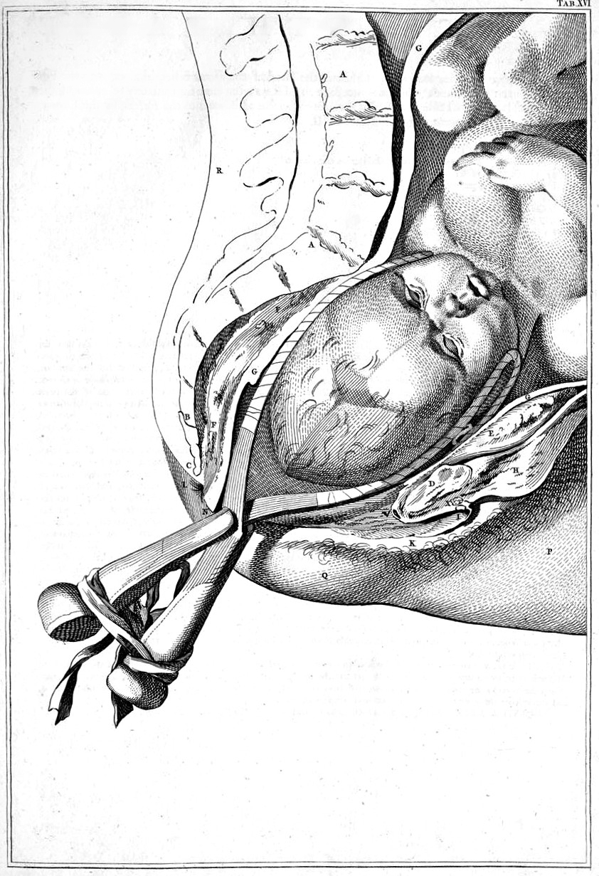
Drawing of childbirth with use of forceps by Smellie
ICD-9-CM72.0-72.4

Obstetrical forceps are a medical instrument used in childbirth. Their use can serve as an alternative to the ventouse (vacuum extraction) method.

Medical uses

[edit]

Forceps births, like all assisted births, should be undertaken only to help promote the health of the mother or baby. In general, a forceps birth is likely to be safer for both the mother and baby than the alternatives – either a ventouse birth or a caesarean section – although caveats such as operator skill apply.[1]

Advantages of forceps use include avoidance of caesarean section (and the short and long-term complications that accompany this), reduction of delivery time, and general applicability with cephalic presentation (head presentation). Common complications include the possibility of bruising the baby and causing more severe vaginal tears (perineal laceration) than would otherwise be the case. Severe and rare complications (occurring less frequently than 1 in 200) include nerve damage, Descemet's membrane rupture,[2] skull fractures, and cervical cord injury.

  • Maternal factors for use of forceps:
  1. Maternal exhaustion.
  2. Prolonged second stage of labour.
  3. Maternal illness such as heart disease, hypertension, glaucoma, aneurysm, or other conditions that make pushing difficult or dangerous.
  4. Hemorrhaging.
  5. Analgesic drug-related inhibition of maternal effort (especially with epidural/spinal anaesthesia).
  • Fetal factors for use of forceps:
  1. Non-reassuring fetal heart tracing.
  2. Fetal distress.
  3. After-coming head in breech delivery.

Complications

[edit]

Baby

[edit]
  • Cuts and bruises.
  • Increased risk of facial nerve injury (usually temporary).
  • Increased risk of clavicle fracture (rare).
  • Increased risk of intracranial hemorrhage - sometimes leading to death: 4/10,000.[3]
  • Increased risk of damage to cranial nerve VI, resulting in strabismus.

Mother

[edit]
  • Increased risk of perineal lacerations, pelvic organ prolapse, and incontinence.
  • Increased risk of injury to vagina and cervix.
  • Increased postnatal recovery time and pain.
  • Increased difficulty evacuating during recovery time.

Structure

[edit]
Obstetrical forceps, by William Smellie (1792)
Wooden forceps c.1800, Hunterian Museum, Glasgow
James Young Simpson's Caesarian forceps, Hunterian Museum, Glasgow

Obstetric forceps consist of two branches (blades) that are positioned around the head of the fetus. These branches are defined as left and right depending on which side of the mother's pelvis they will be applied. The branches usually, but not always, cross at a midpoint, which is called the articulation. Most forceps have a locking mechanism at the articulation, but a few have a sliding mechanism instead that allows the two branches to slide along each other. Forceps with a fixed lock mechanism are used for deliveries where little or no rotation is required, as when the fetal head is in line with the mother's pelvis. Forceps with a sliding lock mechanism are used for deliveries requiring more rotation.[medical citation needed]

The blade of each forceps branch is the curved portion that is used to grasp the fetal head. The forceps should surround the fetal head firmly, but not tightly. The blade characteristically has two curves, the cephalic and the pelvic curves. The cephalic curve is shaped to conform to the fetal head. The cephalic curve can be rounded or rather elongated depending on the shape of the fetal head. The pelvic curve is shaped to conform to the birth canal and helps direct the force of the traction under the pubic bone. Forceps used for rotation of the fetal head should have almost no pelvic curve.[medical citation needed]

The handles are connected to the blades by shanks of variable lengths. Forceps with longer shanks are used if rotation is being considered.[medical citation needed]

Anglo-American types

[edit]

All American forceps are derived from French forceps (long forceps) or English forceps (short forceps). Short forceps are applied on the fetal head already descended significantly in the maternal pelvis (i.e., proximal to the vagina). Long forceps are able to reach a fetal head still in the middle or even in the upper part of the maternal pelvis. At present practice, it is uncommon to use forceps to access a fetal head in the upper pelvis. So, short forceps are preferred in the UK and USA. Long forceps are still in use elsewhere.

Simpson forceps (1848) are the most commonly used among the types of forceps and has an elongated cephalic curve. These are used when there is substantial molding, that is, temporary elongation of the fetal head as it moves through the birth canal.[medical citation needed]

Elliot forceps (1860) are similar to Simpson forceps but with an adjustable pin in the end of the handles which can be drawn out as a means of regulating the lateral pressure on the handles when the instrument is positioned for use. They are used most often with women who have had at least one previous vaginal delivery because the muscles and ligaments of the birth canal provide less resistance during second and subsequent deliveries. In these cases, the fetal head may remain rounder.[medical citation needed]

Kielland forceps (1915, Norwegian) are distinguished by having no angle between the shanks and the blades and a sliding lock. The pelvic curve of the blades is identical to all other forceps. The common misperception that there is no pelvic curve has become so entrenched in the obstetric literature that it may never be able to be overcome, but it can be proved by holding a blade of Kielland's against any other forceps of one's choice. Kielland forceps are probably the most common forceps used for rotation. The sliding mechanism at the articulation can be helpful in asynclitic births (when the fetal head is tilted to the side)[4] since it is no longer in line with the birth canal. Because the handles, shanks, and blades are all in the same plane the forceps can be applied in any position to affect rotation. Because the shanks and handles are not angled, the forceps cannot be applied to a high station as readily as those with the angle since the shanks impinge on the perineum.

Wrigley's forceps, named after Arthur Joseph Wrigley, are used in low or outlet deliveries (see explanations below),[5] when the maximum diameter is about 2.5 cm (0.98 in) above the vulva.[6] Wrigley's forceps were designed for use by general practitioner obstetricians, having the safety feature of an inability to reach high into the pelvis.[6] Obstetricians now use these forceps most commonly in cesarean section delivery where manual traction is proving difficult. The short length results in a lower chance of uterine rupture.

Piper's forceps has a perineal curve to allow application to the after-coming head in breech delivery.

Technique

[edit]

The cervix must be fully dilated and retracted and the membranes ruptured. The urinary bladder should be empty, perhaps with the use of a catheter. High forceps are never indicated in the modern era. Mid forceps can occasionally be indicated but require operator skill and caution. The station of the head must be at the level of the ischial spines. The woman is placed on her back, usually with the aid of stirrups or assistants to support her legs. A regional anaesthetic (usually either a spinal, epidural or pudendal block) is used to help the mother remain comfortable during the birth. Ascertaining the precise position of the fetal head is paramount, and though historically was accomplished by feeling the fetal skull suture lines and fontanelles, in the modern era, confirmation with ultrasound is essentially mandatory. At this point, the two blades of the forceps are individually inserted, the left blade first for the commonest occipito-anterior position; posterior blade first if a transverse position, then locked. The position on the baby's head is checked. The fetal head is then rotated to the occiput anterior position if it is not already in that position. An episiotomy may be performed if necessary. The baby is then delivered with gentle (maximum 30 lbf or 130 Newton[7]) traction in the axis of the pelvis.[8][page needed]

Outlet, low, mid or high

[edit]

The accepted clinical standard classification system for forceps deliveries according to station and rotation was developed by the American College of Obstetricians and Gynecologists and consists of:[medical citation needed]

  • Outlet forceps delivery, where the forceps are applied when the fetal head has reached the perineal floor and its scalp is visible between contractions.[9] This type of assisted delivery is performed only when the fetal head is in a straight forward or backward vertex position or in slight rotation (less than 45 degrees to the right or left) from one of these positions.
  • Low forceps delivery, when the baby's head is at +2 station or lower. There is no restriction on rotation for this type of delivery.
  • Midforceps delivery, when the baby's head is above +2 station. There must be head engagement before it can be carried out.
  • High forceps delivery is not performed in modern obstetrics practice. It would be a forceps-assisted vaginal delivery performed when the baby's head is not yet engaged.

History

[edit]

Abu al-Qasim al-Zahrawi, known in the West as Albucasis, was a pioneering 10th-century physician and surgeon from Al-Andalus. In his comprehensive medical encyclopedia, Al-Tasrif, he introduced over 200 surgical instruments, many of which he designed himself. Notably, al-Zahrawi devised surgical scissors, grasping forceps, and obstetrical forceps.[10]

The obstetric forceps were introduced into the European practice by the eldest son of the Chamberlen family of surgeons. The Chamberlens were French Huguenots from Normandy who worked in Paris before they migrated to England in 1569 to escape the religious violence in France. William Chamberlen, the patriarch of the family, was most likely a surgeon; he had two sons, both named Pierre, who became maverick surgeons and specialists in midwifery.[11] William and the eldest son practiced in Southampton and then settled in London. The inventor was probably the eldest Peter Chamberlen the elder, who became obstetrician-surgeon of Queen Henriette, wife of King Charles I of England and daughter of Henry IV, King of France. He was succeeded by his nephew, Dr. Peter Chamberlen (barbers-surgeons were not doctors in the sense of physician), as royal obstetrician. The success of this dynasty of obstetricians with the royal family and high nobles was related in part to the use of this "secret" instrument allowing delivery of a live child in difficult cases.

Chamberlen forceps as found in Malden
Chamberlen forceps (Maldon)

In fact, the instrument was kept secret for 150 years by the Chamberlen family. Hugh Chamberlen the elder, grandnephew of Peter the eldest, tried to sell the instrument in Paris in 1670, but the demonstration he performed in front of François Mauriceau, responsible for Paris Hotel-Dieu maternity, was a failure which resulted in the death of mother and child. The secret may have been sold by Hugh Chamberlen to Dutch obstetricians at the start of the 18th century in Amsterdam, but there are doubts about the authenticity of what was actually provided to buyers.

The forceps were used most notably in difficult childbirths. The forceps could avoid some infant deaths when previous approaches (involving hooks and other instruments) extracted them in parts. In the interest of secrecy, the forceps were carried into the birthing room in a lined box and would only be used once everyone was out of the room and the mother blindfolded.[12]

Models derived from the Chamberlen instrument finally appeared gradually in England and Scotland in 1735. About 100 years after the invention of the forceps by Peter Chamberlen Sr. a surgeon by the name of Jan Palfijn presented his obstetric forceps to the Paris Academy of Sciences in 1723. They contained parallel blades and were called the Hands of Palfijn.

Palfyn hands in different versions
Palfijn "hands" in different versions

These "hands" were possibly the instruments described and used in Paris by Gregoire father and son, Dussée, and Jacques Mesnard.[13]

Dussee forceps with its two different locks
Dussee French forceps (circa 1725) with two different locks

In 1813, Peter Chamberlen's midwifery tools were discovered at Woodham Mortimer Hall near Maldon (UK) in the attic of the house. The instruments were found along with gloves, old coins and trinkets.[14] The tools discovered also contained a pair of forceps that were assumed to have been invented by the father of Peter Chamberlen because of the nature of the design.[15]

The Chamberlen family's forceps were based on the idea of separating the two branches of "sugar clamp" (as those used to remove "stones" from bladder), which were put in place one after another in the birth canal. This was not possible with conventional tweezers previously tested. However, they could only succeed in a maternal pelvis of normal dimensions and on fetal heads already well engaged (i.e. well lowered into maternal pelvis). Abnormalities of pelvis were much more common in the past than today, which complicated the use of Chamberlen forceps. The absence of pelvic curvature of the branches (vertical curvature to accommodate the anatomical curvature of maternal sacrum) prohibited blades from reaching the upper-part of the pelvis and exercising traction in the natural axis of pelvic excavation.

In 1747, French obstetrician Andre Levret, published Observations sur les causes et accidents de plusieurs accouchements laborieux (Observations on the Causes and Accidents of Several difficult Deliveries), in which he described his modification of the instrument to follow the curvature of the maternal pelvis, this "pelvic curve" allowing a grip on a fetal head still high in the pelvic excavation, which could assist in more difficult cases.

the first illustration of Levret's pelvic curve
First illustration of Levret's pelvic curve – 1747

This improvement was published in 1751 in England by William Smellie in the book A Treatise on the theory and practice of midwifery. After this fundamental improvement, the forceps would become a common obstetrical instrument for more than two centuries.

French forceps, Levret-Baudelocque type (1760–1860)
French forceps, Levret-Baudelocque type (1760–1860) with perforator and hook at the end of the handles

The last improvement of the instrument was added in 1877 by a French obstetrician, Stephan Tarnier in "descriptions of two new forceps." This instrument featured a traction system misaligned with the instrument itself, sometimes called the "third curvature of the forceps". This particularly ingenious traction system, allowed the forceps to exercise traction on the head of the child following the axis of the maternal pelvic excavation, which had never been possible before.

Tarnier forceps with tractor handle (1877) and USA Dewey model (1900)

Tarnier's idea was to "split" mechanically the grabbing of the fetal head (between the forceps blades) on which the operator does not intervene after their correct positioning, from a mechanical accessory set on the forceps itself, the "tractor" on which the operator exercises traction needed to pull down the fetal head in the correct axis of the pelvic excavation. Tarnier forceps (and its multiple derivatives under other names) remained the most widely used system in the world until the development of the cesarean section.

Hodge forceps derived from French Levrret type forceps
Hodge "Eclectic" forceps – US (1833)
American Elliott forceps
Elliott forceps with "pressure regulating" screw at the end of handles – US (1860)

Forceps had a profound influence on obstetrics as it allowed for the speedy delivery of the baby in cases of difficult or obstructed labour. Over the course of the 19th century, many practitioners attempted to redesign the forceps, so much so that the Royal College of Obstetrics and Gynecologists' collection has several hundred examples.[16] In the last decades, however, with the ability to perform a cesarean section relatively safely, and the introduction of the ventouse or vacuum extractor, the use of forceps and training in the technique of its use has sharply declined.

Historical role in the medicalisation of childbirth

[edit]

The introduction of the obstetrical forceps provided huge advances in the medicalisation of childbirth. Before the 18th century, childbirth was thought of as a medical phase that could be overseen by a female relative. Usually, if a doctor had to get involved that meant something had gone wrong. Around this era in the 18th century, there were no female doctors. Since males were exclusively called in under extreme circumstances, the act of childbirth was thought to be better known to a midwife or female relative than a male doctor. Usually the male doctor's job was to save the mother's life if, for example, the baby had become stuck while exiting the mother.

Before the obstetrical forceps, this had to be done by cutting the baby out piece by piece. In other cases, if the baby was deemed undeliverable, then the doctor would use a tool called a crochet. This was used to crush the baby's skull, allowing the baby to be pulled out of the mother's womb. Still in other cases, a caesarean section (c section) could be performed, but this would almost always result in the mother's death. "In addition, women who had forceps deliveries had shorter after-childbirth complications than those who had caesarean sections performed."[17] These procedures came with various risks to the mother's health, along with the death of the baby.

However, with the introduction of the obstetrical forceps, the male doctor had a more important role. In many cases, they could actually save the baby's life if called early enough. Although the use of the forceps in childbirth came with its own set of risks, the positives included a significant decrease in risk to the mother, a decrease in child morbidity, and a decreased risk to the baby. Since the forceps in childbirth were made public around 1720, they gave male doctors a way to assist and even oversee childbirths.

Around this time, in large cities such as London and Paris, some men would become devoted to obstetrical practices. It became stylish among wealthy women of the era to have their childbirth overseen by male midwives. A notable male midwife was William Hunter. He popularised obstetrics. "In 1762, he was appointed as obstetrician to Queen Charlotte."[18] In addition, with the use of forceps, male doctors invented lying-in hospitals to provide safe, somewhat advanced obstetrical care because of the use of the obstetrical forceps.

Historical complications

[edit]

Child birth was not considered a medical practice before the 18th century. It was mostly overseen by a midwife, mother, stepmother, neighbor, or any female relative. Around the 19th and 20th centuries, childbirth was considered dangerous for women.[19] With the introduction of obstetrical forceps, this allowed non-medical professionals, such as the aforementioned individuals, to continue to oversee childbirths. In addition, this gave some of the public more comfort in trusting childbirth oversight to common people. However, the introduction of obstetrical forceps also had a negative effect, because there was no medical oversight of childbirth by any kind of medical professional, this exposed the practice to unnecessary risks and complications for the fetus and mother. These risks could range from minimal effects to lifetime consequences for both individuals.

The baby could develop cuts and bruises in various body parts due to the forcible squeezing of the body through the mother's vagina. In addition, there could be bruising on the baby's face if the forceps' handler were to squeeze too tight. In some extreme cases, this could cause temporary or permanent facial nerve injury or lifelong scarring often along the jawline. Furthermore, if the forceps' handler were to twist their wrist while the grip was on the baby's head, this would twist the baby's neck and cause damage to a cranial nerve, resulting in strabismus. In rare cases, a clavicle fracture to the baby could occur. The addition of obstetrical forceps came with complication to the mother during and after childbirth. The use of the forceps gave rise to an increased risk in cuts and lacerations along the vaginal wall.

This, in turn, would cause an increase in post-operative recovery time and increase the pain experienced by the mother. In addition, the use of forceps could cause more difficulty evacuating during the recovery time as compared to a mother who did not use the forceps. While some of these risks and complications were very common, in general, many people overlooked them and continued to use them.

See also

[edit]

References

[edit]
[edit]
Revisions and contributorsEdit on WikipediaRead on Wikipedia
from Grokipedia
Obstetrical forceps are a pair of curved, spoon-shaped metal instruments designed to grasp and gently guide a baby's head through the birth canal during the second stage of labor, serving as an assisted method to expedite birth when spontaneous delivery is delayed or fetal distress occurs. The invention of obstetrical forceps is attributed to the of French Huguenot origin in the late 16th or early , with Peter Chamberlen the Elder (c. 1560–1631), a to Queen Henrietta Maria, often credited as the primary innovator. For nearly a century, the device remained a closely guarded secret, transported in a locked case and used discreetly during deliveries to maintain exclusivity among the Chamberlens, who were among the first male practitioners in . The secret began to unravel in the late when Hugh Chamberlen attempted to sell the design in and later to Dutch physicians, leading to broader dissemination by the early . Significant advancements followed, including the introduction of the "English lock" mechanism and pelvic curve by Scottish obstetrician William Smellie in the mid-18th century, which improved the instrument's fit and efficacy during delivery. In the 19th century, further refinements by figures such as James Young Simpson and Robert Barnes enhanced safety and versatility, while Christian Kielland's 1916 design featured straight blades with a sliding lock for rotational deliveries in cases of occiput posterior position. These evolutions transformed forceps from a rudimentary tool into a specialized instrument classified by types such as outlet, low, mid, and rotational, based on the fetal head's position relative to the maternal pelvis. In modern practice, forceps delivery requires a fully dilated , a properly positioned , and an empty maternal , with the procedure involving placement of the blades around the baby's head during a contraction followed by gentle traction synchronized with maternal pushing. It is typically performed under regional and is indicated for conditions like prolonged second-stage labor or fetal abnormalities, potentially averting cesarean sections. However, risks include maternal vaginal tears (up to third- or fourth-degree), , and pelvic prolapse, as well as neonatal complications such as palsy, , or rare fractures. Contemporary usage of obstetrical has declined significantly due to advances in monitoring, , and alternatives like or cesarean delivery, accounting for only about 0.4% of births (approximately 0.6% of vaginal births) as of 2023. Despite this, remain a vital skill in training, emphasizing precise technique to minimize complications, and their application is guided by professional guidelines from bodies like the American College of Obstetricians and Gynecologists, which stress and multidisciplinary decision-making.

Overview and Indications

Definition and Purpose

Obstetrical forceps are a double-bladed designed to grasp and extract the during the second stage of labor in vaginal deliveries. These instruments resemble a pair of large, curved , with each fenestrated to securely hold the without causing , allowing for controlled guidance through the birth canal. The primary purpose of obstetrical forceps is to shorten the duration of the second of labor, thereby protecting maternal and fetal health by mitigating risks associated with prolonged pushing, such as maternal exhaustion or fetal distress. They serve as an alternative to cesarean section in cases of , facilitating vaginal birth when immediate delivery is needed but a surgical procedure is not warranted. By applying gentle traction, often synchronized with maternal contractions, forceps enable safer and faster extraction of the . Key anatomical adaptations of obstetrical forceps include the cephalic curve, which conforms to the shape of the for secure grip, and the pelvic curve, which follows the arc of the maternal birth canal to ensure smooth application and minimize tissue trauma. These curves allow the blades to articulate properly around the fetal skull while accommodating the pelvic anatomy, enhancing the instrument's efficacy in operative vaginal births.

Medical Indications

Obstetrical forceps are indicated in situations where is imminent but stalled, typically after full , to facilitate safe and timely birth. Primary indications include maternal exhaustion or ineffective pushing during the second stage of labor, which can prolong delivery and increase risks to both and . Prolonged second stage of labor, defined as more than 3 hours of pushing in nulliparous women or more than 2 hours in multiparous women (or 2 hours and 1 hour, respectively, without epidural) without progress, also warrants forceps use to prevent further complications. Fetal distress, evidenced by nonreassuring fetal patterns suggesting immediate or potential compromise, is another key indication, as forceps can expedite delivery faster than cesarean in appropriate cases. Forceps are recommended for expedited delivery in maternal conditions that contraindicate prolonged pushing, such as cardiac disease, where extended Valsalva maneuvers may exacerbate hemodynamic stress. Similarly, active hemorrhage or severe preeclampsia may necessitate rapid intervention to stabilize the mother while achieving vaginal birth. In cases of glaucoma, assisted vaginal delivery with forceps is advised to minimize intraocular pressure spikes from voluntary pushing efforts. In select breech vaginal deliveries, specialized Piper forceps may be used by experienced practitioners to assist extraction of the aftercoming head. According to American College of Obstetricians and Gynecologists (ACOG) guidelines, forceps-assisted delivery should only proceed when prerequisites like engaged and known position are met, emphasizing its role in shortening the second stage for maternal or fetal benefit. In high-resource settings, such as the , forceps account for approximately 0.5% of all vaginal deliveries as of 2015, reflecting a decline due to increased cesarean rates and vacuum alternatives.

Contraindications

Obstetrical forceps delivery carries significant risks when applied inappropriately, necessitating strict contraindications to protect maternal and fetal well-being. Absolute contraindications include an unengaged at a high station (station < 0), as engagement is essential for safe instrumentation and reduces the risk of failed delivery or trauma. An unknown fetal position or presentation also absolutely contraindicates use, since accurate determination via vaginal examination is required to ensure proper blade placement and avoid malapplication. Fetal malformations, such as hydrocephalus causing macrocephaly or bone demineralization disorders like osteogenesis imperfecta, prohibit forceps due to heightened vulnerability to skull fracture or hemorrhage. Similarly, maternal pelvic abnormalities, including a contracted pelvis or cephalopelvic disproportion, render forceps futile and dangerous, as they impede descent and increase injury risk. Relative contraindications involve scenarios where forceps may be considered with extreme caution but are often outweighed by safer alternatives. Maternal coagulopathy, such as thrombocytopenia or von Willebrand disease, is relatively contraindicated due to elevated bleeding risks during instrumentation. Untreated maternal infections, particularly pelvic or genital tract infections, pose relative risks by potentially worsening with tissue trauma from forceps. Operator inexperience qualifies as a relative contraindication, as unskilled application correlates with higher complication rates, emphasizing the need for proficient providers. Additionally, when cesarean delivery is deemed safer—such as in cases of borderline fetal distress or macrosomia—forceps should be avoided to minimize harm. Diagnostic prerequisites are critical to rule out contraindications prior to forceps application. Cephalic presentation and fetal station must be verified through vaginal examination or ultrasound, with avoidance recommended for high stations (station < 0), as these positions increase failure and injury risks. The cervix must be fully dilated and retracted, membranes ruptured, and maternal pelvis assessed as adequate for vaginal birth. Guidelines from the American College of Obstetricians and Gynecologists (ACOG), International Federation of Gynecology and Obstetrics (FIGO), and World Health Organization (WHO) stress prioritizing non-operative alternatives, such as cesarean section, when any contraindication exists to avert preventable maternal and fetal harm.

Design and Types

Basic Components

Obstetrical forceps consist of two articulated blades that together form the primary structure for grasping and extracting the fetal head during delivery. Each blade, which may be fenestrated or solid, is connected to the handle by a shank that provides the necessary length for application within the birth canal. The handles, typically finger-ring style, allow the operator to securely grip and apply controlled traction. The blades articulate via a lock mechanism located at the junction of the shanks, with common types including the English lock—a fixed groove or slot for rapid engagement—and the sliding lock, which permits relative movement between the blades for rotational adjustments. The blades feature two essential curves: the cephalic curve, with a radius of approximately 10-15 cm designed to gently cradle the fetal head without excessive compression, and the pelvic curve, which aligns the instrument with the maternal pelvic axis to facilitate smooth traction. These curves ensure that force is distributed evenly across the fetal skull while following the natural contours of the birth canal. Shanks may be parallel or overlapping depending on the forceps design, but all contribute to maintaining blade alignment during use. Traditional obstetrical forceps are constructed from stainless steel, valued for its durability, corrosion resistance, and ability to withstand repeated sterilization processes such as autoclaving. More recent innovations include disposable variants incorporating plastic components, often reinforced polymers like polyarylamide, to reduce infection risks in single-use scenarios and simplify waste management. Biomechanically, traction is applied through the handles along the pelvic axis to mimic natural labor forces, with recommended limits typically not exceeding 45 pounds (200 N) to minimize risks of fetal or maternal injury.

Major Classifications

Obstetrical forceps are classified primarily by their design features, including the shape and curvature of the blades, the type of lock mechanism, and overall length, which influence their suitability for specific fetal presentations and pelvic conditions. These classifications extend beyond the American College of Obstetricians and Gynecologists (ACOG) system, which categorizes deliveries by fetal station (outlet, low, or mid) rather than instrument design. Traditional groupings include Anglo-American and French variants, with additional categorizations based on blade fenestration, lock types, and length to accommodate variations in fetal head molding and station. Anglo-American forceps, derived from English and early American modifications, feature parallel or overlapping shanks and are optimized for cephalic presentations in the United States and United Kingdom. The Simpson forceps have fenestrated blades with a short, rounded cephalic curve and an English lock (pinned), making them ideal for unmolded fetal heads in straightforward occiput anterior positions. Elliot forceps, with solid blades, overlapping shanks, and a pivot lock, include a longer, tapered cephalic curve suited for molded heads, providing enhanced grip during traction. Kielland forceps are distinguished by a minimal pelvic curve, sliding lock, and nearly straight blades, enabling rotation of the fetal head from occiput transverse or posterior positions without excessive force. Wrigley forceps are short (approximately 28 cm), with a gentle curve and English lock, designed for low or outlet deliveries where the fetal head is visible at the introitus. Piper forceps feature long shanks with a reverse pelvic curve, specifically for stabilizing and extracting the aftercoming head in breech presentations. French forceps, originating from continental European designs, emphasize a pronounced pelvic curve to align with the maternal pelvis and often include axis-traction mechanisms for higher stations. The Levret forceps, developed in the 18th century, have shorter blades with a significant pelvic curve to minimize maternal trauma during application. Tarnier forceps incorporate an axis-traction handle, allowing traction along the pelvic axis for midforceps deliveries, which reduces lateral pressure on the fetal head. These differ from Anglo-American types in their longer overall length and focus on adapting to the pelvic canal's curvature. Additional classifications organize forceps by lock mechanism, blade design, and length. Locks are typically English (fixed pin for parallel shanks, as in Simpson) or French (sliding for rotation, as in Kielland), with pivot locks (as in Elliot) providing adjustability. Blades may be fenestrated (perforated for better grip on molded heads, e.g., Simpson) or solid (smooth for reduced marking, e.g., Tucker-McLane variant of Elliot). Length categorizes them as short (under 30 cm for low applications, e.g., Wrigley) or long (over 35 cm for mid-pelvic use, e.g., Kielland). Selection of forceps type depends on fetal station (e.g., low for Wrigley, mid for Tarnier) and position (e.g., Kielland for occiput posterior rotation to align with the pelvic axis). For occiput anterior, Simpson or Elliot types are preferred due to their cephalic curves matching the head's shape, while rotational needs favor Kielland's sliding mechanism. Piper is reserved for breech aftercoming heads to prevent entrapment. In recent developments since the 2020s, disposable forceps made from medical-grade plastic have emerged to reduce infection risks in resource-limited settings, though they are less common for obstetrical use than reusable metal types. Sensor-equipped variants, such as those with pressure sensors on blades or adaptable force-monitoring devices, allow real-time measurement of applied force to minimize trauma, with prototypes validated for clinical training and use.

Procedure and Technique

Preparation and Prerequisites

Before attempting an obstetrical forceps delivery, thorough patient preparation is essential to minimize risks and facilitate the procedure. The maternal bladder should be emptied via catheterization to allow for better descent of the fetal head and reduce the risk of trauma during instrumentation. Adequate anesthesia must be ensured, typically through a pudendal nerve block, epidural analgesia, or local perineal infiltration, to provide sufficient pain relief and muscle relaxation. The patient is positioned in the lithotomy stance with legs supported in stirrups to optimize access to the perineum and vaginal canal. Fetal assessment is a critical prerequisite to confirm suitability for forceps application. The fetal presentation must be vertex (cephalic), with the head engaged in the pelvis, verified through abdominal palpation and vaginal examination. The station of the fetal head should be determined relative to the ischial spines, such as +3 cm for an outlet procedure, using digital vaginal exam to assess descent; ultrasound may be employed adjunctively if clinical evaluation is equivocal. The position of the fetal head, such as occiput anterior, must also be accurately identified via palpation of the sagittal suture and fontanelles to guide proper forceps placement. The procedure requires an experienced operator, such as an obstetrician with documented privileges for operative vaginal delivery, who has obtained informed consent from the patient after discussing the indications, alternatives, and potential complications. A backup plan must be in place, including immediate availability of personnel and facilities for cesarean delivery should the forceps attempt fail. If contraindications such as fetal malpresentation or maternal exhaustion precluding pushing are present, forceps delivery should not proceed. Environmental setup ensures a controlled and sterile operating field. The delivery room must be equipped with adequate lighting, suction devices for airway clearance, and sterile instruments, including the selected forceps and supplies for episiotomy or laceration repair if needed. According to ACOG guidelines (as of 2024), a trial of instrumental delivery may be considered for prolonged second stage of labor, defined as no progress after at least 3 hours of pushing in nulliparous women or 2 hours in multiparous women, with additional time appropriate based on individual clinical factors including epidural analgesia, fetal status, and maternal condition.

Application Methods

Obstetrical forceps application is classified by the station of the fetal head in the birth canal, which determines the level of difficulty and appropriate technique. Outlet forceps are used when the scalp is visible at the introitus without separating the labia, corresponding to a +3 station, with the fetal skull on the pelvic floor, sagittal suture in the anteroposterior diameter, and rotation ≤45° from occiput anterior. Low forceps are applied when the leading point of the fetal skull is at station +2 or greater (but not meeting outlet criteria), including cases with rotation ≤45° or >45° but ≤45° from occiput anterior or posterior. Midforceps are indicated when the head is engaged (station 0/3 or deeper) but above +2 station. High forceps, above station 0, are outdated and no longer recommended due to high risks. The technique begins with the operator positioned at the foot of the bed, facing the maternal in the , ensuring proper analgesia and bladder emptying. The right blade is inserted first along the right side of the fetal head, followed by the left blade, using one hand to protect the maternal and the other to guide the blade over the fetal ear. Handles are then locked, confirming correct application by ensuring the is equidistant between the blades, the is one fingerbreadth above the shanks, and no maternal tissue is grasped. An may be performed if needed to provide space. Gentle traction is applied along the pelvic axis using the Saxtorph-Pajot maneuver (pulling with forearms extended), synchronized with and maternal pushing. If rotation is required, such as for occiput transverse or posterior positions, Kielland forceps may be used for 45–90° adjustments between contractions. After the biparietal diameter passes the vulvar ring, the blades are removed in reverse order (left then right) before completing delivery of the head. Traction mechanics emphasize controlled, intermittent pulls to mimic natural descent, avoiding constant or rocking force that could cause . Each pull is limited to a maximum of 30 pounds of force (lbf), applied only during contractions and maternal efforts, with the operator's hands maintaining alignment with the pelvic curve. Easy descent with minimal effort indicates proper application; excessive resistance prompts reassessment or abandonment. are removed immediately after head delivery to prevent complications. Success rates for forceps application vary by station, with 85–90% achievement for low and outlet procedures, reflecting their relative ease and lower fetal head depth. Midforceps deliveries have lower success, often due to greater rotational needs and higher resistance. Failed attempts occur in 10–20% of cases, typically leading to emergent cesarean section to avoid prolonged labor or injury.

Risks and Complications

Fetal Risks

Obstetrical forceps delivery can result in several minor adverse effects on the fetus, primarily due to direct mechanical pressure on the head during application. Facial bruising and marks from the forceps blades occur frequently but typically resolve without intervention within days to weeks. Cephalohematoma, a subperiosteal collection of blood over the fetal skull, affects approximately 1-2% of all deliveries but carries a 4-5 times higher risk with forceps use compared to spontaneous vaginal birth. Transient facial nerve palsy, often caused by compression of the nerve against the ramus of the mandible, is another common minor complication, with most cases resolving spontaneously within days to two months. More serious fetal risks involve deeper tissue or neurological injury, though these are less frequent. , leading to temporary or permanent arm weakness (such as ), has an overall incidence of about 0.15% in newborns and is associated with forceps-assisted delivery, particularly when traction is applied. Skull fractures may occur in cases of difficult extraction, while (ICH) is a notable concern, with an incidence of approximately 1.5 per 1,000 forceps deliveries—about 1.7 times higher than in spontaneous vaginal births (approximately 0.9 per 1,000). Prolonged application or excessive force can contribute to hypoxic-ischemic encephalopathy, exacerbating brain injury from oxygen deprivation during labor. Long-term outcomes are generally favorable, but a rare association exists with , with some studies reporting an increased of 1.2-2.0 for instrumental deliveries compared to unassisted vaginal births; however, this risk appears lower for forceps than for in certain meta-analyses. Neurodevelopmental delays or cognitive impairments are uncommon and often linked more to underlying labor complications than the forceps themselves. Risks are notably higher with midforceps procedures (head at or above +2 station) compared to outlet forceps (scalp visible at introitus), with midforceps conferring up to a fourfold increase in ICH and other traumas due to greater manipulation and traction. Proper operator technique, including accurate blade placement and limited force, significantly mitigates these risks, emphasizing the need for experienced practitioners.

Maternal Risks

The use of obstetrical forceps during vaginal delivery is associated with a heightened risk of perineal trauma, particularly third- and fourth-degree lacerations, which involve the anal sphincter and rectal mucosa, respectively. These severe lacerations occur in approximately 20-30% of forceps-assisted deliveries, often necessitating surgical repair with sutures to restore tissue integrity and function. Forceps delivery is associated with a higher rate of severe perineal lacerations compared to vacuum extraction (odds ratio approximately 1.8). Such injuries can lead to anal sphincter damage, increasing the likelihood of long-term complications like fecal incontinence. Additional maternal complications include vaginal formation, where blood accumulates in the vaginal wall due to vessel trauma from the forceps application, and postpartum hemorrhage, often resulting from lacerations or exacerbated by the procedure. Long-term risks encompass urinary and , affecting 5-10% of women, as well as , stemming from muscle weakening and avulsion associated with forceps use. Recovery from forceps delivery frequently involves prolonged perineal pain, requiring analgesics and extended monitoring, alongside an elevated risk of at the laceration site, which may necessitate antibiotics. Women undergoing forceps delivery typically experience longer hospital stays compared to those with spontaneous vaginal births, due to these complications and the need for wound care. In comparisons, forceps delivery carries a higher rate of severe perineal lacerations than ( 1.5), though the risk profile is similar to that of midpelvic forceps procedures transitioning to cesarean section.

History and Evolution

Origins and Early Development

The origins of obstetrical trace back to ancient civilizations, where crude instruments were occasionally referenced for operative deliveries, though primarily for extracting dead fetuses to save the mother. For instance, Hindu medical texts from the 6th century BC describe instrumental , while Greek and Roman sources around 500 BC to 500 AD, including Hippocratic writings, mention tools like hooks or for similar purposes. The modern form of obstetrical forceps is widely attributed to the Chamberlen family of French Huguenot origin, who emigrated to in the late 16th century. Peter Chamberlen the Elder (1560–1631), a barber-surgeon, is credited with inventing the instrument around the 1620s, designing it as two interlocking metal blades to grasp and extract the during difficult labors, thereby enabling live births in obstructed cases. The family guarded this innovation as a for nearly 150 years, passing it down through generations and using it exclusively in their practice; they transported the in a locked, ornate wooden box and employed dramatic tactics during deliveries, such as blindfolding the mother, covering her with blankets, and making noises to conceal the instrument's application. This secrecy was occasionally challenged, as when Hugh Chamberlen the Elder attempted to sell the design to French obstetrician François Mauriceau in 1670 but failed, though partial leaks occurred, including one blade sold to Dutch practitioner Roger Roonhuysen in the late 17th century. The original Chamberlen , characterized by straight blades without a pelvic curve, were only publicly discovered in 1813 beneath the floorboards of the family's home. The forceps entered wider medical use in the after the secret began to unravel through apprentices and demonstrations. Scottish obstetrician William Smellie (1697–1763) played a pivotal role in publicizing and refining the instrument; in 1752, he published detailed descriptions and illustrations in his Treatise on the Theory and Practice of Midwifery, introducing an "English lock" for secure blade alignment and a pelvic curve based on anatomical measurements to reduce maternal trauma. Concurrently, French obstetrician André Levret (1703–1780) independently advanced the design around 1747, adding a pronounced pelvic curve to better conform to the birth canal's axis, which minimized injury and became a standard feature in . In 1848, Scottish surgeon (1811–1870) further modified the forceps with shorter, oval-shaped, fenestrated blades suited for molded fetal heads, enhancing grip and ease of use; these "Simpson forceps" with an English lock quickly gained popularity in Britain. Key advancements continued into the late 19th and early 20th centuries, focusing on and rotation. In 1877, French obstetrician Étienne Stéphane Tarnier (1828–1897) introduced axis-traction , incorporating a handle extension to apply traction along the pelvic axis, reducing rotational force on the and maternal tissues. This innovation addressed the limitations of earlier straight-traction designs. In 1915, Norwegian obstetrician Christian Kielland (1871–1941) developed rotational with minimal pelvic curve and a sliding lock, specifically for correcting deep transverse arrests by rotating the without excessive force, marking a significant step in handling malpositions.30357-1/fulltext) These milestones shifted from a secretive tool to a refined instrument integral to obstetric practice.

Impact on Childbirth Practices

The introduction of obstetrical forceps in the played a pivotal role in the of , facilitating a transition from midwife-dominated home births to physician-led interventions in clinical settings. Prior to widespread forceps use, deliveries were primarily managed by female midwives in community environments, with male practitioners intervening only in extreme cases using crude or destructive methods. The popularization of forceps by obstetricians like William Smellie enabled male physicians to assume greater authority, promoting hospital-based care and standardizing obstetric training through institutions such as hospitals. This shift, accelerating from the mid- onward, marginalized traditional and entrenched medical oversight in labor and delivery. Forceps also diminished the necessity for more invasive procedures, such as symphysiotomy or fetal dismemberment, which were common in obstructed labors before reliable instrumentation. By allowing controlled traction on the fetal head, forceps supported safer operative vaginal deliveries, contributing to the establishment of formalized obstetric education and the growth of specialized hospitals dedicated to maternity care. These developments professionalized obstetrics as a male-dominated field, integrating surgical techniques into routine practice and fostering a culture of interventionism that prioritized institutional efficiency over traditional birthing support networks. However, the early and often untrained application of forceps resulted in substantial complications, including high incidences of puerperal infection, severe perineal and cervical lacerations, and fetal trauma such as skull fractures or nerve damage. In the , these risks were exacerbated by poor antisepsis and operator inexperience, leading to maternal mortality rates in forceps-assisted deliveries that were markedly elevated—ranging from 2.7% to 3.9% in documented cases, and up to 10% in high-risk institutional settings plagued by outbreaks. Such outcomes underscored the dangers of premature adoption without rigorous protocols, prompting ongoing debates within the medical community about the balance between intervention and natural labor.00962-8/fulltext) Ethically, the forceps' history evolved from the Chamberlen family's secretive monopoly in the , which prioritized profit over dissemination, to greater accessibility following its exposure in the and integration into medical curricula by the late . This openness facilitated broader training but also encouraged overuse, particularly of high forceps applications in the early , raising concerns about unnecessary trauma and iatrogenic harm. By the 1920s, these issues led to regulatory pushes, including discouragement of high forceps by influential obstetricians like Joseph DeLee, who advocated for prophylactic use but highlighted the need for stricter guidelines to curb excesses and protect maternal-fetal well-being. In developed countries, the use of obstetrical forceps has significantly declined over recent decades, dropping from approximately 10% of all vaginal deliveries in the 1990s to less than 1% by the 2020s, primarily due to the parallel rise in cesarean section rates to around 32% and a shift toward as the preferred instrumental method. This trend reflects broader changes in obstetric practices, where operative vaginal deliveries overall have fallen to about 3% of all births, with forceps comprising only a fraction of those procedures. Contemporary innovations aim to address training gaps and enhance safety amid declining proficiency. Simulation-based training, including (VR) models introduced in the , has become integral for teaching application without risking patients, improving procedural skills and reducing errors in simulated scenarios. Recent prototypes incorporate force-monitoring sensors into forceps blades to provide real-time feedback on traction pressure, with developments reported as early as 2023 to minimize fetal and maternal trauma during use. These advancements, often integrated into mixed-reality systems, allow for precise measurement of applied forces, fostering safer techniques in controlled environments. Alternatives to forceps include and cesarean section, each with distinct risk-benefit profiles. is favored for its association with lower maternal perineal trauma compared to forceps, though it carries a higher risk of fetal scalp injuries, such as cephalhematoma and certain intracranial hemorrhages. Cesarean sections offer greater safety for midpelvic deliveries by avoiding instrumental risks but introduce surgical complications, including infection and longer recovery times, contributing to their increased adoption. Globally, forceps usage remains higher in low-resource settings, accounting for 5-10% of deliveries where access to cesareans is limited, though overall operative vaginal rates vary due to equipment and skill constraints. The emphasizes comprehensive training programs to reduce complications from operative vaginal births in these contexts, highlighting the need for skilled providers to maintain efficacy. A 2023 study in the American Journal of Obstetrics and Gynecology found that forceps deliveries were associated with lower rates of in neonates compared to , underscoring potential advantages in select cases despite the overall decline.

References

Add your contribution
Related Hubs
User Avatar
No comments yet.