Hubbry Logo
HLA-DQHLA-DQMain
Open search
HLA-DQ
Community hub
HLA-DQ
logo
8 pages, 0 posts
0 subscribers
Be the first to start a discussion here.
HLA-DQ
HLA-DQ
from Wikipedia

MHC class II, DQ
(heterodimer)
DQ1 binding pocket with ligand
Protein typecell surface receptor
FunctionImmune recognition and
antigen presentation
Subunit name Gene Chromosomal locus
α HLA-DQA1 Chromosome 6p21.31
β HLA-DQB1 Chromosome 6p21.31

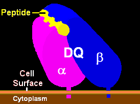
HLA-DQ (DQ) is a cell surface receptor protein found on antigen-presenting cells. It is an αβ heterodimer of type MHC class II. The α and β chains are encoded by two loci, HLA-DQA1 and HLA-DQB1, that are adjacent to each other on chromosome band 6p21.3. Both α-chain and β-chain vary greatly. A person often produces two α-chain and two β-chain variants and thus 4 isoforms of DQ. The DQ loci are in close genetic linkage to HLA-DR, and less closely linked to HLA-DP, HLA-A, HLA-B and HLA-C.

Different isoforms of DQ can bind to and present different antigens to T-cells. In this process T-cells are stimulated to grow and can signal B-cells to produce antibodies. DQ functions in recognizing and presenting foreign antigens (proteins derived from potential pathogens). But DQ is also involved in recognizing common self-antigens and presenting those antigens to the immune system in order to develop tolerance from a very young age.

When tolerance to self proteins is lost, DQ may become involved in autoimmune disease. Two autoimmune diseases in which HLA-DQ is involved are coeliac disease and type 1 diabetes. DQ mediates autoimmunity by skewing the T-cell receptor (TCR) repertoire during thymic selection.[1] Carriers of risk serotypes such as DQ8 have a higher proportion of circulating T-cell receptors that may bind insulin, the primary autoantigen in type 1 diabetes.

DQ is one of several antigens involved in rejection of organ transplants. As a variable cell surface receptor on immune cells, these D antigens, originally HL-A4 antigens, are involved in graft-versus-host disease when lymphoid tissues are transplanted between people. Serological studies of DQ recognized that antibodies to DQ bind primarily to the β-chain. The currently used serotypes are HLA-DQ2, -DQ3, -DQ4, -DQ5, -DQ6, -DQ7, -DQ8, -DQ9. HLA-DQ1 is a weak reaction to the α-chain and was replaced by DQ5 and DQ6 serology. Serotyping is capable of identifying most aspects of DQ isoform structure and function, however sequence specific PCR is now the preferred method of determining HLA-DQA1 and HLA-DQB1 alleles, as serotyping cannot resolve, often, the critical contribution of the DQ α-chain. This can be compensated for by examining DR serotypes as well as DQ serotypes.

Structure, Functions, Genetics

[edit]
HLA DQ Receptor with bound peptide and TCR

Function

[edit]

The name 'HLA DQ' originally describes a transplantation antigen of MHC class II category of the major histocompatibility complex of humans; however, this status is an artifact of the early era of organ transplantation.

HLA DQ functions as a cell surface receptor for foreign or self antigens. The immune system surveys antigens for foreign pathogens when presented by MHC receptors (like HLA DQ). The MHC Class II antigens are found on antigen presenting cells (APC) (macrophages, dendritic cells, and B-lymphocytes). Normally, these APC 'present' class II receptor/antigens to a great many T-cells, each with unique T-cell receptor (TCR) variants. A few TCR variants that recognize these DQ/antigen complexes are on CD4 positive (CD4+) T-cells. These T-cells, called T-helper cells, can promote the amplification of B-cells which, in turn recognize a different portion of the same antigen. Alternatively, macrophages and other megalocytes consume cells by apoptotic signaling and present self-antigens. Self antigens, in the right context, form a regulatory T-cell population that protects self tissues from immune attack or autoimmunity.

Genetics

[edit]
HLA region of chromosome 6

HLA-DQ (DQ) is encoded on the HLA region of chromosome 6p21.3, in what was classically known as the "D" antigen region. This region encoded the subunits for DP,-Q and -R which are the major MHC class II antigens in humans. Each of these proteins have slightly different functions and are regulated in slightly different ways.

DQ is made up of two different subunits to form an αβ-heterodimer. Each subunit is encoded by its own "gene" (a coding locus). The DQ α subunit is encoded by the HLA-DQA1 gene and the DQ β subunit is encoded by the HLA-DQB1 gene. Both loci are variable in the human population (see regional evolution).

Detecting DQ isoforms

[edit]

In the human population DQ is highly variable, the β subunit more so than the alpha chain. The variants are encoded by the HLA DQ genes and are the result of single nucleotide polymorphisms (SNP). Some SNP result in no change in amino-acid sequence. Others result in changes in regions that are removed when the proteins is processed to the cell surface, still others result in change in the non-functional regions of the protein, and some changes result in a change of function of the DQ isoform that is produced. The isoforms generally change in the peptides they bind and present to T-cells. Much of the isoform variation in DQ is within these 'functional' regions.

Serotyping. Antibodies raised against DQ tend to recognize these functional regions, in most cases the β-subunit. As a result, these antibodies can discriminate different classes of DQ based on the recognition similar DQβ proteins known as serotypes.

An example of a serotype is DQ2.

  • Recognize HLA-DQB1*02 gene products which include gene products of the following alleles:
    • HLA-DQB1*02:01
    • HLA-DQB1*02:02
    • HLA-DQB1*02:03

Sometimes DQ2 antibodies recognize other gene products, such as DQB1*03:03, resulting in serotyping errors. Because of this mistyping serotyping is not as reliable as gene sequencing or SSP-PCR.

While the DQ2 isoforms are recognized by the same antibodies, and all DQB1*02 are functionally similar, they can bind different α subunit and these αβ isoform variants can bind different sets of peptides. This difference in binding is an important feature that helps to understand autoimmune disease.

The first identified DQ were DQw1 to DQw3. DQw1 (DQ1) recognized the alpha chain of DQA1*01 alleles. This group was later split by beta chain recognition to DQ5 and DQ6. DQ3 is known as broad antigen serotypes, because they recognize a broad group of antigens. However, because of this broad antigen recognition their specificity and usefulness is somewhat less than desirable.

For most modern typing the DQ2, DQ4 - DQ9 set is used.

DQA1-B1 haplotypes in Caucasian Americans
DQ DQ DQ Freq
Serotype cis-isoform Subtype A1 B1 %[2] rank
DQ2 α5-β2 2.5 05:01 02:01 13. 16 2nd
α2-β2 2.2 02:01 02:02 11. 08 3rd
α3-β2 2.3 03:02 02:02 0. 08
DQ4 α3-β4 4.3 03:01 04:02 0. 03
03:02 04:02 0. 11
α4-β4 4.4 04:01 04:02 2. 26
DQ5 α1-β5.1 5.1 01:01 05:01 10. 85 5th
01:02 05:01 0. 03
01:03 05:01 0. 03
01:04 05:01 0. 71
α1-β5.2 5.2 01:02 05:02 1. 20
01:03 05:02 0. 05
α1-β5.3 5.3 01:04 05:03 2. 03
α1-β5.4 5.4 01:02 05:04 0. 08
DQ6 α1-β6.1 6.1 01:03 06:01 0. 66
α1-β6.2 6.2 01:02 06:02 14. 27 1st
01:03 06:02 0. 03
01:04 06:02 0. 03
α1-β6.3 6.3 01:02 06:03 0. 27
01:03 06:03 5. 66 8th
α1-β6.4 6.4 01:02 06:04 3. 40 10th
α1-β6.9 6.9 01:02 06:09 0. 71
DQ7 α2-β7 7.2 02:01 03:01 0. 05
α3-β7 7.3 03:01 03:01 0. 16
03:03 03:01 6. 45 7th
03:01 03:04 0. 09
03:02 03:04 0. 09
α4-β7 7.4 04:01 03:01 0. 03
α5-β7 7.5 05:05 03:01 11. 06 4th
α6-β7 7.6 06:01 03:01 0. 11
DQ8 α3-β8 8.1 03:01 03:02 9. 62 6th
03:02 03:02 0. 93
DQ9 α2-β9 9.2 02:01 03:03 3. 66 9th
α3-β9 9.3 03:02 03:03 0. 79
DQA1*03:02 & *03:03 not resolved; DQB1*05:01 & *05:05
, and some *03:03 are resolvable by haplotype

Genetic Typing. With the exception of DQ2 (*02:01) which has a 98% detection capability, serotyping has drawbacks in relative accuracy. In addition, for many HLA studies genetic typing does not offer that much greater advantage over serotyping, but in the case of DQ there is a need for precise identification of HLA-DQB1 and HLA-DQA1 which cannot be provided by serotyping.

Isoform functionality is dependent on αβ composition. Most studies indicate a chromosomal linkage between disease causing DQA1 and DQB1 genes. Therefore, the DQA1, α, component is as important as DQB1. An example of this is DQ2, DQ2 mediates Coeliac disease and Type 1 diabetes but only if the α5 subunit is present. This subunit can be encoded by either DQA1*05:01 or DQA1*05:05. When the DQ2 encoding β-chain gene is on the same chromosome as the α5 subunit isoform, then individuals who have this chromosome have a much higher risk of these two disease. When DQA1 and DQB1 alleles are linked in this way they form a haplotype. The DQA1*05:01-DQB1*02:01 haplotype is called the DQ2.5 haplotype, and the DQ that results α5β² is the "cis-haplotype" or "cis-chromosomal" isoform of DQ2.5

To detect these potential combinations one uses a technique called SSP-PCR (Sequence specific primer polymerase chain reaction). This techniques works because, outside of a few areas of Africa, we know the overwhelming majority of all DQ alleles in the world. The primers are specific for known DQ and thus, if a product is seen it means that gene motif is present. This results in nearly 100% accurate typing of DQA1 and DQB1 alleles.

'How does one know which isoforms are functionally unique and which isoforms are functionally synonymous with other isoforms'?. The IMGT/HLA database also provides alignments for various alleles, these alignments show the variable regions and conserved regions. By examining the structure of these variable regions with different ligands bound (such as the MMDB) one can see which residues come into close contact with peptides and those have side chains that are distal. Those changes more than 10 angstroms away generally do not affect binding of peptides. The structure of HLA-DQ8/insulin peptide at NCBI can be view with Cn3D or Rasmol. In Cn3D one can highlight the peptide and then select for amino acids within 3 or more Angstroms of the peptide. Side chains that come close to the peptide can be identified and then examined on the sequence alignments at IMGT/HLA database.

Effects of heterogeneity of isoform pairing

[edit]

As an MHC class II antigen-presenting receptor, DQ functions as a dimer containing two protein subunits, alpha (DQA1 gene product) and beta (DQB1 gene product), a DQ heterodimer. These receptors can be made from alpha+beta sets of two different DQ haplotypes, one set from the maternal and paternal chromosome. If one carries haplotype -A-B- from one parent and -a-b- from the other, that person makes 2 alpha isoforms (A and a) and 2 beta isoforms (B and b). This can produce 4 slightly different receptor heterodimers (or more simply, DQ isoforms). Two isoforms are in the cis-haplotype pairing (AB and ab) and 2 are in the trans-haplotype pairing (Ab and aB). Such a person is a double heterozygote for these genes, for DQ the most popular situation. If a person carries haplotypes -A-B- and -A-b- then they can only make 2 DQ (AB and Ab), but if a person carries haplotypes -A-B- and -A-B- then they can only make DQ isoform AB, called a double homozygote. In coeliac disease, certain homozygotes and are at higher risk for disease and some specific complications of coeliac disease such as Gluten-sensitive enteropathy associated T-cell lymphoma

Homozygotes and double homozygotes
Homozygotes at DQ loci can change risk for disease. In mice for instance, mice with 2 copies of the DQ-like Iab haplotype are more likely to progress toward fatal disease compared to mice that are heterozygotes only for the beta allele (MHC IAαb / IAαb, IAβb / IAβbm12). In humans, celiac disease DQ2.5/DQ2 homozygotes are several times more likely to have celiac disease versus DQ2.5/DQX individuals.[3] DQ2/DQ2 homozygotes are at elevated risk for severe complications of disease.[4] For an explanation of the risk association see:Talk:HLA-DQ#Effects of heterogeneity of isoform pairing-Expanded

Involvement of transhaplotypes in disease
There is some controversy in the literature whether trans-isoforms are relevant. Recent genetic studies into coeliac disease have revealed that the DQA1*05:05:X/Y:DQB1*02:02 gene products explain disease not linked to the haplotype that produces DQ8 and DQ2.5, strongly suggesting the trans-isoforms can be involved in disease. But, in this example, it is known that the transproduct is almost identical to a known cis-'isoform' produced by DQ2.5. There is other evidence that some haplotypes are linked to disease but show neutral linkage with other particular haplotypes are present. At present, the bias of relative isoform frequency toward cis pairing is unknown, it is known that some trans-isoforms occur.

DQ Function in Autoimmunity

[edit]

HLA D (-P,-Q,-R) genes are members of the Major histocompatibility complex (MHC) gene family and have analogs in other mammalian species. In mice the MHC locus known as IA is homologous to human HLA DQ. Several autoimmune diseases that occur in humans that are mediated by DQ also can be induced in mice and are mediated through IA. Myasthenia gravis is an example of one such disease.[5] Linking specific sites on autoantigens is more difficult in humans due to the complex variation of heterologous humans, but subtle differences in T-cell stimulation associated with DQ-types has been observed.[6] These studies indicate that potentially a small change or increase in the presentation of a potential self-antigen can result in autoimmunity. This may explain why there is often linkage to DR or DQ, but the linkage is often weak.

Regional Evolution

HLA DQ- subunit alleles, mature chains, contact variants
Known HLA-DQ Potential
Locus: A1 B1 Combinations
Alleles 33 78 2574
Subunit: α β isoforms
Mature Chains 24 58 1392
Contact Variants* ~9 40 360
Caucasian (USA)
Contact Variants (CV) 7 12 84
CV-haplotypes 30
*Subunits vary within 9Â of peptide in DQ2.5 or DQ8

Many HLA DQ were under positive selection of 10,000s potentially 100,000s of years in some regions. As people moved they have tended to lose haplotypes and in the process lose allelic diversity. On the other hand, on arrival at new distal locations, selection would offer unknown selective forces that would have initially favored diversity in arrivals. By an unknown process, rapid evolution occurs, as has been seen in South Americas indigenous population (Parham and Ohta, 1996, Watkins 1995), and new alleles rapidly appear. This process may be of immediate benefit of being positively selective in that new environment, but these new alleles might also be 'sloppy' in a selective perspective, having side effects if selection changed. The table to the left demonstrates how absolute diversity at the global level translates into relative diversity at the regional level.

-

Heterozygous DQ Combinations and Disease

[edit]
DQ2.5 DQ8 DQ2.5/8
Sweden 15.9 18.7 5.9
Jalisco 11.4 22.8 5.2
England 12.4 16.8 4.2
Kazakh 13.1 11 2.9
Uygur 12.6 11.4 2.9
Finland 9 15.7 2.8
Poland 10.7 9.9 2.1

DQ2.5/DQ8 Heterozygotes

[edit]

The distribution of this genotype is largely the result of admixtures between peoples of eastern or central Asian origin and peoples of western or central Asian origin. The highest frequencies, by random mating, are expected in Sweden, but pockets of high levels also occur in Mexico, and a larger range risk exists in Central Asia.

Diseases that appear to be increased in heterozygotes are celiac disease and type 1 diabetes. New evidence[timeframe?] is showing an increased risk for late onset type 1 diabetes in Heterozygotes (which includes ambiguous type 1/type 2 diabetes). 95% of celiac disease patients are positive for DQ2 or DQ8.[7]

References

[edit]
[edit]
Revisions and contributorsEdit on WikipediaRead on Wikipedia
from Grokipedia
HLA-DQ is a cell-surface and a member of the (HLA) class II family, encoded within the (MHC) on the short arm of at position 6p21.3. It forms a transmembrane heterodimer composed of an alpha chain, encoded by the polymorphic HLA-DQA1 gene, and a beta chain, encoded by the polymorphic HLA-DQB1 gene, with the two genes located adjacent to each other, approximately 4 kilobases apart, separated by a non-coding region. The alpha chain is approximately 34 kDa, while the beta chain is about 29 kDa, and both chains feature extracellular domains that create a peptide-binding groove for antigen presentation. The primary function of HLA-DQ is to present peptides derived from extracellular proteins to CD4+ T lymphocytes, thereby initiating adaptive immune responses against pathogens and foreign antigens. This process occurs on the surface of professional antigen-presenting cells, including B lymphocytes, macrophages, and dendritic cells, where HLA-DQ molecules bind and display antigenic fragments to T-cell receptors. Due to high polymorphism in HLA-DQA1 (966 alleles) and HLA-DQB1 (2924 alleles, as of 2025), HLA-DQ exhibits diverse peptide-binding specificities, influencing immune recognition and response variability across individuals. HLA-DQ plays a critical role in transplantation immunology, as mismatches in alleles are associated with increased risk of in transplants. It is also implicated in autoimmune disorders; for instance, specific HLA-DQ alleles, such as those forming DQ2 and DQ8 heterodimers, confer strong susceptibility to celiac disease by preferentially binding gluten-derived peptides. Additionally, certain HLA-DQ variants are linked to and other immune-mediated conditions, highlighting its influence on disease predisposition through altered .

Structure and Biochemistry

Protein Composition

HLA-DQ is a major complex (MHC) class II molecule that functions as a heterodimer composed of two non-covalently associated polypeptide chains: an α-chain encoded by the DQA1 gene with an approximate molecular weight of 34 kDa, and a β-chain encoded by the DQB1 gene with an approximate molecular weight of 29 kDa. These chains are transmembrane glycoproteins integral to the cell surface of antigen-presenting cells. Each chain features a modular domain architecture typical of MHC class II proteins. The α-chain includes two extracellular domains—α1 (approximately 82 amino acids) and α2—followed by a transmembrane helix and a short cytoplasmic tail. Similarly, the β-chain comprises extracellular β1 (approximately 87 amino acids in common alleles) and β2 domains, a transmembrane region, and a cytoplasmic tail. The α2 and β2 domains adopt an immunoglobulin-like fold, which stabilizes the overall heterodimer structure through non-covalent interactions. Post-translational modifications play a crucial role in the maturation of HLA-DQ. Both chains undergo N-linked glycosylation, with a conserved site at 86 in the α1 domain of the α-chain and at 19 (position 15 in standardized numbering) in the β1 domain of the β-chain. These modifications are vital for facilitating proper in the and ensuring efficient transport through the secretory pathway to the cell surface. The α1 and β1 domains exhibit remarkable evolutionary conservation of key residues across mammalian species, particularly those forming the peptide-binding platform, underscoring their essential function in immune recognition. This conservation highlights the structural integrity preserved from early mammalian divergence to maintain capabilities.

Heterodimer Assembly

The alpha (DQA1) and beta (DQB1) chains of HLA-DQ are synthesized separately in the rough (ER) of antigen-presenting cells, where they undergo initial folding and before associating to form the alpha-beta heterodimer. This association occurs through non-covalent interactions between the immunoglobulin-like domains of the chains, facilitated by chaperone proteins that ensure proper . The invariant chain (Ii), encoded by CD74, plays a crucial role by binding to newly formed alpha-beta dimers in the ER, stabilizing them and preventing premature binding while directing the complex to endosomal compartments for subsequent maturation. Unlike HLA-DR, which requires Ii for efficient dimer formation, HLA-DQ can form initial alpha-beta pairs in the ER even in the absence of Ii, though Ii enhances the stability of these dimers, particularly for SDS-resistant conformations. HLA-DM and HLA-DO serve as additional chaperones in the assembly and quality control process, with their activities extending from the ER to the MHC class II compartments (MIICs). HLA-DM interacts with alpha-beta dimers to edit peptide occupancy, promoting the release of low-affinity peptides like CLIP (class II-associated invariant chain peptide) and facilitating the loading of higher-affinity antigens, which indirectly supports stable heterodimer maturation. HLA-DO acts as a co-chaperone by associating with HLA-DM in the ER and modulating its activity, preferentially aiding the assembly of DM-dependent MHC class II molecules like certain HLA-DQ variants while inhibiting DM's peptide-editing function to fine-tune repertoire selection. In heterozygotes, HLA-DQ heterodimers can form in cis (alpha and beta chains from the same haplotype) or trans (from different haplotypes), but cis pairing is far more efficient due to complementary polymorphisms that enhance chain compatibility, whereas trans pairing yields less stable dimers with reduced surface expression. The stability of the HLA-DQ heterodimer relies on a network of hydrogen bonds and hydrophobic interactions primarily between the alpha1 and beta1 domains, which form the -binding groove. For instance, conserved hydrogen bonds link the main-chain atoms of the chains, while residues like Arg-76α form salt bridges with Asp-57β, bolstering the interface; hydrophobic contacts, such as those involving Trp-48α with the beta2 domain, further reinforce this structure. Heterodimer stability is also pH-dependent, with acidic conditions in endosomes (pH ~5.0-5.5) promoting conformational changes that optimize peptide loading for variants like HLA-DQ3.2, whereas neutral pH in the ER maintains initial dimer integrity but limits binding for less stable alleles like HLA-DQ3.1. Unpaired alpha or beta chains that fail to form proper heterodimers are recognized as misfolded and targeted for degradation via ER-associated degradation (ERAD), a proteasome-dependent pathway that extracts and ubiquitinates them in the ER membrane, preventing their accumulation and ensuring cellular .

Peptide Binding

The peptide binding groove of HLA-DQ is formed by the membrane-distal alpha1 and beta1 domains of the alpha-beta heterodimer, creating a polymorphic cleft that is open at both ends to accommodate elongated peptides typically 10-25 amino acids in length. This open-ended architecture contrasts with the closed grooves of MHC class I molecules and allows flexibility in peptide register, with many HLA-DQ isoforms exhibiting a core binding motif centered on a 9-amino acid segment while permitting overhanging residues at the N- and C-termini. Crystal structures of HLA-DQ2 and DQ8 in complex with peptides confirm that the groove's floor and walls are lined by conserved and variable residues that stabilize peptide hydrogen bonds and side-chain interactions, respectively. Binding specificity within the groove is dictated by five primary pockets—P1, P4, P6, P7, and P9—that engage the side chains of anchor residues protruding downward from the backbone. These pockets provide allele-specific selectivity; for instance, in the celiac disease-associated HLA-DQ2.5 (DQA105:01/DQB102:01), the P1 and P9 pockets favor bulky hydrophobic or polar residues such as , enabling stable accommodation of deamidated peptides like QLQPFPQPELPY, where glutamines occupy these positions. Experimental binding assays and structural analyses further reveal preferences for negatively charged residues (e.g., glutamate) at P4, P6, or P7 in DQ2.5, which enhance affinity through electrostatic interactions with positively charged groove residues. Polymorphisms in the beta chain significantly modulate pocket properties and overall binding repertoire, particularly through alterations in electrostatic environments. A key example is position beta57, which lines the P9 : in (DQB102:01), serine at beta57 results in a neutral that tolerates non-negatively charged anchors at P9, whereas at beta57 in (DQB103:02) imparts a negative charge, favoring positively charged or hydrophobic residues and restricting the repertoire accordingly. This charge variation at beta57 influences P1 accessibility indirectly by affecting groove conformation, thereby altering specificity for in autoimmune contexts. In the late endosomal MIIC compartment, acts as a peptide editor by catalyzing the exchange of low-stability for higher-affinity ligands on HLA-DQ molecules. This process involves binding to the lateral surfaces of HLA-DQ, inducing conformational changes in the peptide binding groove that accelerate CLIP dissociation and promote selective loading of antigenic with optimal half-lives. For disease-associated alleles like DQ2 and DQ8, HLA-DM editing is less efficient due to intrinsic peptide-MHC stability differences, leading to retention of suboptimal in the repertoire.

Genetics and Nomenclature

Gene Loci

The HLA-DQA1 and genes, which encode the alpha and beta chains of the HLA-DQ heterodimer, are located on the short arm of at position 6p21.3, within the ( region. This region spans approximately 1.1 megabases and is situated about 110 kilobases centromeric to the locus. The precise genomic coordinates for HLA-DQA1 are 6:32,637,406-32,655,272 (GRCh38), encompassing roughly 18 kb, while is positioned telomerically at 6:32,659,467-32,666,684, spanning about 7 kb. The HLA-DQA1 gene consists of five exons: exon 1 encodes the leader peptide, exons 2 and 3 encode the alpha1 and alpha2 extracellular domains, respectively, and exon 4 encodes the transmembrane domain and part of the cytoplasmic tail. The HLA-DQB1 gene has a similar organization but includes six exons, with exon 1 for the leader peptide, exons 2 and 3 for the beta1 and beta2 extracellular domains, exon 4 for the transmembrane and partial cytoplasmic regions, exon 5 for the remaining cytoplasmic domain, and exon 6 present in some transcripts. Nearby, the HLA-DQB2 pseudogene is located within the same MHC class II region, approximately adjacent to the DQA2 locus, and shares high sequence homology with functional DQB1 alleles but lacks full transcriptional activity due to structural features that prevent complete expression. The HLA-DQA1 and genes exhibit strong (LD) within extended MHC haplotypes, where specific combinations are inherited together at frequencies far exceeding random assortment. For instance, the DQA105:01 and DQB102:01 form the DQ2.5 haplotype, which is in tight LD with the HLA-DRB1*03:01 on the same . This LD pattern reflects the evolutionary conservation of MHC haplotypes and contributes to the coordinated inheritance of HLA-DQ variants. Humans typically possess one functional copy of the HLA-DQA1 and genes per , resulting in two copies total (one on each homolog). However, rare copy number variations (CNVs), including duplications or deletions affecting these loci, have been documented in certain populations and disease contexts, such as systemic lupus erythematosus, where they may influence thresholds. These CNVs occur at low frequencies and can alter , though the standard diploid configuration predominates.

Allelic Diversity

The allelic diversity of HLA-DQ is governed by the International ImMunoGeneTics information system (IMGT)/HLA , maintained by the Nomenclature Committee for Factors of the HLA System. This standardized system assigns unique identifiers to alleles based on sequencing, using a four-field format such as DQA1*01:02:01, where the first field denotes the gene (e.g., DQA1 or DQB1), the second field specifies the protein-level variation, the third indicates synonymous substitutions in the , and the fourth captures non-synonymous changes outside the coding sequence or in introns. This ensures precise tracking of genetic variants, with new alleles officially named only through submission to the IMGT/HLA database. As of release 3.62.0 (October 2025), the IMGT/HLA database catalogs over 910 alleles for the HLA-DQA1 and 2,910 alleles for the , reflecting extensive polymorphism primarily in the extracellular domains that influence . The DQB1 locus exhibits greater diversity than DQA1, with polymorphisms concentrated in 2, which encodes the β1 domain responsible for peptide binding; this region accounts for the majority of sequence variations observed across alleles. In contrast, the DQA1 shows lower polymorphism, with fewer allelic variants and variations more evenly distributed, though still focused on the α1 domain encoded by 2. Historically, HLA-DQ alleles were classified into serologic types based on reactivity, such as DQ2 (associated with DQB102 alleles) and DQ5 (associated with DQB105 alleles), which provided broad groupings but lacked resolution for functional differences. Modern molecular typing has refined these into protein-level isoforms, exemplified by DQ2.5 (encoded by DQA105:01 and DQB102:01), allowing for more accurate correlation with immunological roles. Population frequencies of HLA-DQ alleles vary significantly worldwide, contributing to differential disease susceptibilities. For instance, the DQ2.5 isoform is present in over 90% of celiac disease cases among Northern Europeans, where its carrier frequency in the general population reaches 20-30%. Global diversity is higher in African and Asian populations, with broader haplotype distributions and rarer alleles reflecting ancient migrations and genetic admixture; for example, unique DQB1 variants are more prevalent in sub-Saharan Africans compared to Europeans. This elevated variability in non-European groups underscores the role of HLA-DQ in shaping population-specific immune responses.

Isoform Pairing

HLA-DQ isoforms are formed by the pairing of alpha (DQA1) and beta (DQB1) chains to create functional αβ heterodimers, with pairing occurring preferentially in cis configuration, where the chains are encoded on the same parental (). However, trans pairing, involving chains from opposite chromosomes, is also possible and contributes to isoform diversity, though it is often limited by the stability of the resulting heterodimer. For instance, stable cis pairings include DQA105:01 with DQB102:01 to form the DQ2.5 isoform, while mismatched trans pairs, such as those between certain DQA101 and DQB102/03/04 alleles, may result in unstable or non-functional molecules that fail to reach the cell surface. Isoform nomenclature follows the HLA serotype system with numeric suffixes to denote specific combinations, such as DQ2.5 (DQA105:01-DQB102:01), DQ2.2 (DQA102:01-DQB102:02), and DQ8.1 (DQA103:01-DQB103:02), reflecting the predominant cis pairings within haplotypes. Over 20 common isoforms have been identified based on frequently occurring pairs across populations, with stability influenced by structural compatibility between the alpha and beta chains; for example, pairings within group G1 (DQA102/03/04/05/06 with DQB102/03/04) or G2 (DQA101 with DQB105/06) are generally more stable than inter-group combinations. These isoforms are distinguished by their unique peptide-binding grooves, which determine specificity. In heterozygous individuals, trans pairing can generate novel isoforms with altered repertoires, expanding the range of presented antigens and potentially influencing immune responses; for example, the trans-encoded DQ2.3 (DQA103:01-DQB102:01) exhibits preferences for negatively charged anchors at specific positions, differing from cis isoforms like DQ2.5. These novel heterodimers are detectable through cell surface expression on antigen-presenting cells, as only stable pairs traffic successfully to the membrane. HLA-DQ genes follow an autosomal codominant inheritance pattern, meaning both parental alleles are expressed, and with heterozygosity exceeding 50% in many populations due to high allelic diversity, individuals often express multiple isoforms—up to four in some cases from cis and trans combinations.

Physiological Functions

Antigen Presentation

HLA-DQ molecules primarily present exogenous antigens derived from extracellular sources through the endocytic pathway in professional antigen-presenting cells such as dendritic cells, macrophages, and B cells. Exogenous antigens are internalized via , macropinocytosis, or and transported to late endosomal-lysosomal compartments, where they are degraded by acid hydrolases and proteases like cathepsins into peptides suitable for binding. These peptides, typically 13-25 long, are loaded onto HLA-DQ in the compartment (MIIC), a specialized acidic vesicular structure enriched with lysosomal enzymes and molecules. The low (around 4.5-5.5) in the MIIC facilitates peptide binding by promoting conformational changes in the HLA-DQ peptide-binding groove. The invariant chain (Ii), also known as CD74, plays a crucial role in this process by associating with newly synthesized HLA-DQ αβ heterodimers in the , preventing premature binding and directing the complex through the Golgi apparatus to the MIIC via endocytic sorting signals in Ii's cytoplasmic tail. Within the MIIC, Ii is proteolytically degraded by cathepsins, leaving a remnant fragment called CLIP (class II-associated invariant chain ) bound in the peptide groove. , a non-polymorphic MHC class II-like molecule, acts as a peptide editor by catalyzing the removal of CLIP and facilitating the exchange for higher-affinity antigenic ; while enhances loading on HLA-DQ, it is not strictly essential for SDS-stable dimer formation, unlike in . This exchange ensures a diverse of stable -HLA-DQ complexes optimized for immune recognition. Once loaded, peptide-HLA-DQ complexes are transported from the MIIC to the plasma membrane, often recycling through early endosomes, to present antigens on the cell surface. The surface of these complexes varies by cell type and activation state, with approximately 5-6 hours in dendritic cells and 10-12 hours in B cells. In contrast, endogenous antigens from intracellular sources are rarely presented by HLA-DQ through autophagy-mediated pathways, where cytoplasmic proteins are sequestered into autophagosomes that fuse with MIIC for processing and loading onto HLA-DQ. This macroautophagy-dependent mechanism contributes to a minor fraction (approximately 10-25%) of the peptidome derived from self-proteins, enabling surveillance of intracellular threats.

T-Cell Interaction

HLA-DQ molecules, as proteins, present antigenic s to + T cells, where the -MHC (pMHC) complex is recognized by the (TCR). The TCR binds diagonally across the HLA-DQ α-helix and the , with (CDR) loops of the TCR engaging both the polymorphic helices of HLA-DQ and the exposed residues, determining the specificity and affinity of the interaction. The co-receptor further stabilizes this engagement by binding to invariant regions on HLA-DQ, enhancing the overall affinity of the trimolecular complex (TCR-pMHC-) by orders of magnitude compared to TCR-pMHC alone, which is crucial for sensitive detection of low-abundance antigens. This affinity is modulated by the , as variations in anchoring and solvent-exposed residues influence TCR docking and T cell activation thresholds. Upon recognition, TCR clustering on the T cell surface initiates by recruiting and activating Src family kinases like Lck, leading to of immunoreceptor tyrosine-based motifs (ITAMs) on the associated CD3 and ζ chains. Phosphorylated ITAMs serve as docking sites for ZAP-70 , which propagates downstream signaling cascades, including of via the CBM complex and AP-1 through the RAS-ERK pathway, ultimately driving transcription of genes for T cell and effector functions. These pathways culminate in production, such as IL-2 for autocrine growth and IFN-γ for promoting and antiviral responses. The nature of peptides presented by HLA-DQ influences + T cell differentiation into helper subsets, with the milieu and co-stimulatory signals determining Th1, Th2, or Th17 polarization. Variations in HLA-DQ isoforms can affect the strength and specificity of these responses. HLA-DQ can also participate in allorecognition, where T cells respond to foreign HLA-DQ molecules, contributing to immune responses against allogeneic cells.

Immune Tolerance

HLA-DQ molecules play a critical role in central tolerance by presenting self-peptides derived from tissue-restricted antigens to developing thymocytes in the , thereby promoting the deletion of autoreactive + T cells. In medullary thymic epithelial cells (mTECs), the (Aire) drives the ectopic expression of peripheral tissue antigens, which are then loaded onto HLA-DQ for display on the cell surface. This ensures negative selection, where thymocytes recognizing self-peptides with high affinity undergo , preventing their maturation into potentially autoreactive T cells. Inefficient peptide editing by on certain HLA-DQ variants can lead to prolonged presentation of invariant peptides like CLIP, potentially impairing this deletion and allowing low-affinity autoreactive clones to escape. In peripheral tolerance, HLA-DQ contributes to the maintenance of self-tolerance outside the thymus by facilitating anergy in autoreactive T cells or inducing regulatory T cells (Tregs) in lymphoid tissues like lymph nodes. Bone marrow-derived antigen-presenting cells (APCs), such as dendritic cells, express HLA-DQ to present self-peptides at low avidity, which promotes the differentiation of FoxP3+ Tregs capable of suppressing inflammatory responses. Self-peptide presentation by HLA-DQ enhances Treg induction, increasing FoxP3 expression and associated markers like CTLA4 and TIGIT, thereby reinforcing peripheral suppression of autoreactivity. Certain HLA-DQ isoforms influence tolerance induction through differences in peptide-binding motifs and dimer stability, affecting the of self-peptides for tolerogenic outcomes. Developmental regulation of HLA-DQ expression ensures its role in tolerance is contextually appropriate, with constitutive low-level expression in professional APCs and minimal presence in non-APCs under steady-state conditions. During inflammation, proinflammatory cytokines such as IFNγ and TNFα upregulate HLA-DQ on non-APCs like endothelial cells, requiring sustained stimulation for surface expression, which may transiently enhance self-peptide to bolster tolerance amid immune activation. This dynamic control, mediated by transcription factors like CIITA, prevents excessive autoantigen exposure while allowing adaptive responses.

Role in Autoimmunity

Molecular Mimicry

Molecular mimicry represents a key mechanism by which HLA-DQ molecules contribute to the onset of , wherein microbial peptides exhibit structural or sequence similarity to self-peptides, allowing them to bind the same HLA-DQ isoform and activate autoreactive CD4+ T cells, thereby breaching . This arises due to the degeneracy of T cell receptors (TCRs), which can recognize both foreign and self-peptide-HLA-DQ complexes with sufficient affinity to initiate an against host tissues. For instance, viral epitopes from pathogens such as have been shown to mimic autoantigens like myelin basic protein when presented by HLA class II molecules. Certain HLA-DQ isoforms, such as DQ2.5, display a binding bias for specific motifs like polyproline-rich sequences found in both microbial antigens and self-peptides, such as those derived from or insulin, facilitating in genetically susceptible individuals. These isoforms preferentially accommodate peptides with residues at key anchor positions (e.g., P4, P6, P7), enabling pathogens harboring similar motifs to exploit the same binding groove and provoke unintended self-reactivity. This preference underscores how allelic variations in HLA-DQ can heighten vulnerability to without altering the core presentation function. Following initial activation through , epitope spreading can occur, where the expands from the mimicking foreign to encompass additional, non-mimicking self-s presented by HLA-DQ, thereby amplifying and perpetuating . This process involves bystander activation and diversification of the T cell repertoire, leading to broader tissue damage over time. Environmental triggers, such as viral infections (e.g., ) or dietary antigens, initiate this in individuals with predisposing HLA-DQ genotypes by providing the initial foreign s that cross-react with self. These triggers exploit the HLA-DQ's role in antigen surveillance, converting a protective response into pathological .

Isoform-Specific Risks

The HLA-DQ2.5 isoform, encoded by DQA105:01-DQB102:01, exhibits high affinity for peptides, which feature a 9-amino acid core with residues at positions P3 and P8, facilitating strong binding and presentation to + T cells in celiac disease pathogenesis. This binding preference arises from the isoform's peptide-binding groove, which accommodates negatively charged residues introduced by 2 deamidation, thereby amplifying gluten-specific immune responses. Additionally, DQ2.5 presents peptides from the insulin B chain, contributing to autoreactive T-cell activation in . In contrast, the isoform (DQA103:01-DQB103:02) shows a strong preference for and other acidic residues at anchor positions P4 and P7 within bound peptides, enabling efficient presentation of insulin-derived autoantigens in and deamidated epitopes in celiac disease. This motif specificity enhances the stability of peptide-MHC complexes, promoting pathogenic T-cell responses in these conditions. However, DQ8 demonstrates weaker associations with compared to other HLA alleles, with limited evidence for a major predisposing role. Certain isoforms confer protection against ; notably, DQ6.2 (DQB1*06:02) inhibits the function of DQ8 through trans-heterodimer pairing, forming complexes with reduced peptide-binding capacity that diminish autoreactive responses. This interaction provides dominant protection in , reducing disease risk by 80-90% even in individuals carrying high-risk DQ8 alleles. Quantitative assessments underscore these isoform-specific effects: for instance, DQ2.5 homozygotes face approximately fourfold higher odds of developing celiac disease compared to heterozygotes, reflecting dose-dependent enhancement of peptide presentation.

Heterodimer Heterozygosity

In heterozygous individuals for HLA-DQ loci, such as those possessing the DQ2.5 (DQA105:01-DQB102:01) and DQ8 (DQA103:01-DQB103:02) haplotypes, alpha and beta chains from different parental alleles can assemble into trans heterodimers, exemplified by the DQA105:01-DQB103:02 pair. These novel trans combinations are functionally expressed on antigen-presenting cells and broaden the peptide-binding repertoire beyond that of cis heterodimers alone, enabling the presentation of a more diverse array of antigens, including self-peptides that may trigger autoreactive T-cell responses. This expanded repertoire in trans heterodimers heightens the potential for autoantigen presentation, contributing to elevated autoimmune risk; for instance, the DQ2.5/DQ8 exhibits additive susceptibility in celiac disease, with a of approximately 14-fold in European populations compared to non-carriers. However, such heterozygosity can modulate risk in opposing directions across diseases. Certain beta chains, such as the DQB1*03:02 allele in DQ8, exhibit dominance in heterodimer assembly, preferentially with available alpha chains and thereby influencing the relative expression levels and functional output of specific DQ isoforms on cell surfaces. This dominance effect stems from structural features in the beta chain that dictate efficiency and stability, leading to skewed isoform representation in heterozygotes. DQ2/DQ8 heterozygosity is prevalent in populations with mixed ancestries, where varying frequencies contribute to heterogeneous ; for example, approximately 28% of patients carry this combination, underscoring its role in modulating autoimmune outcomes across diverse genetic backgrounds.

Disease Associations

Celiac Disease

Celiac disease (CD) is strongly associated with specific HLA-DQ alleles, with approximately 90-95% of patients carrying the and the remainder primarily expressing . In European populations, HLA-DQ2.5 accounts for about 95% of cases, while HLA-DQ8 predominates in the small subset of non-DQ2.5 patients, highlighting the near-universal requirement for these isoforms in disease susceptibility. These alleles encode heterodimers that preferentially bind gluten-derived peptides, initiating an aberrant in the intestinal mucosa. The pathogenesis of CD involves the deamidation of gluten peptides by tissue transglutaminase 2 (TG2), which converts glutamine residues to glutamic acid, enhancing their affinity for the peptide-binding groove of HLA-DQ2.5. Deamidated gliadin peptides anchor primarily at positions P4, P6, or P7 within the DQ2.5 groove, where the negative charge of glutamic acid interacts with positively charged pockets, stabilizing the complex for presentation to CD4+ T cells. This presentation activates gluten-specific CD4+ T cells, leading to cytokine release, B-cell activation, and production of anti-gliadin and anti-TG2 antibodies, which drive villous atrophy and inflammation. Although HLA-DQ alleles confer substantial risk, they account for only 30-40% of the to , with the majority of susceptibility arising from environmental factors such as early exposure after . Individuals carrying HLA-DQ2.5 or DQ8 represent up to 40% of the general population, yet only 3% develop , underscoring the critical role of non-genetic triggers like diet timing and . Recent studies from the 2020s have elucidated additional mechanisms, including the formation of trans-heterodimers such as HLA-DQ2.5α-DQ8β (DQ8.5), which exhibit enhanced binding and presentation of peptides, potentially amplifying T-cell responses in DQ2.5/DQ8 double carriers. Furthermore, the gut influences CD risk in HLA-DQ predisposed individuals, with early —such as reduced and increased —correlating with altered tolerance and immune priming. These findings suggest microbiome modulation as a potential modifier of HLA-DQ-driven .

Type 1 Diabetes

HLA-DQ isoforms play a central role in the genetic susceptibility to (T1D), an autoimmune condition characterized by the destruction of pancreatic beta cells. The high-risk alleles HLA-DQA103:01-DQB103:02 (DQ8) and HLA-DQA105:01-DQB102:01 (DQ2.5) are found in approximately 90% of T1D patients, with DQ8 conferring a particularly strong predisposition in many populations. Heterozygosity for DQ2.5 and DQ8 synergistically elevates risk beyond that of either homozygote alone, as these molecules can form trans-heterodimers capable of presenting a broader repertoire of autoantigenic peptides. In contrast, the HLA-DQB1*06:02 allele (part of the DQ6.2 haplotype) provides dominant protection, nearly eliminating T1D risk in carriers by altering peptide binding and presentation dynamics. Central to T1D , HLA-DQ molecules present autoantigens to + T cells, initiating the autoimmune cascade. Preproinsulin serves as the primary autoantigen, with key epitopes such as the insulin B:9-23 (SHLVEALYLVCGERGFFY) binding preferentially to DQ8 through anchors at P1 (small hydrophobic), P4 (polar), and P9 (Arg residue) positions in the peptide-binding groove. This presentation activates autoreactive T cells that infiltrate the , promoting beta-cell destruction via inflammatory cytokines and cytotoxic mechanisms. For DQ2.5, proinsulin-derived , including those from the region, are similarly presented, contributing to an additive risk when combined with DQ8. These isoform-specific binding motifs explain why DQ2.5/DQ8 heterozygotes exhibit enhanced autoreactivity compared to single-allele carriers. The progression from genetic risk to clinical T1D involves HLA-DQ-mediated antigen presentation triggering beta-cell attack, often linked to earlier disease onset in high-risk genotypes. DQ8 homozygotes or those with the DQ8 haplotype tend to develop T1D at younger ages, with average onset around 13-14 years in some cohorts, reflecting accelerated autoimmunity. Data from the TEDDY study indicate that DQ2.5/DQ8 heterozygosity not only heightens overall risk but also hastens progression from islet autoimmunity to overt diabetes, particularly in the presence of environmental triggers like enteroviral infections that may enhance epitope presentation or mimicry. These gene-environment interactions underscore HLA-DQ's pivotal role in disease timing and severity.

Rheumatoid Arthritis

HLA-DQ alleles exhibit weaker associations with (RA) susceptibility compared to the shared epitope on HLA-DRB1, but they contribute through specific haplotypes and interactions with anti-citrullinated protein antibodies (ACPAs). In particular, HLA-DQB10501 (encoding DQ5) and HLA-DQB10302 have been linked to increased anti-cyclic citrullinated peptide (anti-CCP) antibody positivity in RA patients, with carriers of DQ-DR genotypes containing these susceptibility alleles showing significantly higher rates of anti-CCP production. Similarly, HLA-DQB102 (DQ2) is associated with enhanced presentation of citrullinated antigens in RA contexts, particularly in haplotype combinations with shared epitope alleles. In contrast, (DQB103:02) demonstrates protective effects in some cohorts, potentially modulating disease progression by influencing erosive joint damage. Mechanistically, HLA-DQ molecules promote pathogenesis by binding and presenting citrullinated self-peptides, including those from vimentin, to CD4+ T cells in the synovial tissue. This presentation activates autoreactive T cells, which in turn provide help to B cells for the production of ACPAs such as anti-CCP, exacerbating chronic inflammation and joint destruction. The arginine-to-citrulline increases peptide affinity for HLA-DQ binding pockets—such as positions 4, 6, 7, and 9 in DQ2—creating citrullination-specific motifs that favor neoantigen recognition and breach . These interactions align with broader roles of HLA-DQ in , where altered peptide presentation drives loss of self-tolerance. Quantitatively, HLA-DQ accounts for a modest portion of RA heritability, estimated at 10-15% in haplotype analyses, in contrast to the ~30% attributable to HLA-DR loci, underscoring DQ's secondary but synergistic influence. Ethnic variations modulate these risks, with DQ5-containing haplotypes showing stronger associations in Asian populations, such as Chinese Han, where shared epitope-DQ5 combinations elevate susceptibility to both anti-CCP-positive and -negative RA. Recent investigations, including 2024 reviews, reveal epistatic interactions between HLA-DQ and HLA-DP that influence RA severity, particularly in modulating inflammatory responses and radiographic progression. These findings highlight DQ's role in fine-tuning disease outcomes beyond primary susceptibility.

Clinical and Research Implications

Genetic Testing

Genetic testing for HLA-DQ focuses on the DQA1 and DQB1 loci to identify specific and associated with autoimmune risks. Common techniques include with sequence-specific oligonucleotide probes (PCR-SSOP), which hybridizes probes to amplified DNA for allele detection, and next-generation sequencing (NGS), which provides high-resolution typing by sequencing the full gene regions. For instance, PCR-SSOP or NGS can detect the DQB1*02:01 , a key component of the DQ2.5 , to screen for celiac disease susceptibility. In clinical practice, HLA-DQ genotyping serves as a pre-diagnostic tool for celiac disease, where a negative result for both DQ2.5 (DQA105:DQB102) and DQ8 (DQA103:DQB103:02) haplotypes rules out the disease with over 99% negative predictive value, avoiding unnecessary . For , it enables risk stratification in first-degree relatives, identifying high-risk haplotypes like DR3-DQ2 or DR4-DQ8 to guide monitoring and early intervention. Despite its utility, HLA-DQ testing has limitations, as the presence of risk alleles exhibits low —only a small fraction of carriers develop due to environmental and other genetic factors. Ethical concerns arise in , including potential psychological distress for families and risks of without actionable prevention strategies. Recent advances include portable CRISPR-Cas12a-based platforms for rapid, point-of-care detection of HLA alleles, such as the 2025 method for HLA-B*27, which could extend to DQ isoforms for faster clinical decisions. Additionally, integrating HLA-DQ genotypes with polygenic risk scores improves predictive accuracy for both celiac disease and by incorporating non-HLA loci.

Therapeutic Targeting

Therapeutic strategies targeting HLA-DQ aim to modulate its role in to mitigate autoimmune responses in diseases such as celiac disease and (T1D). Inhibitors, including small molecules and antibodies, seek to block peptide loading or HLA-DQ-peptide complex formation on antigen-presenting cells, preventing activation of autoreactive T cells. For instance, in celiac disease, oral small-molecule inhibitors like IMT-514 from IM Therapeutics target to disrupt peptide binding and presentation, showing preclinical efficacy in reducing T-cell responses to gluten epitopes. Similarly, for T1D, IMT-002, another small-molecule inhibitor, demonstrated safety and pharmacokinetics in a phase 1 multiple ascending dose trial in HLA-DQ8-positive patients, with ongoing development to inhibit islet autoantigen presentation. Antibody-based approaches further exemplify targeted inhibition. A bispecific (DONQ52) specific to HLA-DQ2.5-gluten complexes potently blocked gluten-induced T-cell responses in celiac patients during gluten challenges, reducing interferon-γ production by up to 87% in assays, with a phase 1 trial (NCT05425446) evaluating safety and dosing. In T1D, TCR-like antibodies recognizing HLA-DQ8-insulin complexes have shown preclinical promise in blocking autoreactive + T cells, though clinical translation remains early-stage. Peptide vaccines leverage altered peptide ligands (APLs) to mimic protective HLA-DQ isoforms and induce tolerance. In T1D, APLs derived from insulin B-chain epitopes, presented by HLA-DQ8, have been tested to shift T-cell responses toward regulatory phenotypes; for example, NBI-6024, an APL vaccine, was evaluated in phase 2 trials but did not preserve β-cell function, highlighting challenges in achieving sustained tolerance despite inducing regulatory T cells in preclinical models. More recent antigen-specific approaches, such as nanoparticle-formulated GAD65 or insulin peptides restricted by HLA-DQ, promote Foxp3+ regulatory T-cell expansion in HLA-DQ8 carriers, with phase 1/2 trials demonstrating feasibility for early intervention. Gene editing technologies offer experimental avenues to correct risk-associated HLA-DQ alleles. CRISPR-Cas9 targeting of DQB1 risk variants, such as *02:01 in T1D or *03:01 in (RA), has been demonstrated in stem cells to edit peptide-binding motifs, reducing autoreactive T-cell activation in preclinical models; however, applications remain limited to studies due to off-target risks and delivery challenges. In transplantation contexts, HLA-DQ-matched donor selection improves graft survival by minimizing de novo donor-specific antibodies, with high-resolution typing recommended to avoid mismatches that elevate rejection risk by 20-50% in and transplants. As of 2025, no HLA-DQ-targeted therapies have received FDA approval, though pipelines are advancing. Glutenase enzymes like latiglutenase, designed to degrade gluten peptides before HLA-DQ presentation in celiac disease, completed phase 2 trials showing reduced mucosal damage but await phase 3 confirmation for approval. For T1D, phase 1 trials of DQ8 blockers like IMT-002 continue, with no phase 2 data yet reported, underscoring the need for larger efficacy studies.

Evolutionary Aspects

The HLA-DQ genes, part of the () class II family, originated through gene duplications from primordial MHC ancestors approximately 500 million years ago (MYA) in the common ancestor of jawed vertebrates, coinciding with the emergence of adaptive immunity. These duplications led to the diversification of molecules, including the DQ branch, which is characterized by alpha (DQA1) and beta (DQB1) chains forming heterodimers essential for to + T cells. In early jawed vertebrates like cartilaginous fish, ancestral forms of class II genes exhibited features bridging class I and II structures, suggesting an evolutionary progression where class II pathways preceded and influenced class I development. Balancing selection has been a primary driver maintaining high polymorphism in HLA-DQ loci, primarily through , where individuals carrying diverse DQ alleles can present a broader range of pathogen-derived peptides, enhancing immune and survival. For instance, HLA-DQ heterozygotes show superior resistance to certain viral infections due to expanded peptide-binding repertoires, as evidenced by studies on responses to common human pathogens. This effect, coupled with negative , counters and promotes trans-species polymorphism, with DQ alleles often predating events in . In human evolution, population bottlenecks, such as those during the Out-of-Africa migration around 60,000–70,000 years ago, temporarily reduced HLA-DQ diversity by limiting allelic variation in founding populations. Subsequent migrations and admixture events, including interbreeding with Neanderthals approximately 50,000 years ago, reintroduced archaic alleles, increasing DQ polymorphism in non-African populations; notably, certain DQ variants linked to immune responses show signatures of Neanderthal introgression. Ancient DNA analyses from early Neolithic Europeans reveal elevated frequencies of predisposing HLA-DQ haplotypes (e.g., DQ2) coinciding with the agricultural transition around 8,000–10,000 years ago, likely due to dietary shifts favoring gluten-tolerant immune profiles amid pathogen pressures in farming communities. Ongoing environmental changes, such as altered diets and climate-driven pathogen distributions, may further influence HLA-DQ selection pressures, as genetic adaptations to local diets have shaped allelic distributions across populations. Recent studies, including those leveraging up to 2022, underscore how amplified DQ2 prevalence, potentially setting the stage for heightened autoimmune risks in modern contexts.

References

Add your contribution
Related Hubs
User Avatar
No comments yet.