Hubbry Logo
Motor proteinMotor proteinMain
Open search
Motor protein
Community hub
Motor protein
logo
8 pages, 0 posts
0 subscribers
Be the first to start a discussion here.
Motor protein
Motor protein
from Wikipedia
Kinesin "walking" on a microtubule using protein dynamics on nanoscales. Protein domain dynamics can now be seen by neutron spin echo spectroscopy.

Motor proteins are a class of molecular motors that can move along the cytoskeleton of cells. They do this by converting chemical energy into mechanical work by the hydrolysis of ATP.[1]

Cellular functions

[edit]
The action of myosin along the actin filaments causes the shortening and lengthening of the sarcomere; responsible for muscle contraction and relaxation, respectively.

Motor proteins are the driving force behind most active transport of proteins and vesicles in the cytoplasm. Kinesins and cytoplasmic dyneins play essential roles in intracellular transport such as axonal transport and in the formation of the spindle apparatus and the separation of the chromosomes during mitosis and meiosis. Axonemal dynein, found in cilia and flagella, is crucial to cell motility in spermatozoa, and fluid transport in trachea.[citation needed] The muscle protein myosin "motors" the contraction of muscle fibers in animals.

Diseases associated with motor protein defects

[edit]

The importance of motor proteins in cells becomes evident when they fail to fulfill their function. For example, kinesin deficiencies have been identified as the cause for Charcot-Marie-Tooth disease and some kidney diseases. Dynein deficiencies can lead to chronic infections of the respiratory tract as cilia fail to function without dynein. Numerous myosin deficiencies are related to disease states and genetic syndromes. Because myosin II is essential for muscle contraction, defects in muscular myosin predictably cause myopathies. Myosin is necessary in the process of hearing because of its role in the growth of stereocilia so defects in myosin protein structure can lead to Usher syndrome and non-syndromic deafness.[2]

Cytoskeletal motor proteins

[edit]

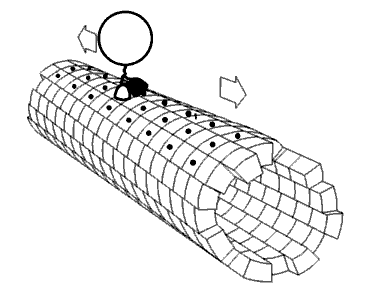
Motor proteins utilizing the cytoskeleton for movement fall into two categories based on their substrate: microfilaments or microtubules. Actin-based motor proteins (myosin) move along microfilaments through interaction with actin, and microtubule motors (dynein and kinesin) move along microtubules through interaction with tubulin.[citation needed]

There are two basic types of microtubule motors: plus-end motors and minus-end motors, depending on the direction in which they "walk" along the microtubule cables within the cell.

Actin motors

[edit]

Myosin

[edit]

Myosins are a superfamily of actin motor proteins that convert chemical energy in the form of ATP to mechanical energy, thus generating force and movement. The first identified myosin, myosin II, is responsible for generating muscle contraction. Myosin II is an elongated protein that is formed from two heavy chains with motor heads and two light chains. Each myosin head contains actin and ATP binding site. The myosin heads bind and hydrolyze ATP, which provides the energy to walk toward the plus end of an actin filament. Myosin II are also vital in the process of cell division. For example, non-muscle myosin II bipolar thick filaments provide the force of contraction needed to divide the cell into two daughter cells during cytokinesis. In addition to myosin II, many other myosin types are responsible for variety of movement of non-muscle cells. For example, myosin is involved in intracellular organization and the protrusion of actin-rich structures at the cell surface. Myosin V is involved in vesicle and organelle transport.[3][4] Myosin XI is involved in cytoplasmic streaming, wherein movement along microfilament networks in the cell allows organelles and cytoplasm to stream in a particular direction.[5] Eighteen different classes of myosins are known.[6]

Genomic representation of myosin motors:[7]

Microtubule motors

[edit]

Kinesin

[edit]

Kinesins are a superfamily of related motor proteins that use a microtubule track in anterograde movement. They are vital to spindle formation in mitotic and meiotic chromosome separation during cell division and are also responsible for shuttling mitochondria, Golgi bodies, and vesicles within eukaryotic cells. Kinesins have two heavy chains and two light chains per active motor. The two globular head motor domains in heavy chains can convert the chemical energy of ATP hydrolysis into mechanical work to move along microtubules.[8] The direction in which cargo is transported can be towards the plus-end or the minus-end, depending on the type of kinesin. In general, kinesins with N-terminal motor domains move their cargo towards the plus ends of microtubules located at the cell periphery, while kinesins with C-terminal motor domains move cargo towards the minus ends of microtubules located at the nucleus. Fourteen distinct kinesin families are known, with some additional kinesin-like proteins that cannot be classified into these families.[9]

Genomic representation of kinesin motors:[7]

Dynein

[edit]

Dyneins are microtubule motors capable of a retrograde sliding movement. Dynein complexes are much larger and more complex than kinesin and myosin motors. Dyneins are composed of two or three heavy chains and a large and variable number of associated light chains. Dyneins drive intracellular transport toward the minus end of microtubules which lies in the microtubule organizing center near the nucleus.[10] The dynein family has two major branches. Axonemal dyneins facilitate the beating of cilia and flagella by rapid and efficient sliding movements of microtubules. Another branch is cytoplasmic dyneins which facilitate the transport of intracellular cargos. Compared to 15 types of axonemal dynein, only two cytoplasmic forms are known.[11]

Genomic representation of dynein motors:[7]

Plant-specific motors

[edit]

In contrast to animals, fungi and non-vascular plants, the cells of flowering plants lack dynein motors. However, they contain a larger number of different kinesins. Many of these plant-specific kinesin groups are specialized for functions during plant cell mitosis.[12] Plant cells differ from animal cells in that they have a cell wall. During mitosis, the new cell wall is built by the formation of a cell plate starting in the center of the cell. This process is facilitated by a phragmoplast, a microtubule array unique to plant cell mitosis. The building of cell plate and ultimately the new cell wall requires kinesin-like motor proteins.[13]

Another motor protein essential for plant cell division is kinesin-like calmodulin-binding protein (KCBP), which is unique to plants and part kinesin and part myosin.[14]

Other molecular motors

[edit]

Besides the motor proteins above, there are many more types of proteins capable of generating forces and torque in the cell. Many of these molecular motors are ubiquitous in both prokaryotic and eukaryotic cells, although some, such as those involved with cytoskeletal elements or chromatin, are unique to eukaryotes. The motor protein prestin,[15] expressed in mammalian cochlear outer hair cells, produces mechanical amplification in the cochlea. It is a direct voltage-to-force converter, which operates at the microsecond rate and possesses piezoelectric properties.

See also

[edit]

References

[edit]
[edit]
Revisions and contributorsEdit on WikipediaRead on Wikipedia
from Grokipedia
Motor proteins are a class of molecular machines that convert chemical energy, typically from ATP hydrolysis, into mechanical work to drive a wide array of cellular processes across prokaryotes and eukaryotes. In eukaryotes, the primary cytoskeletal motor proteins are classified into three main superfamilies: myosins, which interact with actin filaments; and kinesins and dyneins, which move along microtubules. Myosins power diverse functions including muscle contraction and vesicle transport, kinesins typically direct cargo toward the plus ends of microtubules for anterograde transport, and dyneins mediate minus-end-directed movement for retrograde transport. In addition to cytoskeletal motors, other molecular motors include rotary proteins such as F-ATP synthase and linear motors like RNA polymerase and helicases. The structural core of motor proteins consists of a conserved motor domain that binds both the nucleotide (ATP/ADP) and the cytoskeletal track, coupled with tail domains that recognize and bind specific cargoes such as organelles, vesicles, or chromosomes. Mechanistically, ATP binding induces conformational changes that propel the motor forward in a processive or non-processive manner; for instance, kinesin-1 advances in 8-nm steps per ATP hydrolyzed, achieving speeds of 2–3 μm/sec, while myosins employ a power stroke via a lever arm amplification. Dyneins, with their AAA+ ATPase rings, exhibit more complex, stochastic stepping coordinated by linker domains. These mechanisms ensure unidirectional motility, often regulated by phosphorylation or mechanical tension to fine-tune cellular responses. Beyond transport, motor proteins are crucial for cytokinesis, where myosin II drives contractile ring assembly; mitotic spindle dynamics, involving kinesin-5 for bipolar spindle formation; and ciliogenesis, powered by axonemal dyneins for flagellar beating. Dysfunctions in motor proteins are implicated in diseases such as neurodegeneration (e.g., kinesin mutations in Charcot-Marie-Tooth disease) and muscular disorders (e.g., myosin heavy chain defects in cardiomyopathies), underscoring their biomedical significance. With over 40 genes each for myosins and kinesins in humans, the diversity of these proteins reflects their adaptability to specialized cellular needs across eukaryotes.

Overview and Fundamentals

Definition and Classification

Motor proteins are a class of enzymes that convert the chemical energy derived from adenosine triphosphate (ATP) hydrolysis into mechanical work, enabling directed movement along polymeric tracks such as actin filaments or microtubules. This force generation facilitates essential cellular processes by allowing these proteins to "walk" or slide along cytoskeletal elements, producing displacements on the order of nanometers per ATP molecule hydrolyzed. Motor proteins are classified into three main superfamilies based on sequence homology in their motor domains and their association with cytoskeletal filaments: the myosin superfamily, which interacts with actin filaments; and the kinesin and dynein superfamilies, which associate with microtubules. The myosin superfamily encompasses over 35 classes, while the kinesin and dynein superfamilies comprise 14 and 9 classes, respectively. Myosins are actin-based motors, kinesins are mostly plus-end-directed microtubule motors, and dyneins are minus-end-directed microtubule motors; all move unidirectionally along linear tracks through conformational changes coupled to ATP hydrolysis. Key functional characteristics of motor proteins include processivity, velocity, and force generation, which vary across families to suit specific cellular roles. Processivity refers to the number of consecutive steps a motor takes along its track before detaching, often exceeding 100 steps for highly processive motors like conventional kinesin-1, allowing efficient long-distance transport per binding event. Velocities typically range from 10 to 800 nm/s, with kinesins achieving higher speeds under unloaded conditions. Force generation is quantified by stall force, the maximum load a motor can exert before stalling; for example, kinesin-1 produces a stall force of approximately 5-7 pN in vitro, enabling it to overcome viscous drag and other cellular resistances. The classification of motor proteins has evolved from early biochemical observations to modern phylogenetic analyses. In the 1930s, Vladimir Engelhardt and Militsa Lyubimova demonstrated that myosin possesses ATPase activity, linking ATP hydrolysis to muscle contraction and establishing the enzymatic basis of force generation. By the 1950s, Andrew Huxley's sliding filament model integrated these findings, proposing that myosin cross-bridges cyclically interact with actin filaments to produce shortening, which laid the groundwork for recognizing motor proteins as mechanochemical transducers. Contemporary classifications, emerging in the 1990s and refined through genomic sequencing, rely on sequence homology and structural similarities in motor domains to delineate superfamilies, revealing evolutionary divergences and novel family members across eukaryotes.

Biological Importance

Motor proteins play essential roles in eukaryotic cells by generating forces that establish and maintain cell polarity, facilitate cell division, and enable responses to environmental cues. Through their ability to produce directed mechanical forces along cytoskeletal filaments, these proteins organize intracellular components asymmetrically, which is crucial for processes such as directed cell migration and tissue morphogenesis. In cell division, motor proteins drive chromosome segregation and cytokinesis by powering the mitotic spindle and contractile ring, ensuring accurate distribution of genetic material to daughter cells. Additionally, they contribute to sensing and adapting to external signals, such as chemotactic gradients, by remodeling the cytoskeleton to orient cellular protrusions and adhesions. The quantitative impact of motor proteins underscores their efficiency in cellular dynamics; for instance, kinesin motors can transport cargos at speeds up to 800 nm/s, covering distances of 10–100 μm that span typical eukaryotic cell dimensions. This processive movement allows rapid repositioning of organelles and vesicles, maintaining cellular architecture and function over extended periods. Motor proteins exhibit remarkable evolutionary conservation across eukaryotes, with core families like kinesins, dyneins, and myosins present in diverse lineages from unicellular protists to multicellular organisms. This conservation facilitated the transition to multicellularity by enabling precise spindle orientation and tissue organization, as seen in ancient functions like those in Dictyostelium that promote coordinated multicellular development. Beyond their biological roles, motor proteins hold significant promise in biotechnology, particularly for developing nanodevices that harness their motility for targeted drug delivery. Engineered systems using kinesin or dynein can propel nanoparticles along microtubules, mimicking intracellular transport to deliver therapeutics directly to cellular compartments.

Molecular Structure and Mechanisms

Structural Components

Motor proteins exhibit a modular architecture that enables their directed movement along cytoskeletal tracks, typically comprising a motor head domain responsible for ATP binding and track interaction, a neck region that transmits conformational changes into mechanical force, and a tail domain for cargo attachment or multimerization. The motor head, often globular and conserved across families, houses the nucleotide-binding site and interfaces with the filament, while the neck acts as a lever or linker to amplify small structural shifts into larger displacements. In kinesins and myosins, the head domain spans approximately 340–850 amino acids and shares a core fold with a central β-sheet surrounded by α-helices, facilitating ATP hydrolysis-driven conformational dynamics. The neck linker or lever arm connects the head to the tail and is crucial for force transmission; in myosins, this region consists of an extended α-helix stabilized by light-chain binding, forming 1–6 IQ motifs that recruit calmodulin or similar proteins for regulation. Kinesins feature a shorter, flexible neck linker of about 15 residues that docks upon ATP binding to propel the partner head forward. Dyneins diverge with a linker domain arching over an AAA+ ring in the motor domain, which serves an analogous role in coordinating movement. The cargo-binding tail varies widely but often includes domains for specific interactions, such as vesicle adapters in kinesin-1 or actin-crosslinking in certain myosins. Dimerization or multimerization motifs promote processive movement by coordinating multiple motor heads; for instance, kinesins and many myosins form homodimers via coiled-coil stalks that link the tails and ensure alternating head engagement with the track. In myosin II, the coiled-coil tail further assembles into bipolar filaments, while cytoplasmic dyneins homodimerize through their N-terminal tail domains. Track-binding sites are family-specific: myosins engage actin filaments via a cleft between 50-kDa subdomains in the head, closing upon strong binding; kinesins and dyneins interact with microtubules, with kinesins using an α4 helix and L11 loop to grip tubulin dimers, and dyneins employing a stalk-tethered microtubule-binding domain approximately 140 Å from the AAA+ ring. Structural variations include heavy and light chains in myosins and dyneins, where heavy chains (~200–500 kDa) carry the motor core and light chains (~15–20 kDa) modulate the neck or regulate activity, such as the essential and regulatory light chains in myosin II or calmodulin in unconventional myosins like myosin V. Kinesins typically lack dedicated light chains but may associate with accessory proteins like kinesin light chains (KLCs) for cargo specificity in heterodimeric complexes. Regulatory domains, often in the tail or neck, respond to cellular signals; for example, calmodulin-binding IQ motifs in myosins allow calcium-dependent control of motor activity. These elements collectively ensure the structural integrity and functional versatility of motor proteins across eukaryotic cytoskeletal systems.

Mechanochemical Cycle and Energy Conversion

Motor proteins convert chemical energy from adenosine triphosphate (ATP) hydrolysis into mechanical work through a conserved mechanochemical cycle, often modeled as a cross-bridge cycle. In this cycle, the motor domain binds to its filamentous track (such as actin or microtubules), and ATP binding induces a conformational change that weakens this affinity, allowing detachment. Subsequent hydrolysis of ATP to adenosine diphosphate (ADP) and inorganic phosphate (Pi) stores energy in strained conformations, priming the motor for movement. The cycle progresses with Pi release, which triggers the power stroke—a rapid conformational rearrangement that generates force and displaces the motor along the track by 5-10 nm per step. This is exemplified in kinesins, where Pi release follows ATP hydrolysis, leading to neck linker docking and advancement toward the microtubule plus end. ADP release then resets the motor to a high-affinity state, completing the cycle and enabling processive movement. In myosins, the power stroke is similarly initiated by Pi release upon actin binding, amplifying displacement via lever arm rotation. These steps ensure tight coupling between chemical and mechanical events, with hydrolysis occurring post-binding to the track in kinesins and pre-detachment in myosins. Energy conversion in this cycle achieves variable efficiency depending on load; for kinesin, measurements indicate up to ~50% at stall force but approximately 20% under typical working conditions, utilizing a portion of the free energy from (ΔG ≈ -50 kJ/mol under physiological conditions) to perform mechanical work. This powers steps of ~8 nm against loads up to several piconewtons, reflecting an economical use of cellular energy resources compared to macroscopic engines. Directionality in motor movement arises from structural asymmetries that bias conformational changes. In kinesins, the neck linker—a flexible peptide segment—docks asymmetrically upon ATP binding, directing the trailing head forward along the microtubule plus end and preventing backward steps. In dyneins, the AAA+ domains (particularly AAA1 as the primary ATPase site) coordinate linker remodeling, with the linker's swing toward the microtubule minus end driven by allosteric signals from AAA3 and AAA4, ensuring minus-end-directed motility. The cycle is regulated by post-translational modifications and accessory proteins to control activity in response to cellular needs. Phosphorylation by mitogen-activated protein kinases (MAPKs), such as JNK and p38, targets the kinesin-1 motor domain, reducing its processivity and microtubule affinity to fine-tune cargo transport. Accessory proteins can further modulate ATP hydrolysis rates or track binding, enhancing adaptability without altering the core cycle.

Cytoskeletal Motor Proteins

Myosins

Myosins constitute a large superfamily of actin-based molecular motors that convert the chemical energy from ATP hydrolysis into mechanical work to drive a wide array of cellular processes, including muscle contraction, intracellular transport, and cytoskeletal remodeling. The superfamily encompasses more than 35 classes identified across eukaryotes, with significant diversity in structure and function tailored to specific cellular roles. Class II myosins, also known as conventional myosins, are primarily responsible for muscle contraction through their assembly into bipolar filaments that interact with actin in sarcomeres. In contrast, class V myosins function in vesicle and organelle transport, exhibiting processive movement along actin filaments to deliver cargo over long distances within the cell. At the core of all myosins is the conserved head domain, which possesses actin-activated ATPase activity that powers the motor cycle. This domain binds actin and ATP, undergoing conformational changes upon hydrolysis that generate force. The power stroke, a pivotal step in this cycle, involves a ~5-10 nm displacement amplified by the lever arm—a structural extension of the head domain consisting of alpha-helices bound to light chains—which tilts to propel the motor forward along actin. This lever arm mechanism allows myosins to achieve directed movement, with the stroke size and speed varying by class to suit their functions. Myosin II exemplifies the contractile role in sarcomeres, where dimeric molecules form thick filaments that slide actin thin filaments past each other during muscle shortening. Each step of myosin II advances ~5-15 nm with velocities ranging from 1-10 μm/s, depending on the muscle fiber type and load, enabling rapid force generation for locomotion. Myosin V, operating as a processive dimer, employs a hand-over-hand walking mechanism, where the trailing head detaches and swings forward in 36 nm steps to bind the next actin monomer, ensuring efficient cargo transport without dissociation. The diversity of unconventional myosins extends their roles beyond contraction and transport. For instance, myosin VIIA contributes to the maintenance of stereocilia in inner ear hair cells, facilitating mechanotransduction essential for hearing. Similarly, myosin VA supports melanosome distribution in melanocytes and retinal pigment epithelium, ensuring proper pigmentation and visual function through actin-dependent trafficking. This versatility underscores the superfamily's adaptation to specialized cellular needs across tissues.

Kinesins

Kinesins constitute a superfamily of microtubule-based motor proteins primarily responsible for plus-end-directed transport within cells. The superfamily is classified into 14 families, designated KIF1 through KIF14, based on phylogenetic analysis of their conserved motor domains. These families exhibit diverse structures and functions, with most members featuring an N-terminal motor domain for ATP hydrolysis and microtubule binding, a neck linker for force generation, and tail regions for cargo interaction. Conventional kinesin-1, also known as KHC or KIF5, exemplifies the Kinesin-1 family and serves as the primary motor for anterograde transport, moving vesicles and organelles toward microtubule plus ends in axons and other cellular compartments. Kinesin-1 operates via a hand-over-hand mechanism, where its two motor heads alternate between detachment from and reattachment to the microtubule, powered by ATP hydrolysis. Each step advances the center of mass by 8 nm, precisely matching the spacing of tubulin dimers in the microtubule lattice. Kinesin-1 demonstrates high processivity, capable of taking up to 100 consecutive steps along a microtubule before dissociating, enabling efficient long-distance transport. Its unloaded velocity reaches approximately 800 nm/s, though this speed slows under opposing loads due to increased dwell times at ATP-binding steps and higher detachment risk. Kinesins bind microtubules through specialized domains in their motor heads, which recognize tubulin subunits. Certain kinesin variants perform specialized roles beyond transport. Kinesin-5 motors, such as Eg5, crosslink and slide antiparallel microtubules to generate outward forces essential for establishing and maintaining bipolar spindle geometry during mitosis. In contrast, kinesin-13 family members, like MCAK and KIF2C, act as non-motile depolymerases that target microtubule ends, inducing tubulin dimer removal through conformational changes that curl protofilaments and accelerate disassembly.

Dyneins

Dyneins constitute a family of minus-end-directed motor proteins that translocate along microtubules, playing essential roles in intracellular transport and the generation of ciliary and flagellar motility. They are classified into two primary types: cytoplasmic dyneins, which facilitate retrograde transport of cargos within the cell, and axonemal dyneins, which drive the sliding of microtubules to produce the beating waveforms in cilia and flagella. Cytoplasmic dynein-1, the predominant isoform for vesicular and organelle transport, contrasts with dynein-2, which supports intraflagellar transport, while axonemal dyneins are specialized for motile appendages absent in higher plants. The core structure of dynein heavy chains features a large AAA+ (ATPases associated with diverse cellular activities) ATPase ring composed of six domains, AAA1 through AAA6, arranged in a hexagonal configuration. AAA1 serves as the primary site for ATP hydrolysis, with AAA2–AAA4 capable of nucleotide binding but reduced catalytic activity, and AAA5–AAA6 providing structural support without hydrolysis function; this ring is connected to a microtubule-binding domain via a coiled-coil stalk and an N-terminal linker that undergoes conformational changes during the cycle. Each heavy chain, exceeding 500 kDa, forms the force-generating unit within multi-subunit complexes, distinguishing dyneins from the simpler dimeric structures of other microtubule motors. Mechanistically, dyneins harness ATP hydrolysis to produce a power stroke through the swinging of the N-terminal linker, which transitions from a pre-powerstroke (bent) to post-powerstroke (straight) conformation upon microtubule rebinding after ATP cleavage at AAA1. This motion advances the motor in 8–32 nm steps along the microtubule, with cytoplasmic dyneins exhibiting lower processivity—typically 10–20 steps before detachment—compared to kinesins, though processivity can be enhanced by regulatory factors. Velocities for cytoplasmic dynein-1 range from 100–400 nm/s under loaded conditions, enabling efficient retrograde transport in opposition to anterograde kinesin movement. Cytoplasmic dynein-1 achieves cargo specificity and enhanced processivity by forming a multi-subunit complex with dynactin, a large adaptor that links the motor to vesicular membranes and recruits additional regulators like LIS1 for force-sensitive activation. This dynein-dynactin supercomplex adopts a extended conformation for stable microtubule engagement and cargo adaptation, crucial for long-distance intracellular trafficking. In contrast, axonemal dyneins are organized into outer arm dyneins (typically 2–3 heavy chains per arm) and inner arm dyneins (including six single-headed and one two-headed forms), which coordinately generate the asymmetric sliding forces necessary for the bending and propagation of ciliary waveforms.

Plant-Specific Motors

Plant cells, constrained by rigid cell walls and high turgor pressure, have evolved unique motor proteins adapted to their cytoskeletal architecture, which differs from that in animals by lacking cytoplasmic dyneins for microtubule-based transport. Instead, plants rely heavily on actin-based myosins for vesicle trafficking toward the plasma membrane and long-distance organelle movement. This dependence on myosins compensates for the absence of dynein-mediated minus-end-directed transport along microtubules, enabling efficient intracellular dynamics in a sessile lifestyle. Among these, class VIII and XI myosins represent plant-specific innovations, with distinct roles in cytoplasmic streaming and organelle positioning. Myosin XI drives rapid bulk flow of cytoplasm, achieving velocities up to 7 μm/s in Arabidopsis thaliana, which facilitates the distribution of nutrients and organelles across large vacuolated cells. This high-speed motility supports organelle trafficking, such as the movement of Golgi-derived vesicles and peroxisomes, essential for cell expansion and polarized growth. In contrast, myosin VIII, smaller in size and localized primarily to the cell cortex, functions in slower processes like endocytosis and anchoring organelles at specific sites, including the positioning of endocytic compartments near the plasma membrane. Both classes interact with actin filaments to maintain cytoskeletal integrity under turgor stress, but myosin XI's processive stepping along actin enables the fast streaming absent in animal systems. The kinesin-14 family provides another plant-adapted motor, operating as minus-end-directed kinesins on microtubules to organize spindle structures during cell division. In land plants, multiple kinesin-14 members cross-link and slide microtubules, promoting spindle morphogenesis and proper orientation for asymmetric divisions, such as those in stomatal development. Unlike animal kinesin-14 homologs, plant versions have diversified to handle microtubule minus-end transport without dynein, ensuring bipolar spindle assembly and chromosome segregation in walled cells. These motors are particularly crucial in the phragmoplast, a plant-unique structure that forms during cytokinesis to assemble the cell plate under turgor pressure. Kinesin-12 family members, such as POK2, transport vesicles along expanding phragmoplast microtubules toward the midzone, delivering cell wall precursors to fuse into the new cell plate. Myosin VIII contributes by linking actin and microtubule networks at microtubule ends, guiding vesicle secretion and maintaining phragmoplast expansion against the cell wall's rigidity. This coordinated action of myosins and kinesins ensures precise cell plate formation, adapting motor functions to the mechanical demands of plant cell division.

Non-Cytoskeletal Molecular Motors

Rotary Motors

Rotary motors are a class of molecular motors that convert chemical or electrochemical energy into rotational torque, distinct from linear motors that produce displacement along tracks. These non-cytoskeletal assemblies are often embedded in membranes and play critical roles in energy transduction, such as ATP synthesis or ion pumping. Unlike the mechanochemical cycles of linear motors, rotary motors feature a central rotor that turns in discrete angular steps, driven by sequential binding and hydrolysis events or ion flux.81142-3) The F1-ATPase, the soluble catalytic domain of mitochondrial ATP synthase, exemplifies a reversible rotary motor. In its synthesis mode, proton flow through the membrane-embedded Fo sector drives counterclockwise rotation of the central γ subunit relative to the α3β3 stator ring, inducing conformational changes that synthesize ATP from ADP and Pi. This rotation reverses the ATP hydrolysis direction under physiological proton motive force, achieving near-100% efficiency in energy conversion. The motor rotates at biological rates of approximately 100–300 revolutions per second (rps), corresponding to 6,000–18,000 rpm, depending on proton gradient and load.81456-7) The rotary mechanism of F1-ATPase proceeds in 120° steps, with each step associated with the binding or hydrolysis of one ATP molecule during the hydrolysis mode, though synthesis involves reversal without hydrolysis. The γ rotor turns within the stator, generating torque of approximately 40 pN·nm per step, sufficient to overcome viscous drag and drive synthesis against the free energy of ATP formation (ΔG ≈ 50–60 kJ/mol under cellular conditions). High-resolution single-molecule observations confirm these discrete steps, with substeps of 80°–90° and 30°–40° resolving the power and catalytic dwells. This torque arises primarily from ATP binding affinity changes and elastic deformations in the stator, ensuring tight coupling between rotation and catalysis.11456-7) The bacterial flagellar motor represents an ion-driven rotary engine that propels swimming bacteria via high-speed rotation of flagellar filaments. Embedded in the cell envelope, it harnesses the proton motive force (or sodium motive force in some species) to rotate a rotor-switch complex (including the MS ring, C ring, and rod) against multiple stator units (MotA/MotB complexes). Ion influx through the stators interacts electrostatically with rotor proteins like FliG, generating torque that drives counterclockwise rotation for forward motility or clockwise for tumbling. The motor achieves speeds up to 100,000 rpm near zero load, enabling bacterial velocities of 20–50 body lengths per second. In contrast to nucleotide-driven motors, the flagellar motor's torque-speed curve is nonlinear, producing maximal torque (~1,200–1,500 pN·nm) at low speeds and decreasing to zero at stalling speeds, with efficiency exceeding 50% across a wide range. Seminal biophysical studies have quantified up to 11 independent stator units engaging stepwise with increasing load, optimizing performance for varying viscosities. This ion-powered rotation exemplifies evolutionary adaptation for rapid, reversible motility without ATP hydrolysis. V-ATPases are rotary proton pumps responsible for acidifying intracellular compartments like vacuoles, endosomes, and lysosomes in eukaryotes. Structurally homologous to F-ATPases, they consist of a cytosolic V1 sector (ATP hydrolysis motor) and a membrane Vo sector (proton channel), linked by a rotating central stalk (rotor). ATP hydrolysis in the V1 α3β3 head drives 120° clockwise rotations of the rotor (c-ring and stalk), which mechanically translocate protons across the membrane via the Vo a-subunit interface, establishing pH gradients of 2–3 units essential for degradation, secretion, and homeostasis. The rotary catalysis in V-ATPases mirrors F1 but is unidirectional for pumping, with rotation rates around 3–10 rps (180–600 rpm) under physiological conditions, slower than F-ATPases due to tighter coupling and regulatory pauses. Torque generation (~20–30 pN·nm) arises from ATP-driven conformational waves in the β subunits, propagating through the central stalk to rotate the c-ring, with each 36° c-subunit step corresponding to one proton translocation. This mechanism enables reversible disassembly for regulation, preventing excessive acidification, and is conserved across eukaryotes for diverse physiological roles.76823-8)

Linear Nucleic Acid Motors

Linear nucleic acid motors are enzymes that harness the energy from nucleotide triphosphate (NTP) hydrolysis to translocate linearly along DNA or RNA strands, facilitating essential processes such as transcription, replication, repair, and recombination without reliance on cytoskeletal filaments. These motors exhibit high processivity, enabling them to cover substantial distances along nucleic acids while maintaining directionality and generating mechanical force to unwind, translocate, or remodel substrates. Unlike rotary motors that produce torque, linear nucleic acid motors primarily employ inchworm-like or hand-over-hand mechanisms to advance step-by-step, often in 1- to 3-base-pair increments per ATP hydrolyzed. RNA Polymerase exemplifies a linear motor during transcription elongation, where it processively synthesizes RNA by incorporating nucleotides complementary to the DNA template. Bacterial RNA polymerase (RNAP), such as that from Escherichia coli, advances at speeds of 50–100 nucleotides per second, driven by the hydrolysis of NTPs that stabilize a transcription bubble—a transiently unwound DNA-RNA hybrid region of about 12–14 base pairs. This energy conversion powers a power-stroke mechanism, wherein conformational changes in the RNAP clamp domain upon NTP binding and hydrolysis propel forward translocation by one nucleotide, generating forces up to 25–30 pN to overcome DNA barriers. Eukaryotic RNA polymerase II operates more slowly, at 1–4 kb/min (approximately 17–67 nt/s), but shares the core NTP-driven elongation cycle, ensuring faithful gene expression. Helicases function as ATP-dependent motors that unwind double-stranded nucleic acids, separating strands to expose single-stranded templates for replication or repair. These enzymes typically form hexameric rings and translocate unidirectionally along single-stranded DNA or RNA, hydrolyzing one ATP per 1–3 base pairs advanced, consistent with an inchworm or rotary handover model where subunit conformational changes grip and pull the nucleic acid. For instance, the hepatitis C virus NS3 helicase unwinds RNA duplexes in 3-base-pair steps, with each cycle involving ATP binding to induce a 90° rotation of its RecA-like domains, followed by hydrolysis to release and advance, achieving processive unwinding at rates up to 100 bp/s under optimal conditions. Similarly, bacterial RecA protein assembles into helical filaments on single-stranded DNA, where ordered waves of ATP hydrolysis propagate unidirectionally every six monomers, facilitating strand invasion and exchange in recombination by extending and invading homologous duplexes in 1–2 bp increments per hydrolysis event. DNA translocases, such as FtsK in bacteria, actively pump double-stranded DNA to coordinate chromosome segregation during cell division. FtsK forms a hexameric ring that encircles dsDNA and translocates directionally toward the replication terminus, guided by specific DNA sequences (KOPS), at velocities exceeding 5 kb/s—among the fastest known for molecular motors—powered by ATP hydrolysis that drives a pumpjack-like mechanism with 1 bp steps per nucleotide hydrolyzed. This rapid movement, capable of displacing protein roadblocks and generating forces over 20 pN, ensures timely dimer resolution at the dif site by delivering distant DNA segments to the division septum. Chaperone-like motors, including Hsp104 from yeast, extend linear motor principles to protein disaggregation, often in contexts involving nucleic acid-associated aggregates like prions. Hsp104 hexamers use ATP hydrolysis to thread polypeptide substrates through their central pore in a processive manner, unfolding and extracting proteins from amyloid fibrils or amorphous aggregates at rates of about 12–20 amino acids per second, with each cycle involving pore-loop grip and translocation akin to nucleic acid motors. This threading mechanism, requiring 1–2 ATP per residue advanced, reactivates stress-damaged proteins and propagates prions, highlighting convergent evolutionary strategies in linear translocation across substrates.

Cellular Functions

Intracellular Transport and Organelle Movement

Motor proteins play a central role in intracellular transport by moving organelles, vesicles, and other cargoes along cytoskeletal filaments, ensuring efficient distribution within the cell. On microtubules, bidirectional transport is primarily mediated by kinesins for anterograde movement toward the plus end and dyneins for retrograde movement toward the minus end. For example, in neurons, kinesin-1 transports mitochondria from the cell body to synaptic terminals to support local energy demands. This long-range transport allows cargoes to travel distances up to several centimeters or more in long axons, such as those extending up to a meter in humans, maintaining cellular homeostasis. In contrast, short-range transport often occurs on actin filaments using myosin motors, particularly myosin V, which moves cargoes like melanosomes in melanocytes toward the cell periphery. Myosin V adapts to cortical actin tracks near the plasma membrane, facilitating precise positioning after handover from microtubule-based systems. This actin-dependent phase enables fine-tuned dispersion, with myosin V's processive stepping ensuring reliable progression over shorter distances, typically tens of micrometers. Coordination between opposing motors is essential for effective bidirectional transport, involving scaffolds like dynactin that link dynein to cargoes and regulate interactions with kinesins. Dynactin facilitates both anterograde and retrograde motility by serving as a hub for motor recruitment, with its disruption halting organelle movement in both directions. Direction switching occurs through mechanisms such as regulatory pauses at microtubule intersections and tug-of-war models, where competing kinesin and dynein teams generate stochastic reversals based on their relative strengths and numbers. In the tug-of-war, dynein teams often outnumber kinesins (e.g., 4-8 dyneins vs. 1-2 kinesins), allowing net retrograde bias despite kinesin's higher stall force. These dynamics ensure adaptive routing without dedicated coordinators in many cases. This energy investment underscores the scale of intracellular logistics, where multiple motors per cargo (often 1-5 kinesins and several dyneins) collaborate to overcome obstacles and maintain flux.

Force Generation in Motility and Division

Motor proteins play a crucial role in generating the mechanical forces required for cellular motility and division, enabling processes such as muscle contraction, cytokinesis, ciliary beating, and cell migration. These forces arise from the collective action of motor proteins like myosins and dyneins interacting with cytoskeletal filaments, converting chemical energy from ATP hydrolysis into mechanical work. In motility and division, force generation often involves ensembles of motors producing tensions on the order of nanonewtons (nN), which drive structural deformations and oscillations essential for cellular function. In skeletal muscle contraction, myosin II motors slide actin filaments within sarcomeres, the basic contractile units, to produce shortening and force. Each thick filament in a half-sarcomere generates an isometric force of approximately 500-700 pN through the coordinated attachment and power strokes of hundreds of myosin heads to actin, with the full half-sarcomere in a myofibril producing ~100-300 nN overall, as measured in experiments with intact thick filaments. This sliding mechanism, where myosin cross-bridges cycle between attached and detached states, results in filament overlap changes that amplify force across the muscle fiber. During cytokinesis, the final stage of cell division, myosin II assembles into a contractile ring at the cell equator, constricting at a rate of about 0.1–0.2 μm/min to furrow the membrane and separate daughter cells. This constriction is driven by myosin II-mediated sliding of actin filaments, generating tension that reduces the ring circumference while maintaining structural integrity. Coordination with kinesin-6, which localizes to the equatorial cortex via microtubule plus ends, ensures precise positioning and activation of the RhoA signaling pathway that recruits myosin II, preventing premature or misplaced ring assembly. In ciliary and flagellar beating, dynein motors generate oscillatory forces by sliding adjacent microtubule doublets within the axoneme, producing bending waves at frequencies of 10–50 Hz typical for motile cilia and sperm flagella. Outer and inner arm dyneins attach periodically to produce tip-directed forces of ~1 pN per arm, with regulated activation along the axoneme converting linear sliding into planar or helical waves that propel cells or fluids. This rhythmic sliding is modulated by ATP levels and calcium, ensuring efficient oscillation without continuous force application. Cell migration relies on myosin II contractility to generate traction forces in protrusive structures like lamellipodia, where actomyosin networks produce tensions of 10–100 nN to retract the actin meshwork and stabilize adhesions. In lamellipodia of migrating neurons, myosin II contributes to protrusive forces exceeding 20 nN by contracting bundled actin filaments, facilitating forward advancement against substrate resistance. These localized forces integrate with polymerization-driven protrusion to enable directional motility.

Diseases and Pathologies

Defects in Myosins

Defects in myosins disrupt their roles in actin-based transport, contraction, and cellular structure, leading to a range of genetic disorders characterized by impaired muscle function, pigmentation abnormalities, and sensory deficits. These malfunctions often arise from mutations that alter myosin's motor domain, lever arm, or cargo-binding sites, resulting in reduced motility, abnormal force generation, or protein mislocalization. Mutations in myosin II, particularly in the β-cardiac myosin heavy chain gene MYH7, are a primary cause of hypertrophic cardiomyopathy (HCM), an autosomal dominant condition affecting 1 in 500 individuals and increasing the risk of sudden cardiac death. The R403Q missense mutation in the globular head of the myosin motor domain exemplifies this, as it enhances ATPase activity and cross-bridge kinetics while impairing the protein's interaction with regulatory partners like filamin C, leading to hypercontractility and left ventricular hypertrophy. Functional studies in transgenic models show that R403Q alters the force-velocity relationship by accelerating cross-bridge detachment rates, which elevates energy consumption (tension cost) and promotes maladaptive remodeling in cardiomyocytes. Defects in myosin VA, encoded by MYO5A, underlie Griscelli syndrome type 1 (GS1), a rare autosomal recessive disorder marked by hypopigmentation and severe neurological impairment due to failed melanosome transport in melanocytes and neurons. Mutations disrupt the myosin VA-Rab27a-melanophilin complex, preventing actin-dependent delivery of melanosomes to the cell periphery, which causes the characteristic silver-gray hair sheen and uneven skin pigmentation from aggregated melanosomes. Neurologically, these defects impair vesicular trafficking in brain cells, resulting in early-onset hypotonia, developmental delays, intellectual disability, and seizures, often leading to early death with supportive care only. Myosin VIIA mutations in the MYO7A gene cause Usher syndrome type 1B (USH1B), an autosomal recessive condition combining congenital profound deafness, vestibular dysfunction, and progressive retinitis pigmentosa leading to blindness. In inner ear hair cells, myosin VIIA maintains stereocilia integrity by transporting proteins like sans and harmonin to tip-link densities, where it supports mechanotransduction for hearing and balance; pathogenic variants destabilize these structures, causing bundle disorganization and sensory cell degeneration. In the retina, similar transport failures contribute to photoreceptor degeneration, underscoring myosin VIIA's role in anchoring opsins and maintaining ciliary function. Therapeutic strategies for myosin-related myopathies, including HCM, have advanced through gene editing as of 2023, with preclinical studies demonstrating feasibility in correcting MYH7 mutations. Adenine base editing in patient-derived iPSC cardiomyocytes has reversed R403Q-induced hypertrophy by restoring normal myosin function without off-target effects, highlighting potential for CRISPR-based interventions. Similarly, CRISPR/Cas9 approaches targeting MYH7 variants in cellular models have shown promise in preventing sarcomeric disarray, paving the way for clinical translation in myosin II disorders, though trials remain in early phases focused on safety and delivery via AAV vectors. As of 2025, gene therapy approaches for HCM continue to progress, with first-in-human trials underway for related sarcomeric genes like MYBPC3, while MYH7-targeted editing remains in preclinical stages. For MYO5A and MYO7A defects, gene replacement therapies are under investigation in animal models, with challenges in targeting neural and sensory tissues.

Defects in Kinesins and Dyneins

Defects in kinesins and dyneins, which are microtubule-based motor proteins essential for bidirectional intracellular transport, often disrupt axonal trafficking in neurons, leading to protein aggregation, organelle mislocalization, and neurodegeneration. These disruptions manifest in a spectrum of neurological disorders, including motor neuropathies, spastic paraplegias, and cortical malformations, where impaired anterograde (kinesin-mediated) or retrograde (dynein-mediated) transport hinders the delivery of synaptic components, mitochondria, and signaling molecules. Such defects are typically caused by mutations in motor domain genes or regulatory subunits, reducing motor processivity, velocity, or cargo-binding affinity, as evidenced by biophysical studies showing decreased run lengths and force generation in mutant motors. Mutations in kinesin family members, particularly KIF5A (encoding the heavy chain of kinesin-1), are strongly associated with hereditary spastic paraplegia type 10 (SPG10) and Charcot-Marie-Tooth disease type 2 (CMT2). For instance, the N256S missense mutation in the KIF5A motor domain impairs microtubule binding and anterograde transport, resulting in distal axon degeneration and progressive lower limb spasticity. In Alzheimer's disease (AD), kinesin-1 dysfunction, often secondary to amyloid-beta accumulation or tau hyperphosphorylation, disrupts mitochondrial and amyloid precursor protein (APP) transport, exacerbating Aβ plaque formation and synaptic loss; single nucleotide polymorphisms like rs12368653 in KIF5A correlate with reduced mRNA expression in AD brains. Similarly, in amyotrophic lateral sclerosis (ALS), kinesin defects contribute to neurofilament accumulation and "traffic jams" along axons, with KIF5A variants linked to faster disease progression. Dynein defects, primarily in the DYNC1H1 gene encoding the cytoplasmic dynein-1 heavy chain, underlie a range of "dyneinopathies" characterized by compromised retrograde transport and neuronal migration. Dominant mutations such as H3822P and R1962C severely reduce dynein processivity and mechanochemical cycling, causing spinal muscular atrophy with lower extremity predominance (SMA-LED), where patients exhibit distal muscle weakness and foot deformities due to impaired neurotrophic factor retrograde signaling. In malformations of cortical development (MCD), mutations like K3336N disrupt dynein-dynactin interactions, leading to lissencephaly and intellectual disability through defective neuronal positioning during brain development. DYNC1H1 variants are also implicated in Charcot-Marie-Tooth disease (CMT) and ALS, where reduced run lengths of dynein-cargo complexes (by ~2-fold compared to wild-type) promote lysosomal accumulation and motor neuron death. Additionally, mutations in dynactin (DCTN1, e.g., G59S) phenocopy dynein defects in Perry syndrome and ALS by destabilizing the dynein-dynactin complex, resulting in tau inclusions and parkinsonism. In multiple sclerosis (MS), polymorphisms in kinesin genes, such as rs1678542 in KIF5A which increases susceptibility, and rs8702 in KLC1 which may confer protection, along with variants in dynein regulators, are associated with altered risk of MS by affecting myelin component transport in oligodendrocytes and Schwann cells. Overall, these defects highlight the interdependence of kinesin and dynein functions, where imbalances in one motor often exacerbate pathologies in the other, underscoring their role in maintaining neuronal homeostasis.

References

Add your contribution
Related Hubs
User Avatar
No comments yet.