Spermatocyte
Spermatocyte
Main page
2035544

Spermatocyte

logo
Community Hub0 subscribers
Read side by side
from Wikipedia
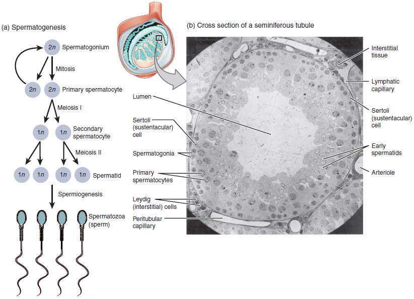
Spermatogenesis as the cells progress from spermatogium, to primary spermatocytes, to secondary spermatocytes, to spermatids and to sperm.

Spermatocytes are a type of male gametocyte in animals. They derive from immature germ cells called spermatogonia. They are found in the testis, in a structure known as the seminiferous tubules.[1] There are two types of spermatocytes, primary and secondary spermatocytes. Primary and secondary spermatocytes are formed through the process of spermatocytogenesis.[2]

Primary spermatocytes are diploid (2N) cells. After meiosis I, two secondary spermatocytes are formed. Secondary spermatocytes are haploid (N) cells that contain half the number of chromosomes.[1]

In all animals, males produce spermatocytes, even hermaphrodites such as C. elegans, which exist as a male or hermaphrodite. In hermaphrodite C. elegans, sperm production occurs first and is then stored in the spermatheca. Once the eggs are formed, they are able to self-fertilize and produce up to 350 progeny.[3]

Development

[edit]
Spermatogonia going through mitosis to form primary spermatocytes in Grasshopper testes.
Spermatocytogenesis

At puberty, spermatogonia located along the walls of the seminiferous tubules within the testis will be initiated and start to divide mitotically, forming two types of A cells that contain an oval shaped nucleus with a nucleolus attached to the nuclear envelope; one is dark (Ad) and the other is pale (Ap). The Ad cells are spermatogonia that will stay in the basal compartment (outer region of the tubule); these cells are reserve spermatogonial stem cells that do not usually undergo mitosis. Type Ap are actively-dividing spermatogonial stem cells which begin differentiation to type B spermatogonia, which have round nuclei and heterochromatin attached to the nuclear envelope and the center of nucleolus.[4] Type B cells will move on to the adluminal compartment (towards the inner region of tubule) and become primary spermatocytes; this process takes about 16 days to complete.[2][5]

The primary spermatocytes within the adluminal compartment will continue on to meiosis I and divide into two daughters cells, known as secondary spermatocytes, a process which takes 24 days to complete. Each secondary spermatocyte will form two spermatids after meiosis II.[1]

Although spermatocytes that divide mitotically and meiotically are sensitive to radiation and cancer, spermatogonial stem cells are not. Therefore, after termination of radiation therapy or chemotherapy, the spermatognia stems cells may re-initiate the formation of spermatogenesis.[6]

Hormones produced by the Pituitary gland. GnRH is secreted by the hypothalamus, which induces anterior pituitary to produce FSH and LH upon puberty.

Role of hormones

[edit]

The formation of primary spermatocytes (a process known as spermatocytogenesis) begins in humans when a male is sexually matured at puberty, around the age of 10 through 14.[7] Formation is initiated upon the pulsated surges of gonadotropin-releasing hormone (GnRH) from the hypothalamus, which leads to the secretion of follicle-stimulating hormone (FSH) and luteinizing hormone (LH) produced by the anterior pituitary gland. The release of FSH into the testes will enhance spermatogenesis and lead to the development of Sertoli cells, which act as nursing cells where spermatids will go to mature after meiosis II. LH promotes Leydig cell secretion of testosterone into the testes and blood, which induce spermatogenesis and aid the formation of secondary sex characteristics. From this point on, the secretion of FSH and LH (inducing production of testosterone) will stimulate spermatogenesis until the male dies.[8] Increasing the hormones FSH and LH in males will not increase the rate of spermatogenesis. However, with age, the rate of production will decrease, even when the amount of hormone that is secreted is constant; this is due to higher rates of degeneration of germ cells during meiotic prophase.[1]

Cell type summary

[edit]

In the following table, ploidy, copy number and chromosome/chromatid counts listed are for a single cell, generally prior to DNA synthesis and division (in G1 if applicable). Primary spermatocytes are arrested after DNA synthesis and prior to division.[1][2]

Cell Type Ploidy/Chromosomes in human DNA copy number/Chromatids in human Process entered by cell Duration
spermatogonium (types Ad, Ap and B) germ cells diploid (2N) / 46 2C / 46 spermatocytogenesis (mitosis) 16 days
primary spermatocyte male gametocyte diploid (2N) / 46 4C / 2x46 spermatocytogenesis (meiosis I) 24 days
secondary spermatocyte male gametocyte haploid (N) / 23 2C / 46 spermatidogenesis (meiosis II) A few hours
spermatids male gametid haploid (N) / 23 1C / 23 spermiogenesis 24 days
spermatozoids sperm haploid (N) / 23 1C / 23 spermiation 64 days (total)

Physiology

[edit]

Damage, repair, and failure

[edit]

Spermatocytes regularly overcome double-strand breaks and other DNA damages in the prophase stage of meiosis. These damages can arise by the programmed activity of Spo11, an enzyme employed in meiotic recombination, as well as by un-programmed breakages in DNA, such as those caused by oxidative free radicals produced as products of normal metabolism. These damages are repaired by homologous recombination pathways and utilize RAD1 and γH2AX, which recognize double strand breaks and modify chromatin, respectively. As a result, double strand breaks in meiotic cells, unlike mitotic cells, do not typically lead to apoptosis, or cell death.[9] Homologous recombinational repair (HRR) of double-strand breaks occurs in mice during sequential stages of spermatogenesis but is most prominent in spermatocytes.[10] In spermatocytes, HRR events occur mainly in the pachytene stage of meiosis and the gene conversion type of HRR is predominant, whereas in other stages of spermatogenesis the reciprocal exchange type of HRR is more frequent.[10] During mouse spermatogenesis, the mutation frequencies of cells at the different stages, including pachytene spermatocytes, are 5 to 10-fold lower than the mutation frequencies in somatic cells.[11] Because of their elevated DNA repair capability, spermatocytes likely play a central role in the maintenance of these lower mutation rates, and thus in the preservation of the genetic integrity of the male germ line.

It is known that heterozygous chromosomal rearrangements lead to spermatogenic disturbance or failure; however the molecular mechanisms that cause this are not as well known. It is suggested that a passive mechanism involving asynaptic region clustering in spermatocytes is a possible cause. Asynaptic regions are associated with BRCA1, kinase ATR and γH2AX presence in pachytene spermatocytes.[12]

Specific mutations

[edit]
Wild-type spermatocyte progression compared to repro4 mutated spermatocytes.

The gene Stimulated By Retinoic Acid 8 (STRA8) is required for the retinoic-acid signaling pathway in humans, which leads to meiosis initiation. STRA8 expression is higher in preleptotene spermatocytes (at the earliest stage of prophase I in meiosis) than in spermatogonia. STRA8-mutant spermatocytes have been shown to be capable of meiosis initiation; however, they cannot complete the process. Mutations in leptotene spermatocytes can result in premature chromosome condensation.[13]

Mutations in Mtap2, a microtubule-associated protein, as observed in repro4 mutant spermatocytes, have been shown to arrest spermatogenesis progress during the prophase of meiosis I. This is observed by a reduction in spermatid presence in repro4 mutants.[14]

Recombinant-defective mutations can occur in Spo11, DMC1, ATM and MSH5 genes of spermatocytes. These mutations involve double strand break repair impairment, which can result in arrest of spermatogenesis at stage IV of the seminiferous epithelium cycle.[15]

History

[edit]
Meiosis in Grasshopper testes (primary spermatocytes in zygotene, pachytene, prophase I).

The spermatogenesis process has been elucidated throughout the years by researchers who divided the process into multiple stages or phases, depending on intrinsic (germ and Sertoli cells) and extrinsic (FSH and LH) factors.[16] The spermatogenesis process in mammals as a whole, involving cellular transformation, mitosis, and meiosis, has been well studied and documented from the 1950s to 1980s. However, during the 1990s and 2000s researchers have focused around increasing understanding of the regulation of spermatogenesis via genes, proteins, and signaling pathways, and the biochemical and molecular mechanisms involved in these processes. Most recently, the environmental effects on spermatogenesis have become a focus as male infertility in men has become more prevalent.[17]

An important discovery in the spermatogenesis process was the identification of the seminiferous epithelial cycle in mammals—work by C.P. Leblound and Y. Clermont in 1952 that studied the spermatogonia, spermatocyte layers and spermatids in rat seminiferous tubules. Another critical discovery was that of the hypothalamic-pituitary-testicular hormone chain, which plays a role in spermatogenesis regulation; this was studied by R. M. Sharpe in 1994.[17]

Other animals

[edit]
Mesostoma ehrenbergii

Primary cilia are common organelles found in eukaryotic cells; they play an important role in development of animals. Drosophila have unique properties in their spermatocyte primary cilia—they are assembled by four centrioles independently in the G2 phase and are sensitive to microtubule-targeting drugs. Normally, primary cilia will develop from one centriole in the G0/G1 phase and are not affected by microtubule targeting drugs.[18]

Mesostoma ehrenbergii is a rhabdocoel flatworm with a distinctive male meiosis stage within the formation of spermatocytes. During the pre-anaphase stage, cleavage furrows are formed in the spermatocyte cells containing four univalent chromosomes. By the end of the anaphase stage, there is one at each pole moving between the spindle poles without actually having physical interactions with one another (also known as distance segregation). These unique traits allow researchers to study the force created by the spindle poles to allow the chromosomes to move, cleavage furrow management and distance segregation.[19][20]

See also

[edit]

References

[edit]
[edit]
Revisions and contributorsEdit on WikipediaRead on Wikipedia
from Grokipedia
A spermatocyte is a type of male germ cell derived from spermatogonia that plays a central role in spermatogenesis, the process of sperm production in the testes.[1] These cells undergo meiosis to reduce the chromosome number from diploid to haploid, ultimately yielding spermatids that mature into spermatozoa.[2] Spermatocytes are essential for male fertility, as their division produces four functional sperm cells per primary spermatocyte, contrasting with oogenesis in females.[3] Spermatocytes exist in two main stages: primary and secondary. Primary spermatocytes are diploid (2n) cells that form after puberty from the mitotic division of spermatogonia and enter the first meiotic division (meiosis I).[2] During meiosis I, each primary spermatocyte divides to produce two haploid (n) secondary spermatocytes, which contain half the genetic material but duplicated chromosomes.[3] Secondary spermatocytes then rapidly undergo meiosis II, a division without DNA replication, resulting in four haploid spermatids per original primary spermatocyte.[1] This meiotic process occurs continuously throughout a male's adult life within the seminiferous tubules of the testes, supported by Sertoli cells and regulated by hormones such as follicle-stimulating hormone (FSH), luteinizing hormone (LH), and testosterone.[2] Spermatocytes are shielded from the immune system by the blood-testis barrier, ensuring the integrity of developing germ cells.[2] Disruptions in spermatocyte function can lead to infertility, highlighting their critical biological importance.[4]

Overview

Definition

Spermatocytes are specialized male germ cells that arise from the mitotic division of type B spermatogonia within the seminiferous tubules of the testes.[5] These cells represent a critical stage in spermatogenesis, transitioning from diploid stem-like cells to those committed to meiotic division.[5] Positioned in the adluminal compartment of the seminiferous epithelium, spermatocytes are protected by the blood-testis barrier, a selective structure formed by tight junctions between adjacent Sertoli cells that segregates them from the systemic circulation and immune surveillance.[6] The primary function of spermatocytes is to undergo meiosis, reducing the chromosome number from diploid to haploid and thereby producing spermatids that mature into spermatozoa.[5] This meiotic reduction is essential for male reproduction, as it ensures the delivery of a single set of chromosomes to the offspring, promoting genetic diversity through mechanisms such as chromosomal recombination during prophase I.[5]

Classification and Types

Spermatocytes are classified into two primary types based on their stage in the meiotic process: primary spermatocytes and secondary spermatocytes. Primary spermatocytes are diploid cells with 2N ploidy, containing 46 chromosomes in humans, and are formed through the mitotic division of type B spermatogonia.[7][8] These cells initiate meiosis I, marking the transition from mitotic proliferation to meiotic reduction division in spermatogenesis.[9] Secondary spermatocytes, in contrast, are haploid cells with N ploidy, possessing 23 chromosomes each consisting of two sister chromatids, resulting from the completion of meiosis I.[10][8] They represent short-lived intermediates that rapidly undergo meiosis II to produce spermatids, ensuring the progression toward haploid gamete formation.[9] Morphologically, primary spermatocytes are the largest germ cells in the seminiferous epithelium, measuring 19–24 μm in diameter, with prominent nuclei featuring coarse, clumped chromatin and a low cytoplasm-to-nucleus ratio.[11] Secondary spermatocytes are smaller, typically 8–12 μm in diameter, and exhibit a more rounded shape with a light, homogeneous nucleus that is often eccentric, distinguishing them from the denser, larger primaries and aiding in their identification during histological analysis.[11]

Development in Spermatogenesis

Spermatocytogenesis

Spermatocytogenesis initiates at puberty within the seminiferous tubules of the testes, where spermatogonial stem cells begin continuous proliferation to support sperm production throughout adult life. Type A spermatogonia, located near the basement membrane of the seminiferous epithelium, undergo self-renewal through mitotic divisions to maintain the stem cell pool, while a subset differentiates into type B spermatogonia in humans. These type B cells then divide mitotically to produce primary spermatocytes, marking the transition from mitosis to meiosis; this proliferative phase from type A to primary spermatocytes spans approximately 16 days in humans.[12] Primary spermatocytes, now committed to gametogenesis, enlarge and migrate toward the lumen of the seminiferous tubule as they prepare for meiotic division. Each primary spermatocyte undergoes meiosis I, a reduction division that yields two secondary spermatocytes, each containing half the chromosomal number (haploid but with duplicated chromosomes); this process is rapid, lasting about 24 hours in humans. Secondary spermatocytes immediately proceed to meiosis II without an intervening S phase, producing four spermatids from each original primary spermatocyte, though the full meiotic sequence occupies specific stages within the epithelial cycle.[5][12] The entire spermatogenic cycle, encompassing spermatocytogenesis and subsequent phases, requires 64–74 days in humans to complete from spermatogonial renewal to mature spermatozoa release, with spermatocytes predominantly appearing in stages IV–VIII of the 16-day epithelial cycle. This temporal organization ensures synchronized progression along the seminiferous tubules, optimizing efficient sperm output. Hormonal signals, such as follicle-stimulating hormone, influence the timing of these transitions to coordinate cellular maturation.[12][5]

Hormonal Regulation

The hormonal regulation of spermatocyte development is primarily governed by the hypothalamic-pituitary-gonadal (HPG) axis, a coordinated endocrine system that ensures the timely progression of spermatogenesis. Gonadotropin-releasing hormone (GnRH), secreted in pulsatile fashion by neurons in the hypothalamus, stimulates the anterior pituitary gland to release follicle-stimulating hormone (FSH) and luteinizing hormone (LH). This pulsatile GnRH signaling is essential for maintaining the rhythmic production of gonadotropins, which in turn support the meiotic divisions characteristic of spermatocytes.[13] FSH exerts its effects primarily on Sertoli cells within the seminiferous tubules, where it binds to FSH receptors to activate signaling pathways such as cAMP/PKA, promoting the nourishment and survival of developing spermatocytes. Through this paracrine support, FSH supports the development and survival of spermatocytes, protects against apoptosis by upregulating factors like galectin-3, and facilitates the transition from spermatogonia to primary spermatocytes. Meanwhile, LH targets Leydig cells in the interstitial tissue of the testes, stimulating the synthesis and secretion of testosterone via upregulation of steroidogenic enzymes. This hormone production is crucial for sustaining the local environment required for spermatocyte maturation.[14][15][13] Testosterone, acting in a paracrine manner at high intratesticular concentrations, binds to androgen receptors predominantly in Sertoli cells to maintain spermatocyte proliferation and meiotic progression, including DNA repair and chromosome segregation during meiosis I and II. This local action is synergistic with FSH, ensuring efficient germ cell development without direct effects on germ cells themselves, as androgen receptors are absent in spermatocytes. Negative feedback mechanisms further regulate the axis: elevated testosterone levels inhibit GnRH release from the hypothalamus and LH secretion from the pituitary, preventing overproduction and maintaining homeostasis. These feedback loops are vital for balancing hormonal inputs during the spermatocyte stages influenced by earlier spermatocytogenesis.[14][15][13]

Physiology and Function

Meiotic Processes

Primary spermatocytes, which are diploid germ cells in the testes, initiate meiosis I following DNA replication during the preceding interphase, resulting in duplicated chromosomes consisting of two sister chromatids each.[16] During prophase I, homologous chromosomes undergo synapsis, a process where they align and pair closely along their lengths, facilitated by the formation of the synaptonemal complex (SC).[17] The SC is a meiosis-specific proteinaceous structure comprising axial elements, transverse filaments, and central elements, which stabilizes the paired homologs and creates a scaffold essential for subsequent chromosomal interactions.[18] This synapsis ensures precise alignment and promotes homologous recombination, initiated by the formation of double-strand breaks (DSBs) catalyzed by the Spo11 enzyme, typically numbering 200–300 per nucleus in mammals.[17] Crossing over, a key event in prophase I, occurs within the SC framework during the pachytene stage, where reciprocal exchanges of genetic material between non-sister chromatids of homologous chromosomes generate chiasmata.[17] These chiasmata, physical manifestations of crossovers, not only increase genetic diversity by shuffling alleles but also enforce proper segregation fidelity by linking homologs until anaphase I.[17] Homologous recombination proceeds through DSB repair pathways involving proteins like RAD51 and DMC1, which form nucleoprotein filaments on single-stranded DNA to invade the homologous chromatid, resolving into crossovers in a subset of events regulated by interference to ensure at least one chiasma per chromosome pair in most organisms.[19] This process is crucial in spermatocytes for producing genetically diverse haploid gametes while maintaining chromosomal stability.[17] Meiosis I culminates in the reduction division, where during metaphase I, the bivalents (paired homologs connected by chiasmata) align at the equatorial plate, and in anaphase I, homologous chromosomes separate and migrate to opposite poles, halving the chromosome number without dividing the sister chromatids.[16] This yields two haploid secondary spermatocytes, each containing 23 replicated chromosomes in humans.[16] Secondary spermatocytes then rapidly enter meiosis II, an equational division resembling mitosis, where no further DNA replication or recombination occurs.[16] In prophase II and metaphase II, the chromosomes condense and align individually, followed by anaphase II, in which sister chromatids separate due to cohesin degradation, migrating to opposite poles.[16] Cytokinesis completes meiosis II, producing four haploid spermatids from each primary spermatocyte, each with unreplicated chromosomes ready for spermiogenesis.[16] This division maintains genetic fidelity by distributing chromatids equally, ensuring the haploid state essential for fertilization.[16]

Damage, Repair, and Apoptosis

During meiosis in spermatocytes, programmed DNA double-strand breaks (DSBs) induced by SPO11 are essential for homologous recombination, and these breaks are primarily repaired through the homologous recombination (HR) pathway. The recombinases DMC1 and RAD51 play critical roles in this process by forming nucleoprotein filaments on resected single-stranded DNA ends at DSB sites, facilitating homology search, strand invasion, and repair to ensure proper chromosome pairing and crossover formation.[20][21][22] Spermatocytes are particularly vulnerable to physiological stresses due to their high rate of DNA replication and metabolic activity during spermatogenesis, which increases susceptibility to oxidative damage from reactive oxygen species (ROS). Unrepaired or improperly repaired DNA lesions, including those from oxidative stress or recombination errors, activate the p53-dependent apoptosis pathway to eliminate damaged cells and maintain genomic integrity in the germline. This programmed cell death is mediated by p53 phosphorylation and upregulation of pro-apoptotic factors like Bax, preventing the transmission of mutations to offspring.[23][24][25] Failure of efficient DNA repair in spermatocytes can lead to spermatogenic arrest, resulting in clinical conditions such as azoospermia (absence of sperm) or oligospermia (low sperm count), which contribute to male infertility. Despite these challenges, the stringent repair and apoptotic mechanisms ensure a low germline mutation rate of approximately 10^{-8} per base pair per generation in humans.[26][27][28]

Genetics and Mutations

Key Genes and Mutations

Mutations in genes critical for meiotic processes in spermatocytes can lead to severe disruptions in spermatogenesis, often resulting in non-obstructive azoospermia (NOA) and male infertility. The SPO11 gene encodes a topoisomerase-like protein essential for initiating meiotic double-strand breaks (DSBs), which are required for homologous recombination and chromosome synapsis. Knockout mutations in Spo11, as observed in mouse models, cause spermatocytes to arrest in early prophase I (zygotene stage) due to failure in DSB formation and synapsis, leading to apoptosis and infertility.[29] Similar effects are seen in human variants, where loss-of-function alleles in SPO11 disrupt recombination timing and reduce sperm counts, contributing to oligospermia.[30] The DMC1 gene is vital for homologous recombination repair during meiosis, functioning as a recombinase that facilitates strand invasion at DSB sites. Mutations in DMC1 result in extensive asynapsis of chromosomes in spermatocytes, preventing proper synaptonemal complex formation and causing arrest at the zygotene stage, as demonstrated in Dmc1-null mice where spermatocytes accumulate DSBs without resolution, triggering apoptosis.[31] In humans, missense variants in DMC1 have been identified in men with NOA, leading to impaired meiotic progression and infertility through defective DNA repair.[32] Other key genes involved include STRA8, which regulates the transition from mitosis to meiosis by promoting pre-meiotic DNA replication and entry into prophase I. Mutations or knockouts in Stra8 block primary spermatocyte progression beyond the pre-leptotene stage, resulting in failure to complete meiosis and germ cell depletion, as shown in Stra8-deficient mice where spermatocytes initiate but do not sustain meiotic division.[33] SYCP2, a component of the synaptonemal complex central element, is crucial for chromosome pairing and synapsis; frameshift or loss-of-function mutations in SYCP2 cause meiotic arrest at zygotene or pachytene stages in spermatocytes, leading to NOA in affected men, with mouse models confirming sterility due to synapsis failure.[34][35] Many of these mutations follow an autosomal recessive inheritance pattern, requiring biallelic variants for phenotypic expression in spermatogenesis genes like SPO11, DMC1, STRA8, and SYCP2.[34] In contrast, deletions in the azoospermia factor (AZF) regions of the Y chromosome, particularly AZFb, directly impact spermatocyte survival and maturation by removing multicopy genes involved in meiosis, leading to arrest at the spermatocyte stage and infertility; these are hemizygous deletions without a recessive requirement.[36]

Epigenetic Regulation

Epigenetic regulation in spermatocytes involves dynamic changes in DNA methylation and histone modifications that are essential for meiotic progression and the establishment of sperm-specific chromatin states. During spermatogenesis, primary spermatocytes undergo a global decline in DNA methylation levels, averaging around 66%, which affects gene bodies, untranslated regions, and transposable elements such as LINEs and SINEs, facilitating chromatin accessibility for meiotic recombination and gene expression.[37] This reprogramming contrasts with earlier erasure of parental imprints in primordial germ cells, as imprinting control regions maintain stable methylation patterns in spermatocytes to ensure proper transmission to gametes.[37] In secondary spermatocytes, methylation partially recovers, with patterns stabilized for inheritance, particularly at key loci that support sperm function and fertility.[37] Histone modifications, including acetylation and deacetylation, play a critical role in chromatin remodeling during meiosis in spermatocytes. In primary spermatocytes at the pachytene stage, increased histone H3 acetylation (e.g., H3K9ac and H3K27ac) correlates with upregulated genes involved in meiotic processes, promoting transcriptional activation and nucleosome dynamics.[38] Histone deacetylase 3 (HDAC3), expressed in late spermatocytes, represses meiotic genes like Rad51 and Stra8 while activating postmeiotic programs, maintaining a balance through deacetylation-independent mechanisms that prevent arrest at meiotic exit.[38] These modifications facilitate the transition to secondary spermatocytes and, in subsequent postmeiotic stages, support histone-to-protamine replacement; for instance, hyperacetylation of histone H4 in elongating spermatids aids histone eviction, enabling protamine incorporation for sperm chromatin compaction.[39] Aberrant epigenetic patterns in spermatocytes are strongly associated with subfertility, as disruptions in DNA methylation and histone modifications impair spermatogenesis and sperm quality. For example, hypomethylation at imprinted loci like H19 and hypermethylation at MEST in spermatocytes from infertile men correlate with reduced sperm concentration, motility, and morphology, leading to lower pregnancy rates.[40] Environmental exposures, particularly endocrine disruptors such as bisphenol A and atrazine, alter these patterns by increasing global DNA methylation or deregulating histone trimethylation (e.g., H3K4me3) during meiosis, resulting in transgenerational effects on fertility.[41] These changes highlight the vulnerability of spermatocyte epigenetics to external factors, contributing to idiopathic male infertility.[41]

Comparative Aspects

In Non-Human Animals

In non-human animals, spermatocyte biology exhibits species-specific adaptations that diverge from the human process, which typically spans about 74 days for complete spermatogenesis. For instance, in mice, spermatogenesis is accelerated, lasting approximately 35 days, allowing for more rapid germ cell production while maintaining structural and functional similarities to human spermatocytes in meiotic progression and hormone responsiveness.[42] Knockout mouse models have been instrumental in elucidating gene functions critical for spermatocyte viability, such as Prdm9 and Tex11, where disruptions lead to meiotic arrest and infertility, mirroring aspects of non-obstructive azoospermia in other mammals.[43] In insects like Drosophila melanogaster, primary spermatocytes contain four orthogonally arranged centrioles, each nucleating a primary cilium, which supports meiotic organization before separation into secondary spermatocytes.[44] These secondary spermatocytes inherit two centrioles each, which do not duplicate during meiosis II, ensuring each resulting spermatid receives one for basal body formation and subsequent flagellar assembly during spermiogenesis.[45] Notably, Drosophila male meiosis proceeds without synaptonemal complex assembly, relying instead on homologous chromosome pairing and alternative mechanisms for segregation, such as ubiquitin ligase-mediated nuclear lamina integrity to prevent anaphase bridges.[46] Among non-mammalian invertebrates, the flatworm Mesostoma ehrenbergii demonstrates unconventional chromosome segregation in spermatocytes, where meiosis I involves distance-based movement of univalents and bivalents toward poles, driven by non-microtubule forces even after microtubule depolymerization with nocodazole.[47] This polar-directed kinetochore motion, averaging 37.7 μm/min, persists independently of spindle microtubules, highlighting a reliance on alternative cytoskeletal elements like actin-myosin interactions for accurate division. In avian species, spermatocytes operate at the elevated core body temperature of 41–42°C due to intra-abdominal testes, but they exhibit heightened sensitivity to further temperature increases, which disrupt signaling pathways and induce damage to spermatogenic cells, reducing overall fertility under heat stress.[48]

Comparison to Oocytes

Spermatocytes are generated in vast quantities during spermatogenesis, with millions produced continuously throughout a male's reproductive life after puberty, enabling ongoing sperm production at rates of approximately 100-200 million sperm per day in humans. In contrast, oogenesis yields a finite pool of oocytes, with around 1-2 million primary oocytes formed by mid-gestation in female fetuses, most of which undergo atresia, leaving only about 300,000 to 400,000 by puberty, and far fewer maturing over a lifetime.[5][49] Meiotic progression in spermatocytes differs markedly from that in oocytes, as primary spermatocytes rapidly complete both meiotic divisions without extended arrest phases, undergoing meiosis I to form secondary spermatocytes and then meiosis II to yield four haploid spermatids in a continuous process. Primary oocytes, however, initiate meiosis during fetal development but arrest in prophase I (diplotene stage) until shortly before ovulation, resuming to complete meiosis I and arrest again at metaphase II, remaining paused until fertilization triggers the final division.[5][49][50] Hormonal regulation of spermatocyte development is predominantly androgen-driven, with testosterone produced by Leydig cells in response to luteinizing hormone (LH) stimulating Sertoli cells to support meiosis and spermatid differentiation, while follicle-stimulating hormone (FSH) aids in initiating spermatogonial proliferation. Oogenesis, by comparison, relies on estrogen and progesterone, where FSH promotes follicular growth and estrogen maintains the ovarian cycle, with a mid-cycle LH surge triggering ovulation and meiosis resumption.[51][49] Apoptosis plays a prominent role in spermatocyte quality control, with elevated rates—often eliminating up to 50-75% of germ cells during development—to remove defective or excess cells and maintain optimal sperm quality amid continuous production. In oocytes, while apoptosis also ensures quality by culling damaged cells, the majority of attrition occurs prenatally through follicular atresia rather than during active meiotic stages, reflecting the finite oocyte reserve.[52][53]

History and Research

Historical Milestones

In the late 19th century, Belgian cytologist Edouard Van Beneden provided the first detailed description of meiosis while studying the roundworm Ascaris megalocephala, observing the process in both oocytes and spermatocytes, where he identified the reduction division that halves the chromosome number to ensure proper inheritance during fertilization.[54] This work, published in 1883, marked a pivotal advancement in understanding gametogenesis, distinguishing meiotic divisions from mitosis and laying the groundwork for modern cell biology.[54] Building on these cytological observations, German biologist August Weismann introduced his germ plasm theory in 1892, proposing that hereditary information is carried exclusively by a continuous germ plasm in germ cells, including spermatocytes, separate from somatic cells, thereby explaining the stability of inheritance across generations without the dilution of traits.[55] Weismann's framework emphasized the role of spermatocytes in transmitting unaltered genetic material, influencing subsequent theories on evolution and heredity by rejecting the inheritance of acquired characteristics.[56] In the mid-20th century, Canadian biologist Charles Philippe Leblond, collaborating with Yves Clermont, defined the stages of the seminiferous epithelium cycle in 1952 through histological analysis of rat testes, identifying a recurring sequence of cellular associations involving spermatocytes that coordinates spermatogenesis.[57] This cyclical model, spanning approximately 12 stages in rodents, highlighted the synchronized progression of spermatocytes through meiosis, providing a structural basis for studying germ cell development.[58] During the 1970s, American endocrinologist Emil Steinberger elucidated the hormonal regulation of spermatogenesis, demonstrating in his 1971 review that follicle-stimulating hormone (FSH) and luteinizing hormone (LH) act synergistically on Sertoli cells to support spermatocyte maturation and meiosis in mammals.[59] Steinberger's in vitro and in vivo studies clarified how testosterone, stimulated by LH, maintains the spermatogenic environment essential for spermatocyte viability and progression.[60] Early genetic insights into meiotic recombination emerged in the 1990s with the identification of the SPO11 gene in budding yeast (Saccharomyces cerevisiae), where Scott Keeney and colleagues showed in 1997 that Spo11 protein initiates double-strand breaks in DNA during prophase I of meiosis in spermatocyte-like cells, essential for crossover formation.[61] By 2000, this discovery was adapted to mammals when Peter J. Romanienko and Paul E. Camerini-Otero disrupted the mouse Spo11 gene, revealing its conserved role in generating meiotic double-strand breaks required for spermatocyte chromosome synapsis and fertility.[62]

Recent Advances

Recent CRISPR/Cas9 studies have advanced the understanding of meiosis regulators in spermatocytes by generating targeted knockouts in mouse models, particularly highlighting the role of TEX11 mutations in azoospermia. In 2021, researchers used CRISPR/Cas9 with homology-directed repair to correct a frameshift mutation in the TEX11 gene within spermatogonial stem cells from a mouse model of non-obstructive azoospermia, restoring normal TEX11 expression and enabling progression through meiosis by alleviating pachytene-stage arrest and chromosome synapsis defects.[63] This approach not only rescued spermatogenesis, leading to the production of functional sperm and fertile offspring, but also demonstrated the potential of gene editing to address genetic causes of meiotic failure in spermatocytes. Complementing this, a 2024 study employed CRISPR/Cas9 to create mice with a partial deletion in the TEX11 SPO22 domain, mimicking a human mutation associated with azoospermia; unexpectedly, these mutants exhibited normal fertility and unaltered meiotic progression, suggesting species-specific differences in TEX11's role as a meiosis regulator.[64] Broader applications of CRISPR/Cas9 in mouse knockouts of genes like Dmrt1 and Zmym3 have further revealed disruptions in germ cell proliferation and meiotic metaphase arrest, underscoring the technology's utility in identifying novel regulators of spermatocyte development.[65] Research in the 2020s has linked environmental exposures, particularly to bisphenol A (BPA) and microplastics, with epigenetic disruptions in spermatocytes that contribute to fertility decline. BPA exposure induces hypomethylation of global DNA and reduced DNMT1 expression in mouse germ cells, alongside altered histone modifications such as decreased H3K9Me3, leading to impaired spermatogenesis, lower sperm motility, and increased abnormal morphology.[66] These changes persist across generations via transgenerational epigenetic inheritance, as evidenced by hypermethylation in rat pup testes following maternal BPA exposure. Similarly, microplastics have been shown to cause testicular toxicity, including oxidative stress and inflammation in spermatocytes, resulting in reduced sperm production and quality in rodent models.[67] Epidemiological data from Danish cohort studies indicate a approximately 50% decline in sperm concentration since the 1970s, correlating with rising environmental plastic use and BPA levels, which exacerbate epigenetic alterations and overall male fertility rates in Western populations. Therapeutic advancements include progress in generating stem cell-derived spermatocytes and leveraging single-cell RNA sequencing (scRNA-seq) for infertility diagnostics. In 2025, methods to initiate meiosis directly from human induced pluripotent stem cells (iPSCs) were established using defined conditions including DNMT1 inhibition, advancing the production of primordial germ cell-like cells toward early spermatocyte stages and offering preclinical promise for in vitro gametogenesis.[68] These approaches build on successful testicular tissue grafting in nonhuman primates from 2019, but remain preclinical with no clinical trials initiated as of November 2025 due to ethical and technical challenges, particularly for non-obstructive azoospermia. In mouse models, organ culture systems have yielded round spermatids capable of fertilization.[69][70] Concurrently, scRNA-seq has mapped spermatocyte transcriptomes in infertile men, identifying diagnostic biomarkers such as upregulated cell cycle genes and downregulated energy metabolism pathways in non-obstructive azoospermia samples, enabling precise classification of spermatogenic arrest subtypes. Studies from 2023 analyzed thousands of cells from patient testes, revealing aberrant Wnt/β-catenin signaling and DNA hypermethylation in leptotene spermatocytes, which inform targeted diagnostics and potential interventions for spermatocyte-related disorders.[71]

References

User Avatar
No comments yet.