Hubbry Logo
MetaphaseMetaphaseMain
Open search
Metaphase
Community hub
Metaphase
logo
8 pages, 0 posts
0 subscribers
Be the first to start a discussion here.
Metaphase
Metaphase
from Wikipedia
The mitotic spindle checkpoint verifies that all the chromosomes are aligned properly on the metaphase plate and prevents premature entry into anaphase.

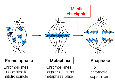
Metaphase (from Ancient Greek μετα- (meta-) beyond, above, transcending and from Ancient Greek φάσις (phásis) 'appearance') is a stage of mitosis in the cell cycle in which chromosomes of eukaryotes are at their second-most condensed and coiled stage (they are at their most condensed in anaphase).[1] These chromosomes, carrying genetic information, align in the equator of the cell between the spindle poles at the metaphase plate, before being separated into each of the two daughter nuclei. This alignment marks the beginning of metaphase.[2] Metaphase accounts for approximately 4% of the cell cycle's duration.[citation needed]

In metaphase, microtubules from both duplicated centrosomes on opposite poles of the cell have completed attachment to kinetochores on condensed chromosomes. The centromeres of the chromosomes convene themselves on the metaphase plate, an imaginary line that is equidistant from the two spindle poles.[3] This even alignment is due to the counterbalance of the pulling powers generated by the opposing kinetochore microtubules,[4] analogous to a tug-of-war between two people of equal strength, ending with the destruction of B cyclin.[5]

In order to prevent deleterious nondisjunction events, a key cell cycle checkpoint, the spindle checkpoint, verifies this evenly balanced alignment and ensures that every kinetochore is properly attached to a bundle of microtubules and is under balanced bipolar tension. Sister chromatids require active separase to hydrolyze the cohesin that bind them together prior to progression to anaphase. Any unattached or improperly attached kinetochores generate signals that prevent the activation of the anaphase promoting complex (cyclosome or APC/C), a ubiquitin ligase which targets securin and cyclin B for degradation via the proteosome. As long as securin and cyclin B remain active, separase remains inactive, preventing premature progression to anaphase.

Metaphase in cytogenetics and cancer studies

[edit]
Human metaphase chromosomes (normal male karyotype)

The analysis of metaphase chromosomes is one of the main tools of classical cytogenetics and cancer studies. Chromosomes are condensed (thickened) and highly coiled in metaphase, which makes them most suitable for visual analysis. Metaphase chromosomes make the classical picture of chromosomes (karyotype). For classical cytogenetic analyses, cells are grown in short term culture and arrested in metaphase using mitotic inhibitor. Further they are used for slide preparation and banding (staining) of chromosomes to be visualised under microscope to study structure and number of chromosomes (karyotype). Staining of the slides, often with Giemsa (G banding) or Quinacrine, produces a pattern of in total up to several hundred bands. Normal metaphase spreads are used in methods like FISH and as a hybridization matrix for comparative genomic hybridization (CGH) experiments.

Malignant cells from solid tumors or leukemia samples can also be used for cytogenetic analysis to generate metaphase preparations. Inspection of the stained metaphase chromosomes allows the determination of numerical and structural changes in the tumor cell genome, for example, losses of chromosomal segments or translocations, which may lead to chimeric oncogenes, such as bcr-abl in chronic myelogenous leukemia.

References

[edit]
[edit]
Revisions and contributorsEdit on WikipediaRead on Wikipedia
from Grokipedia
Metaphase is a critical stage in the processes of and , where replicated align along the metaphase plate—an imaginary equatorial plane at the center of the dividing cell—ensuring precise segregation of genetic material to daughter cells. This alignment is facilitated by the mitotic spindle apparatus, composed of that attach to the kinetochores on each chromosome, positioning them equidistant from the cell's poles. The stage follows , after the has broken down and chromosomes have condensed, and precedes , where the chromosomes separate. In , metaphase occurs in somatic cells to produce two genetically identical daughter cells, with each —consisting of two identical joined at the —aligning individually at the metaphase plate. The centrosomes, having migrated to opposite poles, organize the spindle fibers that pull the chromosomes into alignment, a regulated by the spindle assembly checkpoint to prevent errors in chromosome distribution. This precise orientation is essential for maintaining genomic stability during in multicellular organisms. Metaphase also features prominently in meiosis, the specialized division that generates gametes, occurring twice: once in metaphase I and again in metaphase II. In metaphase I, homologous chromosome pairs (tetrads) align at the metaphase plate, with microtubules attaching to kinetochores such that each homolog faces opposite poles, facilitating independent assortment of chromosomes. Metaphase II, resembling mitotic metaphase, involves the alignment of sister chromatids in the now-haploid cells produced by I, preparing for their separation to yield four unique haploid gametes. These events in introduce crucial for .

Fundamentals

Definition and Characteristics

Metaphase is the second stage of , following , during which the condensed chromosomes align at the metaphase plate, an imaginary equatorial plane equidistant from the two poles of the mitotic spindle. This alignment ensures equitable distribution of genetic material to daughter cells. In , analogous metaphase stages occur in metaphase I and II, where chromosomes align similarly but with variations in and segregation. Key characteristics of metaphase include the full of chromosomes, rendering them compact and visible as distinct structures, and their attachment to spindle fibers through specialized protein complexes called kinetochores located at the centromeres. The , which broke down during , remains absent, allowing direct interaction between chromosomes and the . In mammalian cells, metaphase typically lasts around 20-30 minutes, representing a brief but critical portion of the overall that averages 24 hours in cultured cells. Visually, under light or fluorescence microscopy, metaphase chromosomes appear as aligned, bar-like entities at the cell's midline, with held together and oriented by balanced forces from opposing spindle poles. This hallmark configuration facilitates the subsequent separation in and underscores metaphase's role as a poised state for accurate partitioning.

Historical Context

The discovery of metaphase emerged from pioneering cytological observations in the late , as researchers began to unravel the intricacies of using early microscopic techniques. In 1879, German biologist provided the first detailed description of , including what would later be recognized as metaphase, while studying rapidly dividing cells in salamander embryos. He noted the formation of an "equatorial plate" where thread-like structures—now known as chromosomes—aligned at the cell's midpoint, a critical observation made possible by his innovative use of dyes to stain cellular material for better visibility under the . Flemming's work, published in his 1882 book Zellsubstanz, Kern und Zelle, laid the groundwork for understanding chromosome dynamics during division, though he initially interpreted the process as a continuous transformation rather than discrete stages. Building on Flemming's findings, subsequent researchers extended these observations to reproductive cell division. In 1883, Belgian cytologist Edouard Van Beneden described chromosome behavior during in the roundworm , highlighting how homologous chromosomes paired and aligned similarly to mitotic figures, thus linking metaphase-like arrangements to the reduction of chromosome number in formation. This contribution clarified the role of such alignments in ensuring genetic continuity across generations. The term "metaphase" itself, denoting the "middle phase" between and , was formally coined in 1884 by German botanist Eduard Strasburger in his studies of plant , standardizing for the stage where chromosomes achieve equatorial alignment following condensation in . Advancements in light microscopy during this era were instrumental in these breakthroughs, transforming cytology from vague impressions to precise visualizations. Innovations such as Ernst Abbe's development of oil-immersion lenses in the and improved achromatic objectives in the enhanced resolution, allowing scientists to discern fine details that were previously indistinct. Combined with Flemming's protocols, these tools enabled the foundational of metaphase alignments, establishing cytology as a rigorous discipline and paving the way for later genetic insights.

Role in Mitosis

Process and Mechanism

During metaphase of mitosis, chromosomes achieve stable alignment at the spindle equator, known as the metaphase plate, following initial capture by microtubules in prometaphase. In prometaphase, kinetochores on sister chromatids form attachments to microtubules emanating from opposite spindle poles, enabling congression through microtubule depolymerization-driven movement toward the equator. Improper attachments, such as syntelic or merotelic orientations, are corrected by Aurora B kinase, which phosphorylates kinetochore proteins to destabilize erroneous microtubule-kinetochore interactions, promoting reattachment until bipolar orientation is achieved. The biophysical mechanisms underlying congression involve a balance of forces that position precisely at the metaphase plate. Kinetochore-directed forces pull poleward, while polar ejection forces—generated by chromokinesins interacting with non-kinetochore —push chromosome arms away from the poles, counteracting these pulls to center the chromosomes. This equilibrium is stabilized by inter-kinetochore tension, which arises from bipolar attachments and suppresses further error correction, ensuring bi-orientation. In human cells, metaphase typically lasts 10-20 minutes, during which chromosomes oscillate slightly around the plate to refine alignment. Progression to is triggered by satisfaction of the spindle assembly checkpoint, which monitors attachment and tension status across all .

Key Molecular Components

The serves as a multi-layered proteinaceous structure assembled on the region of chromosomes, essential for mediating attachments to spindle microtubules during metaphase of . At its core, the inner kinetochore incorporates centromere protein A (CENP-A), a variant that forms specialized nucleosomes within , providing a foundational platform for kinetochore assembly and epigenetic marking of centromeres. This inner layer interfaces with the constitutive centromere-associated network (CCAN), which recruits outer kinetochore components, including the KMN network comprising the Knl1, Mis12, and Ndc80 complexes. The Ndc80 complex, in particular, features calponin-homology domains that enable end-on binding to microtubule plus ends, facilitating stable kinetochore-microtubule attachments critical for chromosome alignment. Microtubule-kinetochore interactions during metaphase rely on motor proteins and dynamic properties of polymers to ensure proper positioning. The kinesin-7 family member CENP-E acts as a plus-end-directed motor that initially captures laterally at kinetochores, promoting their transition to end-on attachments and driving congression of to the metaphase plate. Cytoplasmic , a minus-end-directed motor, contributes by generating poleward forces that help correct misaligned attachments and stabilize biorientation. dynamics, involving and at kinetochore-attached ends, are fine-tuned by post-translational modifications such as α-tubulin detyrosination, which reduces catastrophe frequency and enhances attachment stability to support oscillatory movements. These interactions collectively allow kinetochores to track dynamic while resisting detachment under tension. Regulatory complexes orchestrate the timing and fidelity of metaphase progression by controlling protein stability and cohesion. The anaphase-promoting complex/cyclosome (/C), a large E3 ubiquitin ligase, remains inactive during metaphase until chromosomes achieve proper alignment, at which point it ubiquitinates substrates like securin and to trigger onset. , a ring-shaped multi-subunit complex consisting of SMC1, SMC3, SCC1/RAD21, and SCC3, encircles and holds together from through metaphase, generating the tension necessary for bipolar attachments and preventing premature separation. This pairing is maintained until APC/C-mediated cleavage of cohesin's kleisin subunit in , ensuring equitable chromatid distribution.

Role in Meiosis

Metaphase I

In metaphase I of meiosis, homologous chromosomes, paired as bivalents or tetrads, align at the equatorial metaphase plate of the cell. Each bivalent consists of two homologous chromosomes, each comprising two sister chromatids, held together at chiasmata—crossover points formed during prophase I that physically link the homologs and facilitate their co-orientation on the spindle. The spindle microtubules attach to the kinetochores of the homologous chromosomes such that the kinetochores of sister chromatids within each chromosome face the same spindle pole, establishing a monopolar attachment characteristic of this reductional division. This alignment process differs fundamentally from metaphase in , where individual chromosomes align independently with sister kinetochores bi-oriented toward opposite poles. In meiosis I, bivalent formation arises from prior , ensuring that entire homologous pairs segregate rather than , which remain cohesive due to protected centromeric . The monopolar orientation of sister kinetochores, combined with chiasmata-mediated tension, promotes stable bivalent congression and error-free segregation of homologs to opposite poles in I. The random orientation of these bivalents at the metaphase plate underlies Mendel's law of independent assortment, where each homologous pair aligns independently, leading to the equal probability of any parental combination in daughter cells and thus enhancing in gametes. For instance, in humans with 23 pairs, this randomness can generate over 8 million unique gamete genotypes from assortment alone, excluding recombination effects. Chromosome condensation during metaphase I mirrors that in , achieving maximum compaction for alignment.

Metaphase II

Metaphase II is the stage of the second meiotic division where, in each of the two haploid daughter cells produced after of I, the of every align individually at the metaphase plate, without any of homologous chromosomes. This alignment occurs in the absence of an intervening phase, maintaining the haploid number (1n, 2c) from the end of I. The process parallels mitotic metaphase in its equational nature but operates within haploid cells, ensuring precise positioning for subsequent separation. The mechanism of alignment in metaphase II involves the formation of a bipolar in each cell, with emanating from centrosomes at opposite poles attaching to the s of in an amphitelic configuration—meaning each sister kinetochore binds to from opposing spindle poles, generating tension that stabilizes bi-orientation. This attachment ensures that the centromeres of the paired congress to the equatorial plate, a process regulated by the spindle assembly checkpoint to verify proper -kinetochore interactions before progression. Unlike metaphase I, which involves bivalent tetrads, metaphase II treats each replicated as a unit, with attachments focused solely on ; this stage is typically shorter in duration than metaphase I, often proceeding rapidly without extended arrest in many . Following successful alignment, metaphase II transitions directly to anaphase II, an equational division where at the centromeres is cleaved, allowing to separate and migrate to opposite poles, ultimately yielding four haploid gametes (1n, 1c) upon completion of telophase II and . This outcome preserves the introduced by independent assortment during I, while ensuring equitable distribution of chromatids in the final gametic cells.

Biological Significance

Checkpoint Regulation

The spindle assembly checkpoint (SAC) serves as a critical surveillance mechanism during metaphase of both mitosis and meiosis, ensuring that all chromosomes achieve proper bipolar attachment to the mitotic spindle before progression to . Unattached kinetochores act as signal generators, recruiting checkpoint proteins such as Mad1 and Mad2 to initiate SAC activation. This leads to the formation of the mitotic checkpoint complex (MCC), a diffusible inhibitor composed of Mad2, BubR1 (also known as Bub1-related or Mad3 homologue), Bub3, and the APC/C co-activator Cdc20. The MCC binds directly to the anaphase-promoting complex/cyclosome (APC/C), preventing its ubiquitination of key substrates like securin and B1, thereby inhibiting sister chromatid separation until all kinetochores are properly attached. SAC activation occurs rapidly at unattached s through a conformational change in Mad2 from its open (O-Mad2) to closed (C-Mad2) form, which captures Cdc20 and facilitates MCC assembly in conjunction with BubR1's pseudokinase domain and KEN-box motifs that sterically block /C substrate recognition. Silencing of the SAC is triggered by occupancy and bi-orientation-generated tension at kinetochores, which stabilizes attachments and promotes MCC disassembly via factors like p31^{comet} and the stripping of checkpoint proteins from kinetochores. According to the diffusion-based inhibition model, the MCC acts as a soluble inhibitor that propagates the checkpoint signal throughout the , rather than requiring direct kinetochore-/C contact, allowing even a single unattached kinetochore to delay . This process typically concludes within 10-15 minutes in mammalian cells once attachments are established, balancing fidelity with timely progression. The core SAC machinery is highly conserved between and , with homologous proteins like Mad2 and BubR1 performing analogous roles in monitoring attachments across eukaryotes from to mammals. However, I incorporates additional safeguards to accommodate chiasmata, the physical manifestations of crossovers that hold homologous chromosomes together and generate unique tension signals for their segregation. These include enhanced protection against monopolar attachments of sister kinetochores on bivalents, mediated by chiasmata-induced stabilization that reinforces SAC sensitivity to tension, thereby preventing premature separation of homologues and reducing the risk of .

Errors and Consequences

During metaphase, erroneous -microtubule attachments can disrupt proper alignment and segregation. Merotelic attachments occur when a single binds to from both spindle poles, potentially leading to lagging chromosomes that fail to segregate correctly during . Syntelic attachments, in which both sister kinetochores attach to emanating from the same pole, are less common but can result in both chromatids moving to one daughter cell. Lagging chromosomes often arise as a downstream effect of unresolved merotelic errors, where chromosomes trail behind the segregating mass without proper poleward movement. In , these errors frequently cause , an abnormal number in daughter cells, which can trigger through proteotoxic stress or p53-mediated . Uncorrected promotes genomic instability and is a hallmark of cancer, contributing to tumor and resistance in up to 86% of solid tumors. In , nondisjunction resulting from metaphase attachment failures leads to gametes with extra or missing chromosomes, causing aneuploid embryos; for instance, failure to separate homologous chromosomes in metaphase I or in metaphase II underlies 21 (), characterized by , congenital heart defects, and increased malignancy risk. Such errors occur in approximately 1% of normal mitotic divisions, primarily due to merotelic attachments, though rates can rise to 1-5% in chromosomally unstable cells like those in cancer.00894-2) While the spindle assembly checkpoint (SAC) detects and corrects many syntelic errors by delaying , merotelic attachments often evade SAC surveillance, leading to uncorrected segregation failures. Persistent errors may prolong SAC activation or induce to prevent propagation of aneuploid cells.

Applications in Research

Cytogenetic Analysis

Cytogenetic analysis relies on metaphase chromosomes due to their condensed and aligned state, which facilitates clear visualization of chromosomal structure. In karyotyping, cells are treated with to disrupt formation and arrest at metaphase, allowing accumulation of well-spread chromosomes for analysis. This technique enables the visualization of the normal human consisting of 46 chromosomes arranged in 23 pairs. Following hypotonic swelling and fixation, chromosomes are stained using with Giemsa dye after treatment, producing a pattern of 400-800 bands across the that highlights structural details and aids in identifying abnormalities such as deletions, duplications, or rearrangements. Karyotyping detects structural chromosomal variants, including translocations, by revealing altered banding patterns; for instance, the , a t(9;22) translocation, is identifiable in chronic cases through G-banded metaphase spreads. In prenatal diagnosis, amniotic fluid cells obtained via are cultured and arrested at metaphase for karyotyping, enabling detection of fetal aneuploidies or structural anomalies in high-risk pregnancies. This approach provides a resolution sufficient for identifying changes at the level of whole chromosomes or large segments, typically greater than 5-10 Mb. Advancements in include (FISH) applied to metaphase chromosome spreads, where fluorescent probes hybridize to specific DNA sequences for locus-specific detection. Metaphase FISH enhances resolution to approximately 1-5 Mb, allowing precise mapping of loci or submicroscopic rearrangements not visible with standard banding. This method complements karyotyping by targeting repetitive or unique sequences, improving diagnostic accuracy for genetic disorders. Recent developments as of 2025 have integrated (AI) and for automated analysis of metaphase images, enhancing efficiency in chromosome detection and karyotyping. These tools, such as deep neural networks for metaphase finding and chromosome segmentation, achieve high accuracy (e.g., 98.88% in identification) and reduce manual processing time, supporting applications in prenatal screening and cancer diagnostics. Open datasets of annotated metaphase cells have facilitated these AI models, while studies demonstrate their superiority over traditional methods in speed and precision.

Cancer Studies

Chromosomal instability (CIN) during metaphase, particularly errors in chromosome alignment and segregation at the metaphase plate, is a primary driver of aneuploidy observed in over 90% of solid tumors. This instability arises from defects in the spindle assembly checkpoint (SAC), which monitors kinetochore-microtubule attachments to prevent premature anaphase onset. Mutations or deficiencies in SAC components, such as BubR1 (also known as BUB1B), weaken checkpoint signaling, leading to increased rates of chromosome missegregation and promoting tumorigenesis by generating genomic heterogeneity that favors oncogenic adaptations. For instance, hypomorphic BubR1 mutations in mouse models result in elevated CIN and accelerated tumor formation, mirroring observations in human cancers where such alterations correlate with poor prognosis. Therapeutically, targeting metaphase dynamics has proven effective through microtubule-stabilizing agents like taxanes, exemplified by , which bind β-tubulin to suppress microtubule and induce prolonged metaphase arrest. This arrest activates the SAC, halting progression to and triggering in rapidly dividing cancer cells via pathways involving activation and chromosomal instability-induced stress. is a cornerstone treatment for and ovarian cancers, where it achieves response rates of approximately 30-50% in advanced cases by exploiting the high of tumors. In , metaphase chromosome spreads provide critical insights into tumor karyotypes, enabling visualization of patterns and structural aberrations that reflect underlying CIN. These spreads, prepared from colchicine-arrested tumor cells, reveal recurrent gains (e.g., s 7, 8q) and losses that drive oncogenesis, aiding in the of tumor subtypes. Furthermore, studies from the highlighted how SAC hyperactivation, particularly in the context of dysfunction, contributes to chemoresistance by promoting mitotic slippage and survival rather than following taxane exposure. This interaction underscores the need for combined therapies targeting both SAC components and pathways to overcome resistance in CIN-high tumors.

References

Add your contribution
Related Hubs
User Avatar
No comments yet.