Hubbry Logo
search
logo
1622953

Vermilion border

logo
Community Hub0 Subscribers
Read side by side
from Wikipedia
Vermilion border
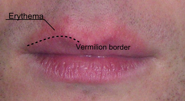
Human lips with the vermilion border outlined. A patch of erythema above the upper lip makes it more difficult to distinguish the vermilion border.
Details
Identifiers
Latinpars intermedia labiorum oris
THH3.04.01.0.00010
Anatomical terminology

The vermilion border (sometimes spelled vermillion border), also called margin or zone, is the normally sharp demarcation between the lip and the adjacent normal skin. It represents the change in the epidermis from highly keratinized external skin to less keratinized internal skin. It has no sebaceous glands, sweat glands, or facial hair.[1][2]

It has a prominence on the face, creating a focus for cosmetics (it is where lipstick is sometimes applied) and is also a location for several skin diseases. Its functional properties, however, remain unknown.[3]

Structure

[edit]

The lips are composed wholly of soft tissue. The skin of the face is thicker than the skin overlying the lips where blood vessels are closer to the surface. As a consequence, the margin of the lips shows a transition between the thicker and thinner skin, represented by the vermilion border. It therefore has the appearance of a sharp line between the coloured edge of the lip and adjoining skin.[4]

It has been described as a pale, white rolled border and also as being a red line.[1][5]

This fine line of pale skin accentuates the colour difference between the vermilion and normal skin. Along the upper lip, two adjacent elevations of the vermilion border form the Cupid's bow.[6]

Microanatomy

[edit]

The vermilion border represents the change in the epidermis from highly keratinized external skin to less keratinized internal skin. It has no sebaceous glands, sweat glands, or facial hair.[2][1]

There are two reasons that the border appears red in some people:

  1. The epithelium is thin and therefore the blood vessels are closer to the surface.[7]
  2. This epithelium contains eleidin which is transparent and the blood vessels are near the surface of the papillary layer, revealing the "red blood cell" color. At the angles of the mouth, there are sebaceous glands, without hair follicles, which are called Fordyce spots.[2][8]
A Cupid's bow on the upper lip.

Clinical significance

[edit]

The vermilion border is important in dentistry and oral pathology as a marker to detect disease, such as in actinic cheilitis.[9]

Associated diseases

[edit]
  • Perioral dermatitis is a rash typically around the mouth, that spares the vermilion border.[10]
  • Cheilitis glandularis may present with a burning sensation over the vermilion border. This chronic progressive condition is associated with thinning of the skin of the lips and ulceration.[11]
  • Infections may involve the vermilion border. Cold sores are one common infection.[12] Impetigo is another.[13]
  • Skin cancer can also occur at the vermilion border.[9]
  • Fetal alcohol syndrome causes facial abnormalities which include a thin vermilion border with a smooth philtrum.[14]

Cosmetic appearance

[edit]
  • Sunlight exposure can blur the junction between the vermilion border and the skin. Applying lip balm and sunscreen moisturizer protects it from sunlight.[15]

Surgery

[edit]

A vermilionectomy (sometimes spelled vermillionectomy) is the surgical removal of the vermilion border. It is sometimes performed to treat carcinoma of the lip.[16]

Close attention is given when repairing any injury to the vermilion border. Even 1 mm of vermilion misalignment could be noticeable.[5]

See also

[edit]

References

[edit]
Revisions and contributorsEdit on WikipediaRead on Wikipedia
from Grokipedia
The vermilion border is the junction between the vermilion zone—the reddish, exposed portion of the lips—and the surrounding facial skin, marked by a 2- to 3-mm pale ridge known as the white roll, formed by the underlying orbicularis oris muscle. This border consists of a thin, non-keratinized stratified squamous epithelium, typically 3 to 5 cell layers thick, which is highly vascularized and lacks hair follicles, sweat glands, or sebaceous glands. The characteristic pink to reddish hue of the vermilion zone is a normal anatomical variation in both men and women, resulting from the thin, translucent epithelium allowing the color of oxygenated blood in underlying capillaries to show through, influenced by genetic factors such as melanin distribution, skin thickness, and vascular density. This mechanism is the same in both sexes and is not typically linked to any specific medical condition unless accompanied by other symptoms (e.g., ruddy complexion in polycythemia).[1] Anatomically, the vermilion border outlines key features of the lips, including the Cupid's bow on the upper lip and the oral commissures. Embryologically, it develops from the fusion of the frontonasal and maxillary prominences during the sixth to seventh weeks of gestation, with basic lip structures formed by the end of the seventh week; misalignment is a feature in congenital anomalies like cleft lip, which affects approximately 1 in 700 births.[1]

Anatomy

Gross anatomy

The vermilion border represents the mucocutaneous junction between the cutaneous skin of the lip and the oral mucosa, manifesting as a sharply defined reddish line attributable to its thin epithelial layer and prominent underlying vascularity.[1] This transitional zone encircles the oral orifice, distinguishing the dry, exposed vermilion surface from the adjacent skin and inner mucosa.[1] Positioned on both the upper and lower lips, the vermilion border extends laterally from one commissure to the other, with greater prominence and definition observed on the upper lip due to its anatomical contouring.[1] Key macroscopic features include the white roll, a subtle pale ridge measuring 2 to 3 mm in width located immediately superior to the border on the upper lip, resulting from the underlying orbicularis oris muscle prominence.[1] The upper lip vermilion border further features the Cupid's bow, a distinctive M-shaped downward arch at the base of the philtrum that enhances facial aesthetics and symmetry.[1] Separating the vermilion from the internal moist mucosa is the wet-dry line, a fine demarcation where salivary gland openings contribute to the transition between dry external exposure and wet internal lubrication.[1] In adults, the exposed vermilion typically measures 7 to 10 mm in vertical height, varying slightly by sex and ethnicity, with the upper lip often shorter than the lower.[2] This dimension plays a critical role in lip projection, providing the everted, rounded contour that defines the lips' forward prominence and contributes to overall facial harmony.[1]

Histology and microanatomy

The vermilion border is covered by a stratified squamous epithelium that exhibits a transitional character, shifting from keratinized on the cutaneous side to non-keratinized on the mucosal side. This epithelium lacks the robust keratinization seen in typical skin, with the stratum corneum reduced to a thin layer of only 3 to 5 cell layers, which contributes to the region's translucency and vulnerability to environmental factors.[3][4] The keratinocytes in this layer produce eleidin, a clear intracellular protein that enhances the reddish appearance by allowing visibility of underlying structures.[5] A defining feature of the vermilion border's microanatomy is the absence of adnexal structures, including hair follicles, sebaceous glands, and sweat glands, which distinguishes it from adjacent facial skin and mucosal tissues. This lack of appendages results in reduced lubrication and increased susceptibility to dryness and irritation, as there are no mechanisms for sebum or sweat production to maintain hydration.[1][6] Beneath the epithelium lies the dermis, composed of dense, regularly arranged collagen fiber bundles that provide structural support and occupy much of the layer's volume. The dermis features prominent superficial vasculature, with blood vessels positioned close to the dermo-epidermal junction. The characteristic red hue of the vermilion border results from the thin, translucent nature of the overlying epithelium, which enables the red color of oxygenated hemoglobin in these superficial capillaries to show through. This coloration is a normal anatomical variation in both men and women, influenced by genetic factors such as melanin distribution, epithelial thickness, and vascular density. Naturally red lips in men are thus the result of the same physiological mechanism as in women and are not typically indicative of any pathological condition absent other accompanying symptoms (e.g., ruddy complexion in polycythemia). Melanocytes are present but sparse in the basal layer compared to surrounding skin, contributing minimally to pigmentation and further enhancing the region's lighter, reddish tone.[7][1]00179-2/abstract) The submucosal layer underlying the vermilion consists of loose connective tissue that transitions toward the oral mucosa, housing minor salivary glands known as labial glands. These glands, primarily serous and mucous in composition, are situated beneath the vermilion and provide some moistening to the inner aspects of the lip, though their influence on the border itself is limited due to the absence of direct ductal openings in this zone.[1][8]

Vascular and neural supply

The arterial supply to the vermilion border arises primarily from the superior labial artery, a branch of the facial artery, which nourishes the upper lip vermilion, and the inferior labial artery, which supplies the lower lip vermilion.[9] These arteries form extensive anastomoses across the midline, ensuring robust perfusion to the region.[9] Venous drainage from the vermilion border is facilitated by the superior and inferior labial veins, which empty into the ipsilateral facial veins.[1] In certain individuals, particularly older adults, dilated venous structures known as venous lakes—benign varicosities—may appear as soft, dark blue papules along the vermilion border due to ectatic venules.[10] Lymphatic drainage of the vermilion border directs primarily to the submandibular lymph nodes for the upper lip and lateral aspects of the lower lip, while the central portion of the lower lip drains to the submental nodes; the middle portion of the upper lip may also contribute to submental drainage.[1] Sensory innervation to the vermilion border is provided by the trigeminal nerve (CN V), with the infraorbital nerve (maxillary division, V2) supplying the upper lip and the mental nerve (mandibular division, V3) innervating the lower lip, resulting in a high density of nerve endings that confer acute sensitivity to touch and pain.[1] Motor innervation derives from the facial nerve (CN VII), with buccal branches controlling the orbicularis oris and lip elevators of the upper lip, and marginal mandibular branches innervating the lip depressors of the lower lip to facilitate movements such as puckering and smiling.[1]

Embryology and development

Embryonic origins

The vermilion border of the lips originates from the stomodeal ectoderm and underlying mesenchyme during early embryogenesis, specifically around weeks 4 to 7 of gestation. The stomodeum, a primitive oral depression lined by ectoderm, serves as the central site where facial prominences develop, with the ectodermal layer contributing to the epithelial covering of the future lips and the neural crest-derived mesenchyme providing structural support and growth factors for morphogenesis.[11][12] The formation of the lips, including the vermilion border, results from the fusion of embryonic facial prominences. For the upper lip, the maxillary prominences grow medially and fuse with the medial nasal prominences around the stomodeum during weeks 6 to 7, creating a continuous structure where the vermilion border emerges at the ectodermal junction of these fused elements. In the lower lip, the paired mandibular prominences merge at the midline through mesenchymal proliferation, establishing the inferior vermilion margin. This fusion process involves epithelial adhesion via molecules like nectins and E-cadherin, followed by programmed cell death in the epithelial seam to allow mesenchymal confluence.[11][12][13] The philtrum, a key feature of the upper vermilion border, arises from the median nasal prominence, which contributes the central portion of the upper lip. During week 6, the bilateral medial nasal processes fuse in the midline, forming the philtral columns and groove that define the superior contour of the vermilion border.[12][14] Incomplete fusion of these prominences can lead to developmental anomalies such as cleft lip, which disrupts the continuity of the vermilion border. In unilateral cleft lip, failure of the maxillary and medial nasal prominences to merge results in a gap across the vermilion, while bilateral forms affect both sides; remnants of partial fusion, known as Simonart's band, may appear as a thin tissue bridge spanning the defect. These anomalies highlight the precise timing and cellular coordination required for vermilion border integrity.[12][15] By the end of week 7, lip fusion is typically complete, with vermilion border differentiation occurring by week 8 as the ectodermal epithelium specializes into its transitional, non-keratinized form.[11][13]

Postnatal changes

During infancy, the vermilion border exhibits initial fullness and relative definition, with its growth occurring proportionally to overall facial development as the child matures.[16] By early childhood, the border begins to refine, achieving sharper demarcation by adolescence through continued tissue maturation and skeletal growth.[16] In adulthood, the vermilion border reaches peak prominence and volume during the 20s and 30s, characterized by optimal collagen density and hyaluronan content that support its plump, well-defined appearance.[7] Starting in the 40s, gradual thinning and flattening ensue, primarily due to progressive loss of collagenous fibers in the dermis (correlation with age: r = -0.721, P = 0.003).[7] Aging further accentuates these changes, with vermilion height reducing by up to 50% by age 80, accompanied by blurring of the white roll, increased perioral lines, and diminished vascular supply in the dermis (vessel area decrease: r = -0.626, P = 0.017; vessel number decrease: r = -0.716, P = 0.004).[16][17] These effects are exacerbated by extrinsic factors such as chronic sun exposure, which degrades collagen and hyaluronan via UV-induced mechanisms, and smoking, which accelerates dermal atrophy and barrier dysfunction.[7][17] Gender variations influence vermilion border characteristics, with females typically displaying relatively fuller and more projected borders compared to males, who exhibit greater overall lip height but less vermilion prominence.[18] Ethnic differences also play a role, as individuals of Asian descent often have fuller vermilion borders than those of Caucasian descent, reflecting variations in cutaneous upper lip height and tissue volume.[19]

Physiological functions

Sensory roles

The vermilion border of the lips exhibits a high density of mechanoreceptors, including Meissner corpuscles, simple corpuscles, and Merkel cell-neurite complexes (Tastcheiben), enabling fine tactile discrimination such as touch and pressure; these specialized endings number approximately 10–20 corpuscular receptors per mm² in primates, contributing to the region's exceptional sensitivity compared to surrounding facial skin.[20] In humans, similar mechanoreceptive structures, such as Meissner and glomerular corpuscles expressing mechanosensitive channels like PIEZO2 and ASIC2, maintain densities of 3–5 per mm², with higher concentrations in the upper lip than the lower.[21] This dense innervation supports precise sensory feedback during activities like eating and speaking, where subtle lip deformations must be detected rapidly. Nociceptors, primarily in the form of free nerve endings, are abundant in the vermilion border's epithelium and underlying connective tissue, providing acute pain detection to protect against mechanical injury or irritants; their proximity to the surface enhances responsiveness, making the area one of the most pain-sensitive regions of the body.[20] Temperature sensation is mediated by these same free nerve endings, which respond to thermal changes and aid in regulating oral intake by signaling extremes of heat or cold; thermal thresholds in the vermilion are notably low, with the upper lip showing heightened sensitivity to both warm and cool stimuli compared to adjacent areas. Motor functions of the vermilion border involve coordinated contractions of the orbicularis oris muscle, which forms the structural core of the lips and enables actions like closure, puckering, and protrusion; sensory feedback from trigeminal afferents integrates with motor output to refine these movements, ensuring precision through oligosynaptic reflexes that adjust for perturbations during dynamic tasks. Efferent signals originate from the facial motor nucleus in the brainstem, projecting via the facial nerve to innervate the orbicularis oris, while afferent sensory information from the vermilion travels via the trigeminal nerve's maxillary and mandibular divisions to the trigeminal sensory nuclei, facilitating closed-loop control.[22] In communication, the vermilion border plays a critical role in speech articulation, particularly for labial consonants such as /p/, /b/, and /m/, where bilabial closure requires precise sensorimotor synchronization to produce plosive or nasal sounds; this integration also supports non-verbal expressions like smiling and whistling, relying on sensory cues to modulate muscle tension for emotional conveyance.[23]

Barrier and protective roles

The vermilion border functions as a transitional barrier between the keratinized skin and non-keratinized oral mucosa, characterized by incomplete keratinization that permits limited moisture retention while rendering it vulnerable to transepidermal water loss (TEWL). This TEWL is substantially higher—up to three times that of facial skin—due to the underdeveloped stratum corneum, yet lower than in the oral mucosa, reflecting its partial barrier role.[24] The incomplete formation of corneocytes in this region compromises the overall integrity of the barrier, contributing to a reduced capacity for water retention and necessitating frequent external hydration to prevent desiccation.[25] In terms of pathogen protection, the stratified squamous epithelium of the vermilion border acts as a primary physical barrier, supported by its proximity to minor salivary glands that secrete saliva rich in antimicrobial lysozyme. This enzyme hydrolyzes bacterial cell walls, providing chemical defense against microbial invasion on the exposed surface.[26] Additionally, antimicrobial lipids, such as lauric and sapienic acids derived from sebaceous glands near the vermilion, further enhance resistance to pathogens in this vulnerable interface.[27] The vermilion border's thin epithelial layer heightens susceptibility to ultraviolet (UV) radiation, increasing the risk of photodamage and conditions like actinic cheilitis due to minimal melanin pigmentation compared to adjacent facial skin.[28] Mechanically, the vermilion border exhibits elasticity that supports resilience during activities like chewing and speaking, enabling the lips to maintain oral competence and adapt to repetitive movements. However, its poor water-holding capacity from inadequate corneocyte formation makes it prone to trauma exacerbated by dryness or mechanical habits such as lip-biting, which can disrupt the epithelial integrity.[6][25]

Clinical significance

Pathological conditions

The vermilion border, as the exposed junction between the lip mucosa and skin, is particularly susceptible to pathological changes due to its thin epithelium, high keratinocyte turnover, and chronic exposure to environmental stressors. Pathological conditions affecting this region often manifest with symptoms such as erythema, scaling, ulceration, or fissuring, and may require biopsy for definitive diagnosis to differentiate benign from malignant processes. Actinic cheilitis represents a common precancerous lesion primarily resulting from cumulative ultraviolet (UV) radiation exposure, especially in fair-skinned individuals with prolonged sun exposure. It typically presents as rough, scaly, or hyperkeratotic patches on the lower lip vermilion border, with potential for induration or leukoplakia in advanced cases. This condition carries a significant risk of progression to squamous cell carcinoma, estimated at 10-30% over time, underscoring the need for early intervention through topical therapies or excision.[29] Various forms of cheilitis directly impact the vermilion border, leading to chronic inflammation and structural alterations. Exfoliative cheilitis is characterized by persistent peeling and desquamation of the vermilion epithelium, often linked to psychological factors or irritants, resulting in a shiny, atrophic appearance. Angular cheilitis involves painful fissures and cracks at the oral commissures extending to the vermilion border, commonly associated with moisture, nutritional deficiencies, or denture-related trauma. Glandular cheilitis, a rarer variant, features swelling, eversion, and atrophy of the vermilion border due to hyperplasia of underlying minor salivary glands, potentially progressing to ulceration if untreated. Infectious etiologies frequently target the vermilion border's mucosal transition zone. Herpes simplex virus (HSV) infection causes recurrent vesicular eruptions along the border, progressing to painful ulcers and crusting, with primary episodes often triggered by stress or immunosuppression. Oral candidiasis presents as adherent white plaques or erythematous patches on the vermilion, particularly in immunocompromised patients, and can lead to angular involvement if untreated with antifungals. Autoimmune disorders can erode the vermilion border's integrity. Discoid lupus erythematosus, a cutaneous manifestation of systemic lupus, often affects the lower lip border with well-demarcated erythematous plaques, hyperkeratosis, and central atrophy, potentially causing scarring and ulceration; biopsy reveals interface dermatitis. Other autoimmune conditions, such as lichen planus, may present with lacy white lesions or erosions confined to the vermilion. Neoplastic processes are a critical concern for the vermilion border owing to its UV-exposed surface. Actinic keratosis may evolve into squamous cell carcinoma (SCC), the most common malignancy here, appearing as persistent nodules, ulcers, or indurated plaques with a propensity for local invasion and metastasis in neglected cases. Basal cell carcinoma occurs less frequently but can erode the border with pearly, telangiectatic borders. Malignant melanoma, though rare on the lips (accounting for <1% of oral melanomas), arises de novo or from preexisting nevi and exhibits aggressive behavior with rapid vertical growth and poor prognosis if not excised early. Additional pathological entities include allergic contact dermatitis, triggered by lip balms, cosmetics, or metals, manifesting as pruritic, eczematous changes with edema and vesiculation along the border; patch testing aids identification of allergens. Factitial cheilitis results from repetitive self-inflicted trauma, such as lip biting, leading to irregular erosions and scarring; management involves behavioral intervention alongside topical care. In all these conditions, histopathological examination via biopsy remains essential for confirming diagnosis and guiding therapy, often revealing epithelial dysplasia, inflammation, or atypical cellular features specific to the etiology.

Cosmetic aspects

The vermilion border, the distinct junction between the lip's red vermilion zone and the adjacent skin, is central to lip aesthetics, as it delineates lip shape and contributes to perceptions of fullness and facial harmony. A well-defined border enhances the visual prominence of the lips, signaling youthfulness and attractiveness in facial proportions. Studies indicate that ideal lip aesthetics often follow a golden ratio, with ratios such as 1:1.6 or 1:2 for upper to lower lip volume or height, promoting balanced symmetry and eversion.[30][31][32] Aging impacts the vermilion border's appearance through progressive volume loss in the lip tissue and flattening of the border, leading to a less defined contour and reduced lip projection. These changes result from collagen and elastin degradation, causing the lips to appear thinner and less vibrant over time. Smoking accelerates this process by promoting perioral wrinkles and pigmentation irregularities that blur the border's sharpness, further diminishing aesthetic appeal. Ethnic variations influence baseline prominence; for instance, individuals of African descent often exhibit more pronounced vermilion borders and fuller lips compared to Caucasians, who may show greater age-related flattening due to thinner initial lip structure.[33][34][35][2] Non-surgical enhancements target the vermilion border to restore definition and volume without invasive procedures. Hyaluronic acid fillers, injected precisely along the border, augment its prominence and create a sharper outline, often yielding natural-looking results lasting 6-12 months. The BOTOX lip flip involves injecting botulinum toxin into the upper lip's orbicularis oris muscle near the border to relax it, causing slight eversion for enhanced fullness and a subtle pout. Topical retinoids, such as tretinoin, can improve border definition by stimulating collagen production and smoothing perioral fine lines, though application requires caution to avoid irritation.[36][37][38][39][40] Makeup techniques offer temporary enhancement by emphasizing the vermilion border's natural line. Lip liners in shades matching the lip color are applied to trace and slightly overline the border, creating an optical illusion of greater fullness and symmetry without altering the underlying structure. Blending the liner into lipstick or gloss ensures a seamless finish, particularly effective for those with subtle age-related blurring.[41][42] Culturally, the vermilion border holds significance in beauty standards, with Western ideals often favoring fuller, well-defined borders as symbols of sensuality and vitality, influencing trends in cosmetics and enhancements. This emphasis contrasts with some Asian aesthetics, where subtler lip proportions may be preferred, highlighting how ethnic backgrounds shape perceptions of ideal lip prominence.[43][2]

Surgical interventions

Surgical interventions for the vermilion border primarily address trauma, reconstruction following oncologic resection, congenital anomalies, and cosmetic enhancement, with techniques emphasizing precise alignment to maintain aesthetic and functional integrity. In cases of trauma, such as lacerations crossing the vermilion border, repair begins with meticulous approximation using interrupted sutures to align the border, as even 1 mm of misalignment can result in a noticeable cosmetic defect.[44] For small defects or gaps, V-Y plasty advancement is employed to mobilize tissue and restore continuity without distorting the lip's contour.[45] Reconstructive procedures are essential for defects arising from cancer excision, where up to one-third of the lip may be involved, requiring flaps to rebuild the vermilion while preserving oral competence. The Abbé flap, a pedicled cross-lip transposition from the contralateral lip, is used for central defects near the commissure, providing well-vascularized tissue that matches the color and texture of the vermilion.[46] The Estlander flap, a variant for commissural defects, rotates tissue from the upper to lower lip or vice versa, effectively reconstructing the border but potentially requiring secondary revision for oral opening.[47] Vermilion advancement flaps, such as mucosal V-Y plasty, are suitable for superficial defects, advancing inner lip mucosa to recreate the border with minimal donor site morbidity.[48] For congenital deformities like unilateral cleft lip, the Millard rotation-advancement technique reconstructs the vermilion border by rotating the medial segment downward and advancing the lateral flap to align the white roll and Cupid's bow, achieving symmetry in the mucocutaneous junction.[49] This repair is typically performed between 3 and 6 months of age to optimize growth and minimize scarring, with attention to layering the vermilion mucosa for a natural transition.[50] Cosmetic surgery targeting the vermilion border includes the direct lip lift, which involves an incision just above the border to excise a strip of skin, shortening the philtrum and increasing vermilion show for enhanced lip projection.[51] This procedure risks visible scarring along the border and asymmetry if tissue tension is uneven, necessitating careful suture placement and postoperative monitoring.[52] Throughout these interventions, the vermilion border's rich vascular supply from the superior and inferior labial arteries facilitates rapid healing and reduces necrosis risk, though meticulous hemostasis is required to prevent hematoma.[46] Precise preservation of sensory nerves, including branches of the mental and infraorbital nerves, is critical to avoid numbness or dysesthesia, with regional blocks preferred over local infiltration to minimize distortion during alignment.[53] Postoperative care emphasizes wound protection, suture removal at 5-7 days, and serial evaluations to ensure border alignment and function.[54]

References

User Avatar
No comments yet.