Hubbry Logo
FlippaseFlippaseMain
Open search
Flippase
Community hub
Flippase
logo
8 pages, 0 posts
0 subscribers
Be the first to start a discussion here.
Flippase
Flippase
from Wikipedia
Structure of a flippase, showing the two major subunits of the enzyme

Flippases are transmembrane lipid transporter proteins located in the cell membrane. They are responsible for aiding the movement of phospholipid molecules between the two layers, or leaflets, that comprise the membrane. This is called transverse diffusion, also known as "flip-flop" transition. Flippases move lipids to the cytosolic layer, usually from the extracellular layer. Floppases do the opposite, moving lipids to the extracellular layer. Both flippases and floppases are powered by ATP hydrolysis and are either P4-ATPases or ATP-Binding Cassette transporters. Scramblases are energy-independent and transport lipids in both directions.[1][2][3]

Lateral and transverse movements

[edit]

In organisms, the cell membrane consists of a phospholipid bilayer. Phospholipid molecules are movable in the bilayer. These movements are categorized into two types: lateral movements and transverse movements (also called flip-flop). The first is the lateral movement, where the phospholipid moves horizontally on the same side of the membrane. Lateral movement is fast, with an average speed of up to 2 mm per second.[4] Transverse movement is the movement of the phospholipid molecule from one side of the membrane to the other. Transverse movement without the assistance of enzymes is slow, occurring once a month.[4] This is because the polar head groups of phospholipid molecule cannot easily pass through the hydrophobic center of the bilayer, limiting their diffusion in this dimension.

Lateral and transverse movements of lipids

Although flip-flop is slow, this movement is necessary to continue the cell's normal function of growth and mobility.[5] The possibility of active maintenance of the asymmetric distribution of molecules in the phospholipid bilayer was predicted in the early 1970s by Mark Bretscher.[6]  Lipid asymmetry has broad physiological implications, from cell shape determination to critical signaling processes like blood coagulation and apoptosis.[7] Many cells maintain asymmetric distributions of phospholipids between their cytoplasmic and exoplasmic membrane leaflets. The loss of asymmetry, in particular the appearance of the anionic phospholipid phosphatidylserine on the exoplasmic face, can serve as an early indicator of apoptosis[8] and as a signal for efferocytosis.[9]

Different classes of lipid transporters

[edit]

Lipid transporters transport lipids across the bilayers. There exist three major classes of lipid transporters:

Three major classes of lipid transporters and different functions to lipid of each enzyme[10]
  1. P-type Flippase
  2. ABC Floppase
  3. Scramblases

P-type Flippase and ABC Floppase are energy-dependent enzymes that can create lipid asymmetry and transport specific lipids. Scramblases are energy-independent enzymes that can dissipate lipid asymmetry and have broad lipid specificity.[11]

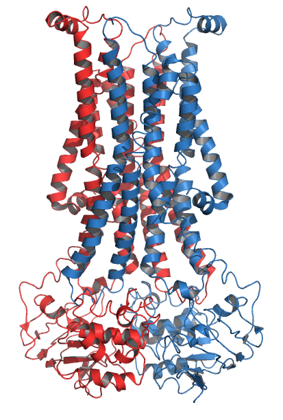
Structure and domains of P4-type flippases

[edit]
Domains of flippase

The P4-type flippase contains a large transmembrane segment and two major subunits, a catalytic domain called the alpha-subunit and an accessory domain named the beta-subunit.[5] Transmembrane segments contain 10 transmembrane alpha helices and this domain together with the beta-subunit plays an important role in the stability, localization, and recognition of the lipid substrate of flippase.[5] Alpha-subunits include A, P, and N domains and each of them corresponds to a different function of flippase. The A-domain is an actuator segment of flippase that facilitates phospholipid binding through conformational change of the complex, although it does not bind the phospholipid itself. The P-domain is responsible for binding phosphate, a product of ATP hydrolysis. The next domain is the N-domain, whose job is to bind the substrate (ATP).[5] Finally, a C-terminal autoregulatory domain has been identified, whose function differs between yeast and mammalian P4-type flippases.[12]

Mechanism of P4-type flippases

[edit]
Schematic of flippase mechanism: EP stands for phosphorylated flippases, PL represents the phospholipid substrate

To bind specific lipid on the outer layer of membrane, P4-type flippase needs to be phosphorylated by ATP on its P-domain. After ATP hydrolysis and phosphorylation, P4-type flippases undergo conformational change from E1 to E2 (E1 and E2 stand for different conformations of flippases).[5] Further conformational change is induced by the binding of a phospholipid, resulting in the E2Pi.PL conformation.[12] The flippase in its E2 conformation can then be dephosphorylated at its P-domain, allowing the lipid to be transported to the inner layer of membrane, where it diffuses away from the flippase. As the phospholipid dissociates from the complex, a conformational change on flippase occurs from E2 back to E1 readying it for the next cycle of lipid transportation.[5]

The A-domain binds to the N-domain after that domain releases ADP. The A-domain can bind to the N-domain by a TGES four-amino-acid motif when the P-domain is phosphorylated. The release of ADP from the N-domain transitions the complex from the E1P-ADP state to the E2P state, which might be further stabilized by binding of the C-terminal regulatory domain. Binding of a phospholipid to the first two transmembrane segments induces a conformational change that rotates the A domain outward by 22 degrees, allowing dephosphorylation of the P domain. Dephosphorylation of the P-domain is energetically coupled to translocation of the polar phospholipid head across the membrane leaflets.[12]

See also

[edit]

References

[edit]
Revisions and contributorsEdit on WikipediaRead on Wikipedia
from Grokipedia
Flippases are a family of ATP-dependent transmembrane proteins, primarily belonging to the P4-ATPase subfamily of P-type ATPases, that actively translocate specific phospholipids—such as (PS) and (PE)—from the exofacial (extracellular or luminal) leaflet to the cytosolic leaflet of lipid bilayers, thereby establishing and maintaining asymmetric lipid distribution across cellular membranes. This unidirectional transport counters the natural tendency of lipids to diffuse spontaneously across bilayers, which has a high barrier and half-time of approximately 100 hours without catalysis. In eukaryotic cells, flippases play a crucial role in biogenesis, vesicle trafficking, and signaling by ensuring that aminophospholipids like PS and PE are sequestered in the inner leaflet under normal conditions, while choline-containing lipids such as (PC) predominate in the outer leaflet. Mammals express 14 P4-ATPase flippases (ATP8A1 through ATP11C), which are divided into P4A and P4B subfamilies; P4A members typically form heterodimers with accessory β-subunits from the CDC50 (TMEM30) family to stabilize the complex and enhance activity, whereas some P4B flippases, like the Neo1, function as monomers. The transport mechanism involves coupled to a phosphorylation-dephosphorylation cycle, creating alternating access conformations (E1 and E2 states) that facilitate binding at an extracellular entry site, passage through a hydrophobic gate, and release to the . Flippases are distinguished from other lipid translocators: floppases (e.g., ABC transporters like ) move lipids outward to the exoplasmic leaflet, while scramblases (e.g., TMEM16 family members) enable bidirectional, ATP-independent scrambling that dissipates asymmetry, often activated during processes like or blood clotting. Dysregulation of flippase activity is implicated in diseases, including (e.g., ATP8B1 mutations in progressive familial intrahepatic ), neurological disorders (e.g., ATP8A2 defects in ), and hemolytic anemias (e.g., ATP11C variants affecting erythrocyte PS exposure). In and other model organisms, flippases like Drs2p are essential for endosomal sorting and Golgi-to-plasma membrane trafficking, highlighting their conserved role in eukaryotic membrane dynamics.

Membrane Lipid Organization

Lateral Mobility

Lateral diffusion refers to the rapid, energy-independent movement of phospholipids within the same leaflet of the , occurring parallel to the membrane plane. This process is a fundamental aspect of membrane dynamics, driven by and the fluid nature of the bilayer. In fluid bilayers, the lateral coefficients of phospholipids typically range from 1 to 2 μm²/s, corresponding to effective displacement rates of up to 1-2 μm/s under physiological conditions. These rates are modulated by several factors, including , which increases with higher temperatures or unsaturated chains; content, which can reduce by rigidifying the bilayer; and protein crowding, which impedes mobility through steric hindrance and can decrease coefficients by up to two orders of magnitude at high protein densities. Experimental measurement of lateral mobility commonly employs (FRAP), where a region of fluorescently labeled is photobleached and the recovery of due to from surrounding areas is quantified to derive diffusion coefficients. Single-particle tracking (SPT) provides complementary spatiotemporal resolution by visualizing the trajectories of individual labeled molecules, revealing heterogeneous patterns at the nanoscale. Biologically, lateral diffusion maintains membrane fluidity essential for cellular processes, facilitates dynamic protein-lipid interactions that regulate signaling and transport, and contributes to the formation of ordered domains such as lipid rafts, where sphingolipids and cholesterol cluster to influence protein localization. In contrast, transverse diffusion across leaflets is a much slower, energy-dependent process.

Transbilayer Asymmetry and Flip-Flop

Flip-flop, or transverse , refers to the movement of lipid molecules from one leaflet of the to the other, requiring the polar headgroup to traverse the hydrophobic core of the membrane. This process faces a substantial energetic barrier due to of the headgroup and disruption of bilayer packing, with activation energies typically ranging from 20 to 50 kcal/mol for phospholipids. Without enzymatic catalysts, spontaneous flip-flop of phospholipids occurs extremely slowly, with half-times on the order of hours to days—such as approximately 24 hours on average or up to 25 days at . Biological membranes maintain strict transbilayer in composition, which is essential for cellular function and is actively preserved by energy-dependent mechanisms. The cytosolic (inner) leaflet is enriched in anionic (PS) and zwitterionic (PE), while the extracellular (outer) leaflet predominantly contains zwitterionic (PC) and (SM). This distribution creates distinct biophysical properties between leaflets, such as differences in charge, thickness, and fluidity, which influence , protein , and signaling. Loss of transbilayer asymmetry, particularly the exposure of PS on the outer leaflet, has profound physiological consequences. In apoptotic cells, PS externalization serves as an "eat-me" signal that promotes recognition and by macrophages, preventing from cellular debris. Similarly, PS exposure on activated platelets accelerates blood coagulation by providing a binding platform for clotting factors like prothrombinase. Disruption of asymmetry can thus trigger uncontrolled signaling or pathological . The concept of membrane lipid asymmetry was first proposed by Mark Bretscher in 1972, based on experiments showing that aminophospholipids like PS and PE in erythrocytes were inaccessible to chemical labeling from the external medium, indicating their confinement to the inner leaflet. This seminal observation laid the foundation for understanding how cells maintain leaflet-specific lipid distributions despite the inherent tendency for lateral mobility within leaflets to promote mixing.

Classification of Lipid Translocators

Flippases, Floppases, and Scramblases

Flippases, floppases, and scramblases constitute the primary classes of proteins responsible for transbilayer movement in cellular membranes, each playing distinct roles in establishing, maintaining, or disrupting the asymmetric distribution of phospholipids between the cytosolic and exoplasmic leaflets. Flippases and floppases are ATP-dependent transporters that actively generate and sustain , while scramblases facilitate passive, bidirectional translocation to randomize distribution. Flippases translocate specific phospholipids from the exoplasmic leaflet to the cytosolic leaflet in an ATP-dependent manner, primarily enriching the inner leaflet with aminophospholipids such as (PS) and (PE). These transporters, often belonging to the P4-ATPase family, utilize to drive uphill lipid movement against concentration gradients, thereby contributing to the sequestration of charged lipids like PS in the to prevent unwanted interactions with extracellular components. A representative example is ATP8A1, a flippase expressed in erythrocytes that maintains PS asymmetry in the plasma membrane to support integrity and function. In contrast, floppases move lipids in the opposite direction, transporting them from the cytosolic leaflet to the exoplasmic leaflet using ATP hydrolysis as an energy source. These transporters, frequently members of the ATP-binding cassette (ABC) superfamily, preferentially handle choline-containing lipids like phosphatidylcholine (PC), sphingomyelin, and cholesterol, thereby populating the outer leaflet with more neutral and bulkier species to enhance membrane stability and signaling capacity. ABCA1 serves as a key example, functioning as a floppase in cholesterol efflux from cells to apolipoprotein A-I, a process critical for high-density lipoprotein formation and reverse cholesterol transport. Scramblases, unlike flippases and floppases, operate in an energy-independent fashion to enable bidirectional and non-specific movement across the bilayer, effectively dissipating transbilayer asymmetry. These proteins are typically activated by signals such as elevated intracellular calcium or during , allowing rapid randomization that exposes inner leaflet lipids on the cell surface for processes like or clearance. TMEM16F exemplifies this class as a calcium-activated scramblase in platelets, where it promotes PS externalization to assemble procoagulant platforms during . The functional interplay among these translocators ensures dynamic control of organization: flippases and floppases work antagonistically to establish and preserve under steady-state conditions, whereas scramblases counteract this by promoting equilibration during membrane remodeling, vesicle , or . This coordinated activity is essential for cellular , with imbalances potentially altering membrane properties and signaling.

ATP-Dependent vs. Independent Transporters

Lipid translocators are categorized based on their energy requirements for facilitating the movement of phospholipids across bilayers. ATP-dependent transporters harness the from to drive vectorial transport of lipids against their concentration gradients, thereby maintaining or establishing membrane asymmetry. These include P4-ATPases, which function as flippases to translocate aminophospholipids such as (PS) and (PE) from the exoplasmic to the cytosolic leaflet, and ABC transporters, which act as floppases to move phospholipids, , and other lipids in the opposite direction. In contrast, ATP-independent transporters, primarily scramblases, enable bidirectional and non-specific movement down electrochemical gradients without requiring energy input, often triggered by factors such as lipid headgroup charge, membrane curvature stress, or signaling ions like calcium. Prominent examples include proteins from the Xkr family (e.g., Xkr8), which are activated during to expose PS on the outer leaflet, and TMEM16 family members (e.g., TMEM16F), which respond to elevated intracellular calcium to scramble lipids rapidly. The XK family, related to Xkr, also contributes to this ATP-independent scrambling in various cellular contexts. From an evolutionary perspective, P4-ATPases diverged from ancestral cation-transporting P-type ATPases (such as P2-ATPases), adapting their scaffold to accommodate bulky lipid substrates through modifications in substrate-binding sites and the development of accessory subunits like Cdc50 for lipid specificity. Scramblases, however, arise from more diverse evolutionary lineages, with TMEM16 proteins evolving through and selection in metazoans to balance and lipid scrambling functions, while Xkr and XK families represent distinct clades adapted for regulated bidirectional transport. The activity and specificity of these transporters are commonly assessed using assays with nitrobenzoxadiazole (NBD)-labeled analogs incorporated into reconstituted proteoliposomes or native membranes, where translocation rates are quantified by dequenching upon lipid flip-flop, often revealing ATP-dependent rates of approximately 10 to 100 lipids per second for selective substrates like aminophospholipids in P4-ATPases. ATP-dependent systems exhibit high substrate selectivity, with flippases preferentially handling aminophospholipids to preserve inner leaflet enrichment, whereas scramblases show broad specificity across classes. This energy distinction underpins the directional roles of flippases and floppases in asymmetry maintenance versus the disruptive, equilibrating action of scramblases.

P4-ATPase Flippases

Molecular Structure and Domains

Most P4-ATPase flippases, particularly those in the P4A subfamily, are heteromeric complexes composed of a catalytic α-subunit and a regulatory β-subunit from the CDC50 family. The α-subunit exhibits a conserved topology typical of P-type ATPases, featuring ten transmembrane helices (TM1–10) that span the and form a central cavity serving as the lipid-binding pocket for substrate recognition and translocation. This pocket is primarily delineated by TM2 through TM5, which create a hydrophilic groove for interacting with headgroups, while the acyl chains remain embedded in the hydrophobic environment. The β-subunit, such as CDC50A in humans, consists of two transmembrane helices and an extracellular domain, stabilizing the α-subunit, facilitating its exit from the , and enhancing enzymatic activity through direct interactions at the interface. The cytosolic portion of the α-subunit comprises three principal domains: the nucleotide-binding (N) domain, the (P) domain, and the (A) domain. The N domain houses the ATP-binding site, characterized by conserved Walker A (GxxxxGK[T/S]) and Walker B (hhhD, where h denotes hydrophobic residues) motifs that coordinate magnesium ions and facilitate nucleotide . Adjacent to it, the P domain contains the aspartate residue that undergoes autophosphorylation and the conserved DKTGT motif, which positions the phosphate group transferred from ATP during the catalytic cycle. The A domain, located at the N-terminal end of the cytosolic region, modulates the phosphorylation-dephosphorylation transitions by interfacing with the P domain; structural analyses indicate it undergoes a substantial —approximately 35° relative to the E2P state—to propagate conformational changes across the protein. Examples of P4-ATPases include ATP8A1, which exists in at least three isoforms generated by , each potentially varying in tissue-specific expression and lipid specificity. Recent 2025 cryo-EM structures of monomeric ATP9A and ATP11C mutants (at ~2.5-3.0 Å) have further elucidated gating mechanisms and substrate specificity alterations in P4B ATPases. Regulatory features of P4-ATPases include a C-terminal autoinhibitory tail in the α-subunit, which interacts with the A, P, and N domains to suppress basal activity until relieved by regulatory factors or phosphorylation. Recent cryo-electron microscopy (cryo-EM) structures from 2020 to 2023, resolved at 2.4–3.3 Å, have elucidated the E1 (ATP-bound, inward-open) and E2 (phosphorylated, outward-open) conformational states, highlighting dynamic lipid headgroup interactions within the TM2–5 cavity. For instance, in ATP8A1-CDC50A and ATP8B1-CDC50A complexes, polar residues in TM4 and TM5, such as glutamine and serine, form hydrogen bonds with phospholipid headgroups like those of phosphatidylserine or phosphatidylcholine, enabling selective substrate engagement while maintaining membrane asymmetry. These structures underscore the role of the heterodimeric assembly in stabilizing the lipid translocation pathway.

Catalytic Mechanism

The catalytic mechanism of P4-ATPase flippases follows a modified Post-Albers scheme, involving cyclic transitions between conformational states driven by ATP binding, , and to achieve translocation from the exoplasmic to the cytosolic leaflet of the . The cycle begins in the E1 state, characterized by an inward-open conformation that facilitates initial binding from the exoplasmic leaflet into a formed primarily by transmembrane helices TM2 through TM5. Subsequent ATP binding to the nucleotide-binding domain triggers autophosphorylation at a conserved aspartate residue in the phosphorylation domain, transitioning to the E1P state where the becomes occluded within the , sealing the through movements of the actuator domain. This occlusion in E1P is followed by a major conformational shift to the E2P state, an outward-open form that positions the for release toward the , with the domain rotating to propagate changes across the transmembrane helices. , facilitated by the and repositioning of the domain toward the cytosolic side, drives the transition to the E2 state, where the is fully released into the cytosolic leaflet, completing the translocation. The cycle returns to E1 through ATP binding, resetting the for another round while maintaining directionality against the concentration gradient. Key steps include the initial binding of the headgroup in the exoplasmic pocket, cavity closure by domain movement upon , and the flip driven by dephosphorylation-induced that opens the cytosolic exit. Energy coupling in this mechanism relies on , which provides approximately 10-15 kcal/mol under cellular conditions to power the uphill translocation of against their , with one ATP molecule hydrolyzed per flipped. Substrate specificity is determined by interactions between the headgroups and conserved residues in the binding pocket; for example, () and () headgroups engage with a conserved residue in TM4, enabling selective recognition and transport by enzymes like ATP8A1. Recent structural and functional studies have expanded this specificity to include glucosylceramide (GlcCer) for flippases such as ATP10B, highlighting broader roles in glycosphingolipid and mitochondrial homeostasis. The CDC50 beta-subunit (e.g., CDC50A in humans) is essential for stabilizing the complex and enabling full catalytic activity in those ATPases that require it, as it modulates the transmembrane domain for proper lipid entry and occlusion. Fluorescence-based assays utilizing quenching of NBD-labeled lipids in reconstituted liposomes have quantified translocation rates on the order of 1-100 lipids per minute under various conditions, providing precise measurements of turnover under physiological conditions.

Physiological Roles and Regulation

Cellular Functions

Flippases play essential roles in maintaining membrane lipid , which is critical for various cellular processes including vesicle trafficking and signaling pathways. By translocating aminophospholipids such as (PS) and (PE) from the exoplasmic to the cytosolic leaflet of membranes, flippases generate local lipid gradients that influence membrane curvature and protein recruitment. This , primarily established by P4-ATPase family members, supports the dynamic organization of cellular compartments and facilitates processes like and . In vesicle formation, flippases contribute to membrane curvature generation necessary for and . For instance, the flippase Drs2p, localized to the trans-Golgi network (TGN) and endosomes, promotes the formation of clathrin-coated vesicles by flipping PS to the cytosolic leaflet, which recruits adaptor proteins and stabilizes vesicle budding. Studies in demonstrate that Drs2p activity is required for protein transport in the secretory and endocytic pathways, with mutants exhibiting defects in vesicle production and cargo delivery. Similarly, human flippases like ATP9A and ATP9B form complexes that support vesicular (VSVG) trafficking from the Golgi to the plasma membrane, highlighting conserved mechanisms across eukaryotes. Flippases also underpin cell polarity and signaling by sustaining PS asymmetry in the plasma membrane, which modulates (PKC) activation and other pathways. The inner leaflet enrichment of PS creates a platform for the recruitment and activation of PKC isoforms, influencing downstream signaling events such as cytoskeletal reorganization and . In immune cells, the flippase ATP11A maintains this asymmetry to regulate mechanosensitive channels like , which are involved in Ca²⁺ influx critical for T-cell activation and immune responses. During apoptosis and phagocytosis, flippase inhibition is a key event that exposes PS on the outer leaflet, serving as an "eat-me" signal for . Caspase-3-mediated cleavage inactivates flippases like ATP11A and ATP8A1, while activating scramblases such as XKR8, leading to rapid PS externalization on apoptotic cells. This process ensures efficient clearance by , preventing and ; for example, in mammalian cells, disrupted flippase activity results in persistent PS exposure that triggers engulfment via receptors like TIM-4. Flippase-deficient models confirm that this PS flip-flop reversal is essential for apoptotic cell recognition and resolution of . Organelle-specific functions of flippases highlight their role in intracellular trafficking and sorting. In endosomes, ATP8A1 translocates PS to the cytosolic leaflet of recycling endosomes, fine-tuning late endosome maturation and sorting; knockout studies show impaired protein trafficking and accumulation of undegraded material in lysosome-related organelles. At the ER and Golgi, flippases such as Drs2p in and ATP9A/ATP9B in mammals facilitate sorting by generating asymmetry that directs partitioning into transport vesicles at the TGN. This activity ensures proper delivery of and proteins to the plasma membrane, with disruptions leading to missorted glycoproteins. Recent advances underscore the expanding roles of flippases in specialized cellular contexts. In 2024-2025 studies, reconstituted Drs2p-Cdc50p complexes in hybrid polymer-lipid vesicles demonstrated flippase-mediated division, mimicking asymmetric fission in synthetic membranes to model vesicle biogenesis. In , the Arabidopsis flippase ALA3 regulates growth and guidance by maintaining asymmetry at the tip, ensuring directed elongation toward ovules; mutants exhibit widened tubes and reduced fertility. These findings illustrate flippases' conserved yet diverse functions across kingdoms. Specific examples further illustrate flippase involvement in cellular signaling. ATP11A supports T-cell activation by preserving plasma asymmetry, enabling PIEZO1-mediated Ca²⁺ signaling that drives immune synapse formation and release. In , ATP8B3 functions in capacitation, where it flips aminophospholipids in the acrosomal to prepare for fertilization; mouse knockouts show premature PS exposure and impaired hyperactivated motility. These roles emphasize flippases' integration into dynamic cellular events.

Regulation of Activity

Flippases, particularly those in the P4-ATPase family, are subject to autoinhibition through their N- and C-terminal tails, which interact with catalytic sites and domain interfaces to block lipid access and prevent premature activity. In the yeast flippase Drs2p, the C-terminal domain occupies a regulatory groove on the cytosolic face, inhibiting translocation until relieved. This autoinhibition is alleviated by phosphorylation of regulatory motifs, which destabilizes inhibitory interactions, as observed in several P4-ATPases, or by binding of accessory proteins such as the beta-subunit CDC50, which stabilizes the complex and promotes activation. For instance, in ATP8B1, truncation of the C-terminal tail enhances basal activity, underscoring its role in maintaining latency. Lipid regulators, especially phosphoinositides, play a key role in activating flippases by binding to specific sites that disrupt autoinhibitory conformations. ATP8B1 is stimulated by PI(4,5)P2 and PI(3,4,5)P3, with the latter inducing a conformational shift that exposes the lipid-binding cavity and boosts up to fourfold. This activation mechanism is conserved, as PI(4)P similarly enhances activity in homologs like Drs2p by coordinating with the domain. Recent structural studies from 2023 have elucidated how phosphoinositides interact with the regulatory domain of ATP8B1, facilitating substrate occlusion and , though direct links to PI4K enzymes remain under exploration for generating activating lipids like PI(4)P. Protein partners further modulate flippase localization and function, with CDC50 family beta-subunits acting as essential chaperones that ensure export, membrane trafficking, and catalytic competence. These subunits form stable heterocomplexes with P4-ATPase alpha-subunits, preventing degradation and directing them to target membranes such as the plasma membrane or endosomes. Additionally, Arf influence recruitment; for example, PS flipping by ATP8A1-Drs2p complexes generates membrane asymmetry that facilitates Arf effector binding, indirectly stabilizing flippase positioning at trafficking hubs like the trans-Golgi network. Post-translational modifications, including ubiquitination, regulate flippase turnover and degradation via the pathway, ensuring precise control of cellular levels. In , the E3 PUB11 ubiquitinates the flippase ALA10, targeting it for 26S degradation and modulating lipid asymmetry during stress responses. In eukaryotes, endosomal of P4-ATPases depends on the retromer complex, which recognizes sorting signals in the C-terminal tails to retrieve flippases from late endosomes back to the trans-Golgi network, preventing lysosomal loss and maintaining compartmental function. Tissue-specific regulation arises from isoform expression patterns, with ATP10A predominantly enriched in the , where it supports synaptic maintenance through flipping. This neural bias contrasts with broader expression of other isoforms like ATP8B1 in liver and intestine, allowing tailored lipid asymmetry in specialized compartments such as synapses. Recent findings highlight dynamic tuning of flippase activity in endosomal compartments; in 2025 studies, ATP8A1 was shown to translocate (PS) in Rab7-positive late endosomes, fine-tuning multivesicular body formation and endo-lysosomal trafficking by enriching the cytosolic leaflet with PS for cargo sorting. Evolutionarily, regulation via N- and C-termini has diversified in , where autoinhibitory motifs in these domains control flippase activation in response to environmental cues, as seen in Arabidopsis ALA1, which uses terminal segments for trafficking without animal-like beta-subunit dependence.

Pathological Implications

Associated Diseases

Mutations in the P4-ATPase flippase ATP8B1 are a primary cause of progressive familial intrahepatic cholestasis type 1 (PFIC1), a severe liver disorder characterized by impaired transport across canalicular membranes, leading to , pruritus, and progressive in infancy or childhood. These mutations disrupt the protein's ability to maintain phospholipid asymmetry in membranes, resulting in defective biliary phospholipid excretion and bile salt toxicity. The same is implicated in benign recurrent intrahepatic cholestasis type 1 (BRIC1), a milder allelic disorder with episodic but no progression to cirrhosis, due to heterozygous or less severe variants that partially impair ATP8B1 function. Defects in ATP8A2, another P4-ATPase flippase, underlie , mental retardation, and dysequilibrium syndrome type 4 (CAMRQ4), a recessive featuring non-progressive , , and gait abnormalities. Pathogenic variants, such as missense mutations in the domain, reduce the enzyme's flippase activity for aminophospholipids like (), leading to disrupted membrane asymmetry in neuronal cells and . Loss-of-function mutations in ATP11C cause congenital hemolytic anemia, characterized by premature red blood cell destruction due to defective phosphatidylserine (PS) flippase activity, which results in aberrant PS exposure on the outer leaflet of the erythrocyte membrane. This exposure triggers macrophage-mediated clearance, reducing red cell lifespan and causing mild to moderate anemia. Recent reports, including cases from 2023-2024, highlight additional ATP11C variants associated with hereditary hemolytic anemia and elevated PS surface exposure. In neurodegenerative disorders, mutations in ATP10B are linked to , where loss-of-function variants compromise the flippase's transport of (PC) and glucosylceramide (GlcCer) in lysosomes, leading to lipid accumulation, dopaminergic neuron loss, and parkinsonian motor deficits. A 2025 study emphasizes how these mutations disrupt lysosomal lipid export, exacerbating pathology. For , dysregulation of ATP8A1 impairs endosomal PS flipping, contributing to vesicular traffic defects, enlarged early endosomes, and amyloid-beta accumulation as an early pathological event. In cancer, elevated flippase activity correlates inversely with constitutive PS exposure on surfaces across various tumor types, promoting immune evasion and tumor survival. Additionally, emerging 2024 case reports document tied to novel flippase deficiencies, underscoring the role of P4-ATPases in erythrocyte membrane integrity.

Therapeutic Potential

Flippases have emerged as promising therapeutic targets in cholestatic liver diseases, particularly progressive familial intrahepatic cholestasis type 1 (PFIC1) caused by ATP8B1 mutations. To compensate for ATP8B1 loss-of-function, strategies focus on enhancing the activity of related transporters like ABCB4 and ABCB11, which are floppases that export and salts to maintain bile flow. For instance, pharmacological chaperones and derivatives have been explored to stabilize and activate ABCB11, indirectly alleviating cholestatic in ATP8B1-deficient models. approaches, including (AAV)-mediated delivery of ATP8B1, remain in as of 2025, showing restored asymmetry and reduced liver in murine models of PFIC1. In , targeting ATP8A2 flippase activity holds potential for treating and related neurodevelopmental disorders linked to its mutations. For cancer therapy, inhibiting P4-ATPase flippases to promote (PS) externalization on tumor cells represents a to enhance immune clearance. Reduced flippase activity correlates with increased constitutive PS exposure, sensitizing cancer cells to by macrophages and natural killer cells; 2023 studies in and models validated small-molecule flippase inhibitors that boosted antitumor immunity without systemic toxicity. This approach leverages PS as an "eat-me" signal, synergizing with checkpoint inhibitors to reprogram the . In congenital associated with ATP11C , chaperone therapies aim to correct defects that impair erythrocyte membrane integrity. Complementary 2024 in silico modeling of ATP11C missense predicted binding sites for pharmacological correctors, guiding the design of isoform-specific stabilizers to prevent progression. Endosomal flippase modulation offers therapeutic potential in by addressing lipid asymmetry disruptions that impair amyloid-beta trafficking and endosomal maturation. Dysregulation of ATP8A1 leads to aberrant PS exposure in endosomes. Key challenges in flippase-targeted therapeutics include achieving isoform specificity amid the 14 human P4-ATPases, as off-target inhibition could disrupt normal membrane asymmetry.

References

Add your contribution
Related Hubs
User Avatar
No comments yet.