Hubbry Logo
MeningismMeningismMain
Open search
Meningism
Community hub
Meningism
logo
8 pages, 0 posts
0 subscribers
Be the first to start a discussion here.
Meningism
Meningism
from Wikipedia
Meningism
Other namesMeningismus,[1][2] pseudomeningitis[1][3]
Positive Kernig's sign in cerebrospinal meningitis
Differential diagnosisMeningitis

Meningism is a set of symptoms similar to those of meningitis but not caused by meningitis.[1][3][4] Whereas meningitis is inflammation of the meninges (membranes that cover the central nervous system), meningism is caused by nonmeningitic irritation of the meninges, usually associated with acute febrile illness,[1][2] especially in children and adolescents.[2] Meningism involves the triad (3-symptom syndrome) of nuchal rigidity (neck stiffness), photophobia (intolerance of bright light) and headache. It therefore requires differentiating from other CNS problems with similar symptoms, including meningitis and some types of intracranial hemorrhage. Related clinical signs include Kernig's sign and three signs all named Brudzinski's sign.

Although nosologic coding systems, such as ICD-10 and MeSH, define meningism/meningismus as meningitis-like but in fact not meningitis, many physicians use the term meningism in a loose sense clinically to refer to any meningitis-like set of symptoms before the cause is definitively known. In this sense, the word implies "suspected meningitis". The words meningeal symptoms can be used instead to avoid ambiguity, thus reserving the term meningism for its strict sense.

Signs and symptoms

[edit]

The main clinical signs that indicate meningism are nuchal rigidity, Kernig's sign and Brudzinski's signs. None of the signs are particularly sensitive; in adults with meningitis, nuchal rigidity was present in 30% and Kernig's or Brudzinski's sign only in 5%.[5]

Nuchal rigidity

[edit]

Nuchal rigidity is the inability to flex the neck forward due to rigidity of the neck muscles; if flexion of the neck is painful but full range of motion is present, nuchal rigidity is absent.[citation needed]

Kernig's sign

[edit]

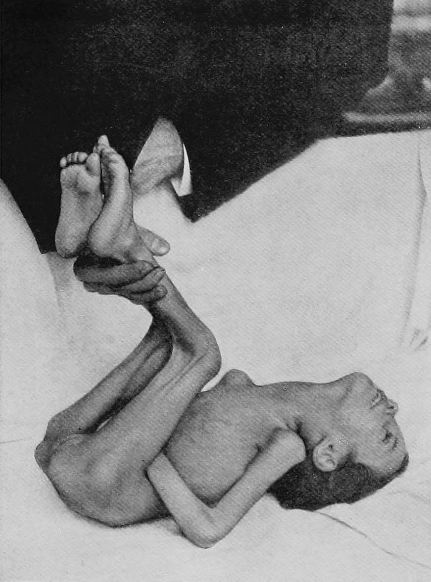
Kernig's sign (after Waldemar Kernig (1840–1917), a Russian neurologist) is positive when the thigh is flexed at the hip and knee at 90 degree angles, and subsequent extension in the knee is painful (leading to resistance).[6] This may indicate subarachnoid hemorrhage or meningitis.[7] Patients may also show opisthotonus—spasm of the whole body that leads to legs and head being bent back and body bowed forward.[citation needed]

Brudzinski's signs

[edit]

Jozef Brudzinski (1874–1917), a Polish pediatrician, is credited with several signs in meningitis. The most commonly used sign (Brudzinski's neck sign) is positive when the forced flexion of the neck elicits a reflex flexion of the hips, with the patient lying supine.[5][8]

Other signs attributed to Brudzinski:[9]

  • The symphyseal sign, in which pressure on the pubic symphysis leads to abduction of the leg and reflexive hip and knee flexion.[10]
  • The cheek sign, in which pressure on the cheek below the zygoma leads to rising and flexion in the forearm.[10]
  • Brudzinski's reflex, in which passive flexion of one knee into the abdomen leads to involuntary flexion in the opposite leg, and stretching of a limb that was flexed leads to contralateral extension.[11]

See also

[edit]

References

[edit]
[edit]
Revisions and contributorsEdit on WikipediaRead on Wikipedia
from Grokipedia
Meningism, also known as meningism or meningeal syndrome, is a clinical syndrome characterized by a constellation of symptoms and signs indicative of irritation of the —the protective membranes surrounding the and —without necessarily involving actual or of these structures. The hallmark features of meningism include nuchal rigidity (stiff neck due to resistance to passive flexion), severe headache often exacerbated by movement or coughing, and (sensitivity to light), frequently accompanied by nausea, vomiting, and sometimes fever or altered consciousness. Physical signs such as (pain or resistance upon attempted extension of the knee with the hip flexed) and Brudzinski's sign (involuntary flexion of the hips and knees in response to passive neck flexion) are classic indicators, though their sensitivity varies by age and underlying condition, with higher reliability in children over one year but limited utility in infants. These manifestations mimic those of but can arise from non-infectious causes, prompting urgent clinical evaluation to differentiate true meningeal pathology from pseudomeningitis. Meningism most commonly results from infectious etiologies like bacterial or , but it can also stem from non-infectious irritants such as , intracranial tumors, carcinomatous meningitis, autoimmune disorders, or drug-induced reactions (e.g., to ibuprofen or other nonsteroidal drugs). In diagnostic contexts, the presence of meningism signs increases the likelihood of bacterial (positive likelihood ratios ranging from 1.39 to 13.3), while their absence reduces it (negative likelihood ratio of 0.41), though evidence quality is low due to study biases and imprecision; thus, these signs should be integrated with analysis, , and patient history for accurate .

Definition and Epidemiology

Definition

Meningism is a clinical characterized by a constellation of symptoms and signs indicative of meningeal irritation, including , nuchal rigidity (), and , occurring without the presence of actual meningeal as seen in . This condition represents a state of reflex irritation affecting the , often mimicking the presentation of infectious but arising from non-inflammatory or transient causes. The term "meningism" was introduced by French neurologists in the early to differentiate symptom complexes resembling from true intracranial , particularly in the context of febrile illnesses among children. It was recognized as a distinct entity to avoid misdiagnosis in cases where meningeal symptoms appeared without pathological evidence of or other inflammatory processes, such as during acute fevers or intoxications. This historical framing emphasized its occurrence at the onset of various acute conditions, aiding clinicians in distinguishing irritation from more severe pathologies. The core triad of meningism—headache, nuchal rigidity, and —serves as the hallmark for clinical recognition, often accompanied by and . Fever may also be present, particularly in pediatric cases associated with systemic illnesses, but the syndrome generally does not involve focal neurological deficits in uncomplicated presentations. These features arise from heightened sensitivity and in meningeal structures, underscoring the syndrome's basis in rather than structural damage.

Epidemiology

Meningism, characterized by signs of meningeal irritation such as nuchal rigidity, is a syndromic presentation that occurs infrequently as an isolated entity but is more commonly observed in the context of acute illnesses. In pediatric emergency settings, signs of meningeal irritation are identified in a subset of children presenting with fever or , with bacterial confirmed in approximately 30% of such cases among 326 children aged 1 month to 15 years evaluated over a decade. Approximately 5% of children presenting to the with have serious secondary causes, such as , often prompting further evaluation to rule out underlying . The condition predominantly affects children and adolescents, with a peak incidence in those aged 2-5 years, reflecting the vulnerability of young pediatric populations to infectious and inflammatory processes that irritate the . Incidence is notably higher in developing regions, where infectious exposures contribute to a disproportionate global burden; as of 2021, there were approximately 1.02 million incident cases of globally in children under 5 years, many of which manifest with meningism. There is no strong gender bias reported in epidemiological data. Key risk factors include immunocompromised states, which increase susceptibility to meningeal from opportunistic infections or non-infectious causes. Recent travel to endemic areas for pathogens like heightens risk due to exposure in high-burden settings such as sub-Saharan Africa's meningitis belt. A history of trauma can also precipitate meningism through direct or secondary . Seasonal variations are evident, with peaks during winter-spring viral outbreaks that amplify febrile illnesses in temperate regions. Meningism remains underreported owing to its frequent overlap with confirmed , complicating syndromic . Non-infectious causes include drug reactions, such as antibiotic- or NSAID-induced (e.g., 329 documented instances in the French database from 1985-2017), and rare post-COVID-19 complications, including vaccine-associated events.

Pathophysiology

Mechanisms of Meningeal Irritation

The , consisting of the , , and , form a protective tri-layered membrane enveloping the and . of these structures, particularly the pia and arachnoid, stimulates densely innervated receptors and mechanosensitive afferents embedded within the meningeal layers, leading to nociceptive signaling and reflexive muscle spasms as a protective mechanism. This anatomical arrangement allows even minor perturbations to elicit widespread symptoms by activating sensory pathways that interface with the . Inflammatory processes contribute to meningeal through the release of chemical mediators such as prostaglandins, cytokines, and other inflammatory molecules derived from breakdown products, purulent exudates, or exogenous toxins. These mediators induce local , increased , and perimeningeal , which heighten sensitivity of nociceptors and provoke reflexive contraction of paraspinal muscles, manifesting as (nuchal rigidity). The resulting serves to limit further stretching of irritated meningeal attachments to the and roots during movement. Prostaglandins, in particular, sensitize meningeal nociceptors, amplifying transmission and contributing to the intensity of associated symptoms. Neurological pathways underlying meningeal irritation primarily involve the trigeminovascular system, where activation of meningeal nociceptors projects via the to the trigeminocervical complex in the and upper cervical . This pathway accounts for through central sensitization and release of neuropeptides like (CGRP), while arises from convergent thalamic projections linking meningeal input to visual processing areas. Reflex arcs, mediated by spinal , drive involuntary flexion responses and muscle guarding in response to meningeal stretch, preventing exacerbation of irritation. Non-inflammatory triggers elicit meningeal irritation via mechanical means, such as stretch or distortion of meningeal tissues due to elevated (ICP) or direct contact from hemorrhage. Raised ICP displaces brain structures, tensioning meningeal attachments and stimulating sensory afferents without leukocyte infiltration or overt inflammation. Similarly, introduces blood into the , causing chemical irritation through osmotic effects and clot formation that mechanically tugs on pain-sensitive dura, but distinct from immune-mediated responses. These mechanisms highlight how physical perturbations alone can replicate the clinical features of meningism.

Distinction from Meningitis

Meningism refers to the clinical syndrome characterized by signs and symptoms of meningeal irritation, such as and , without underlying histological inflammation of the or cerebrospinal fluid (CSF) pleocytosis exceeding 5 per microliter. In contrast, involves true inflammatory infiltration of the , typically evidenced by CSF pleocytosis greater than 5 cells/μL, often accompanied by elevated protein levels and reduced glucose concentration. This fundamental pathological distinction underscores that meningism represents functional irritation—often due to mechanical, chemical, or vascular factors—rather than an infectious or inflammatory process. There is notable clinical overlap between the two conditions, as meningeal signs suggestive of meningism appear in a substantial proportion of cases, with reported in 65–83% of adults with bacterial meningitis. Conversely, among patients presenting with signs of meningeal irritation (meningism), bacterial is confirmed in only about 30% of pediatric cases, implying that meningism arises in approximately 70% of non-meningitic scenarios, such as where meningeal signs occur in up to one-third of patients due to blood irritating the subarachnoid space. This overlap complicates initial assessment, as CSF analysis remains essential to differentiate the entities, revealing normal findings in meningism versus abnormal pleocytosis in . Clinically, meningism serves as a provisional in patients with suggestive signs but without confirmed , necessitating vigilant monitoring since underlying untreated infections could progress to full . Misclassification risks inappropriate , such as withholding antibiotics in evolving bacterial cases or overtreating benign . The term "meningism" was coined by French physician Dupré in 1894 to describe benign meningeal syndromes lacking inflammatory changes, helping to avert unnecessary interventions like repeated punctures in presumed bacterial during an era before routine CSF diagnostics. This nomenclature evolved to promote precise , reducing overtreatment in non-inflammatory presentations.

Etiology

Infectious Causes

Infectious causes are the predominant etiologies of meningism, a clinical of meningeal irritation characterized by signs such as nuchal rigidity, , and without overt meningeal inflammation or full-blown . These infections often manifest in early or partially treated stages, where bacterial exotoxins or irritate meningeal nociceptors prior to significant inflammatory response in the . Globally, infectious agents account for the majority of meningism cases, with viral pathogens being particularly prevalent in pediatric populations during seasonal outbreaks. Bacterial infections can lead to meningism through early dissemination or partial antibiotic treatment, where exotoxins from pathogens like Neisseria meningitidis and Streptococcus pneumoniae trigger meningeal irritation before widespread inflammation develops. N. meningitidis, a leading cause of bacterial meningitis, often presents with initial meningism accompanied by fever and petechial rash in adolescents and young adults, particularly in unvaccinated or crowded settings. Similarly, S. pneumoniae may cause transient meningism in the context of otitis media or pneumonia, with exotoxin-mediated activation of sensory neurons contributing to neck stiffness and headache. In the pre-Haemophilus influenzae type b (Hib) vaccine era, invasive Hib disease was a major contributor to pediatric meningism, with incidence rates of approximately 40 to 100 cases per 100,000 children under 5 years in unvaccinated populations, about 20,000 annual cases in the pre-vaccine US; vaccination has since reduced these cases by over 99% in vaccinated cohorts. Viral infections are the most frequent infectious triggers of meningism, especially in aseptic presentations without bacterial involvement, and often resolve spontaneously. Enteroviruses, such as and , account for 55-70% of cases with associated meningism, commonly causing transient irritation in children during summer and fall epidemics; these are linked to up to 90% of pediatric episodes in temperate regions. (HSV), particularly HSV-2, can induce recurrent or isolated meningism with and , though it rarely progresses to severe in immunocompetent hosts. In pediatric viral fevers, such as those from enteroviral infections, meningism occurs in a substantial proportion of cases, emphasizing the need for clinical differentiation from bacterial disease. Fungal and parasitic infections rarely cause isolated meningism but can do so in immunocompromised individuals through direct mechanical irritation or low-grade inflammation. is a key fungal pathogen, primarily affecting those with or on immunosuppressive therapy, where it leads to subacute meningism with and minimal fever in up to 10% of advanced HIV cases before full cryptococcal meningitis develops. Parasitic agents like may produce meningism via focal lesions or rupture in the , predominantly in immunocompromised patients such as those with AIDS, though such presentations are uncommon outside endemic areas. Overall, infectious meningism represents approximately 60% of cases in clinical series, with higher rates in unvaccinated or resource-limited settings where bacterial and viral pathogens circulate unchecked.

Non-Infectious Causes

Non-infectious causes of meningism encompass a range of conditions that lead to meningeal irritation without microbial involvement, primarily through mechanical, chemical, or compressive mechanisms. Vascular events, such as (SAH), are prominent triggers, where blood and its breakdown products, including , directly irritate the , often resulting in classic signs of meningism. SAH can arise from aneurysmal rupture or trauma, with traumatic SAH occurring in 33% to 60% of patients with moderate to severe (TBI). This irritation typically manifests hours after the bleed as blood extends into the subarachnoid space and irritates surrounding tissues. Traumatic injuries to the head represent another key category, where direct contusion, laceration, or formation causes meningeal irritation independent of . In severe TBI, such non-infectious meningeal responses contribute to meningism, often linked to epidural or subdural that exert pressure or release irritants into the meningeal space. These events highlight the role of physical disruption in provoking the syndrome, distinct from secondary infectious complications. Neoplastic processes induce meningism through compression or infiltration of the , with brain tumors like meningiomas or metastatic lesions causing localized irritation via . Leptomeningeal carcinomatosis, the spread of malignant cells to the leptomeninges, is particularly notable, affecting 5% to 15% of patients with advanced solid tumors such as , , or cancers. This condition leads to diffuse meningeal involvement, mimicking inflammatory causes but driven by tumor cells obstructing pathways and eliciting irritation. Other non-infectious etiologies include drug-induced reactions, toxic exposures, and metabolic derangements. Nonsteroidal anti-inflammatory drugs (NSAIDs), such as ibuprofen, and certain antibiotics like rifampin can provoke aseptic meningeal inflammation, particularly in patients with underlying autoimmune conditions, leading to recurrent meningism. causes chemical irritation of the through neurotoxic accumulation, presenting with meningeal signs alongside . Additionally, raised from or metabolic disturbances like can stretch and irritate meningeal layers, contributing to the syndrome via non-microbial pathways. These diverse triggers underscore the importance of targeted diagnostic evaluation to differentiate them from infectious origins.

Signs and Symptoms

General Symptoms

Meningism typically manifests with a range of subjective symptoms reflecting meningeal irritation, most prominently severe that is diffuse and often localized to the frontal or occipital regions. This worsens with movement or jarring and is reported in approximately 90% of cases, serving as a hallmark complaint due to the stretching or of meningeal structures. Photophobia, or sensitivity to light, and phonophobia, sensitivity to sound, are common accompanying features arising from meningeal sensitization, with occurring frequently alongside and . These sensory sensitivities are often more pronounced in children, contributing to discomfort and avoidance behaviors. Systemic symptoms include fever, which may be low-grade in non-infectious cases, as well as and , the latter sometimes projectile in instances associated with raised . In pediatric patients, or may predominate, reflecting altered comfort and energy levels without more specific verbalized complaints. The onset of these symptoms is typically acute, developing over hours to days, and they generally resolve with treatment of the underlying cause, though benign forms may occasionally mimic patterns with throbbing quality and sensory auras.

Specific Physical Signs

Nuchal rigidity, also known as , is characterized by resistance to passive flexion of the neck due to of the extensor muscles, resulting from of the inflamed and nerve roots when the cervical region is stretched during flexion. This sign arises as a protective response to meningeal , where flexion tugs on the and sensitive structures, eliciting muscle guarding. In adults, its sensitivity for detecting ranges from 40% to 60%, with specificity of 65% to 75%, while in children it demonstrates a higher sensitivity of approximately 65% to 70%. Kernig's sign involves eliciting pain or resistance upon passive extension of the knee while the hip is flexed to 90 degrees, with the patient in a ; it reflects hypersensitivity of inflamed lumbosacral nerve roots and stretched by the maneuver. The test is positive when extension beyond 135 degrees (or 90 degrees in severe cases) provokes discomfort in the hamstrings or lower back. Its sensitivity is low, typically 5% to 30% across studies, but specificity exceeds 90%, making it a reliable indicator when present, particularly for bacterial . Brudzinski's signs encompass reflexive responses to meningeal irritation. The superior (cervical) sign occurs when passive flexion of the induces involuntary flexion of the s and s, as this compensates for dural tension by relaxing the irritated and reducing pain. The inferior sign is elicited by passive flexion of one and , prompting flexion of the contralateral due to interconnected meningeal stretching. Both have low sensitivity (5% to 43%) but high specificity (80% to 100%), with greater utility in detecting severe meningeal irritation rather than mild cases. In infants, opisthotonos—a severe arching of the back and neck due to intense extensor muscle spasms—may manifest as a sign of advanced , often linked to bacterial , though it is uncommon and indicates significant meningeal involvement.

Diagnosis

Clinical Assessment

The clinical assessment of suspected begins with a detailed history to identify risk factors and guide risk stratification. Key elements include the onset and progression of symptoms, such as acute , , or , often accompanied by fever history to differentiate infectious from non-infectious causes. Inquiry into recent trauma exposure, such as or neurosurgical procedures, is essential, as these can precipitate meningeal irritation through or direct contamination. Vaccination status should be assessed, particularly for vaccines against type b, pneumococcus, and meningococcus, as unvaccinated individuals are at higher risk for bacterial etiologies. Red flags in the history, including seizures or focal neurological deficits, suggest a non-benign cause such as bacterial or complications like increased , warranting urgent evaluation. The physical examination follows a systematic sequence, starting with vital signs to detect tachycardia, fever, or hypotension indicative of systemic involvement. A comprehensive is then performed, assessing mental status using the (GCS) to identify altered consciousness, along with evaluation for focal deficits or cranial nerve abnormalities. Meningeal signs are tested subsequently, beginning with neck stiffness or resistance to passive flexion, followed by (pain on knee extension with hip flexed) and Brudzinski's sign (involuntary hip flexion on neck flexion), which, if positive, support meningeal irritation. Scoring tools aid in risk stratification, particularly in pediatric patients. The Bacterial Meningitis Score (BMS), derived from five predictors—positive cerebrospinal fluid Gram stain, cerebrospinal fluid absolute neutrophil count ≥1000 cells/μL, cerebrospinal fluid protein ≥80 mg/dL, peripheral blood absolute neutrophil count ≥10,000 cells/μL, and seizure at or before presentation—identifies children aged 29 days to 19 years at very low risk for bacterial meningitis if the score is zero, with a negative predictive value of 99.9% (95% CI, 99.6%-100%) and sensitivity of 98.3% (95% CI, 94.2%-99.8%) for detecting bacterial cases. Age-specific considerations are critical, as manifestations vary. In infants, particularly neonates, classic meningeal signs are often absent or subtle; a bulging occurs in approximately 20% of cases and signals potential increased , alongside nonspecific signs like or poor feeding. In adults, signs may be subtler, with reliance on history and altered mental status rather than overt rigidity, especially in immunocompromised individuals.

Diagnostic Investigations

The diagnosis of meningism relies on confirmatory investigations to identify or exclude underlying causes, particularly to differentiate it from true . The 2025 World Health Organization guidelines on meningitis diagnosis, treatment, and care provide evidence-based recommendations for clinical management, emphasizing (LP) for (CSF) analysis in suspected cases unless contraindicated, alongside integration of for rapid identification. The gold standard procedure is LP for CSF analysis, which is indicated in patients with meningeal signs unless contraindicated. LP allows direct examination of CSF parameters, including cell count, protein levels, glucose concentration, , and culture. In cases of pure meningism without infection, CSF findings are typically normal, with fewer than 5 white blood cells (WBC) per microliter (predominantly mononuclear cells), protein levels of 15-45 mg/dL, and glucose exceeding 60% of simultaneous blood glucose. Abnormalities such as pleocytosis (>5 WBC/μL), elevated protein (>45 mg/dL), or reduced glucose suggest an infectious or inflammatory , guiding further management. and culture identify bacterial pathogens in up to 70-80% of bacterial cases when performed before antibiotics, while (PCR) on CSF enhances detection of viral or atypical pathogens like enteroviruses or . Contraindications to LP include (e.g., platelet count <50,000/μL or international normalized ratio >1.5), focal neurological deficits, ( <10), or papilledema indicating raised intracranial pressure, as these increase the risk of cerebral herniation. Imaging studies complement LP by assessing for structural complications or alternative diagnoses. Non-contrast computed tomography (CT) of the head is recommended prior to LP in patients with red flags such as focal signs, seizures, or altered mental status to rule out mass effect, hydrocephalus, or abscess that could precipitate herniation. CT findings in meningism are often normal early on but may reveal subtle meningeal thickening or effusions in complicated cases. Magnetic resonance imaging (MRI) with gadolinium contrast is more sensitive for detecting meningeal irritation in non-acute presentations, showing linear leptomeningeal enhancement or basal exudates, particularly useful when LP is deferred or to evaluate for encephalitis or vasculitis. Peripheral laboratory tests provide supportive evidence of systemic involvement. Complete blood count (CBC) often reveals leukocytosis (>11,000 WBC/μL) suggestive of bacterial , while normal counts support non-infectious meningism. C-reactive protein (CRP) and (ESR) are elevated in inflammatory states, with CRP >10 mg/L indicating possible , though these are nonspecific. Blood cultures are positive in 50-90% of bacterial meningitis cases if obtained before antibiotics and are essential to detect disseminated . CSF PCR for specific pathogens, combined with these blood tests, refines in 80-95% of viral cases. Overall, LP alters management in approximately 70% of patients with suspected meningism by confirming or excluding , allowing de-escalation of antibiotics when CSF is normal despite clinical signs, thus establishing benign meningism.

Management

Initial and Supportive Care

Initial management of meningism focuses on stabilizing the patient and addressing immediate life-threatening issues, beginning with assessment and support of airway, breathing, and circulation (ABC). Secure the airway if compromised, provide supplemental oxygen for hypoxia, and ensure adequate circulation through vascular access for intravenous (IV) administration of fluids and medications. In cases of dehydration or shock, administer an initial IV bolus of 0.9% sodium chloride at 20 mL/kg over 5-10 minutes, with reassessment and repeat boluses as needed up to 40-60 mL/kg if hemodynamic instability persists. Supportive care emphasizes symptom relief to improve patient comfort while awaiting diagnostic confirmation. For headache and fever, administer acetaminophen at 15 mg/kg IV or orally every 4-6 hours as needed, not exceeding five doses in 24 hours. Antiemetics such as (0.15 mg/kg IV, maximum 4 mg per dose) can alleviate and . To manage , place the patient in a dark, quiet environment to reduce sensory stimuli. Ongoing hydration with isotonic IV fluids at maintenance rates (or two-thirds maintenance in children to mitigate syndrome of inappropriate secretion [SIADH]) helps prevent complications like . Close monitoring is essential, with serial neurological examinations conducted every 1-2 hours to detect changes in level of , focal deficits, or signs of raised . , fluid balance, electrolytes, and glucose should be checked frequently, particularly in the first 48 hours. Defer lumbar puncture if the patient is hemodynamically unstable or has . If infectious etiology is suspected, implement droplet precautions, including masking and isolation, for at least the first 24 hours.

Cause-Specific Treatment

For infectious causes of meningism, treatment targets the underlying pathogen once identified through cerebrospinal fluid analysis or other diagnostics. In suspected bacterial meningitis, empiric intravenous therapy typically includes at 2 g every 12 hours for adults combined with to cover potential penicillin-resistant , particularly in regions with high resistance rates. Empiric therapy should align with current guidelines, such as the 2025 WHO recommendations for community-acquired bacterial meningitis. For herpes simplex virus (HSV)-associated meningitis, intravenous acyclovir is administered at 10 mg/kg every 8 hours until clinical improvement and negative HSV results confirm resolution. In cryptococcal meningitis, induction therapy preferably consists of liposomal (3-4 mg/kg/day IV) plus (100 mg/kg/day orally, divided into four doses) for 14 days, particularly in immunocompromised patients (as per 2024 guidelines); deoxycholate (0.7-1.0 mg/kg/day) plus (100 mg/kg/day orally) may be used in resource-limited settings. For non-infectious etiologies, interventions address the specific structural or toxic insult. causing meningeal irritation requires urgent neurosurgical intervention to secure the bleeding source, such as or surgical clipping of the , along with supportive measures to manage complications like or rebleeding. In cases of tumor-related meningeal compression, such as leptomeningeal carcinomatosis, high-dose dexamethasone (0.15 mg/kg every 6 hours) is used to reduce peritumoral and associated . presenting with meningeal symptoms, often as part of , is managed with using agents like succimer (dimercaptosuccinic acid) at 10 mg/kg orally three times daily for 5 days, followed by twice daily for 2 weeks, to enhance lead . Adjunctive therapies enhance outcomes in select scenarios. Corticosteroids, such as dexamethasone (0.15 mg/kg every 6 hours for 4 days), are recommended alongside antibiotics in bacterial meningitis, particularly due to , reducing mortality by approximately 20% in adults through mitigation of inflammatory complications. If seizures occur, antiepileptic drugs like (20-40 mg/kg intravenously) are initiated for acute management and short-term prophylaxis to prevent recurrence during the inflammatory phase. De-escalation of therapy occurs once cultures are negative or the etiology is clarified, minimizing unnecessary broad-spectrum exposure. For culture-negative cases initially treated empirically for bacterial causes, antibiotics are narrowed to pathogen-specific agents or discontinued if viral etiology is confirmed, typically after 48-72 hours of negative results. In viral meningitis without complications, patients may transition to outpatient follow-up after initial stabilization, with monitoring for symptom resolution within 7-10 days. Supportive analgesics may be referenced briefly for residual discomfort during this phase.

Prognosis

Outcomes by Etiology

Outcomes for meningism vary significantly depending on the underlying , with infectious causes generally carrying a more favorable when treated promptly compared to non-infectious etiologies, which often involve structural or oncologic complications. In cases of viral meningism, the condition typically resolves within 7-10 days, with most patients achieving complete recovery in 1-2 weeks and sequelae being uncommon outside the neonatal period. is excellent, with nearly all immunocompetent individuals experiencing full recovery without long-term neurologic deficits. For bacterial meningism, which overlaps with acute bacterial , mortality rates range from 10-15% with appropriate antibiotic therapy, though delays in treatment of 3-6 hours can substantially worsen outcomes by increasing and cerebral complications. Non-infectious etiologies present more guarded prognoses. Subarachnoid hemorrhage (SAH)-related meningism is associated with high mortality, approximately 40-50% overall, driven by initial bleeding, rebleeding in 9-17% of cases (with up to 50% fatality in those instances), and complications like vasospasm. In traumatic meningism, outcomes are severity-dependent; mild cases often yield good functional recovery, while severe traumatic brain injury with associated post-traumatic meningitis carries high mortality, exacerbated by factors such as low Glasgow Coma Scale scores and recurrent episodes. Neoplastic meningism, arising from leptomeningeal metastases in solid tumors, confers a poor prognosis, with median survival of 3-6 months under treatment, extending from 4-6 weeks without intervention; breast cancer cases may achieve 7-12 months in select instances, but overall 1-year survival is limited to 11-25%. Autoimmune and drug-induced meningism generally have good prognoses with resolution upon treatment of the underlying cause, often without long-term sequelae. Key predictors of outcomes include the timing of and patient age. Early and initiation of within 24 hours significantly improve survival, with late increasing in-hospital mortality risk by approximately 70% (from 18% to 36%) in community-acquired bacterial cases. Pediatric patients generally fare better than adults, with lower mortality rates (5-10% in children compared to 15-20% in adults) and reduced risk of long-term neurodevelopmental impairments for bacterial etiologies. Long-term sequelae in meningism survivors are relatively rare but can include chronic headaches in about 5-10% of cases, particularly following viral or bacterial episodes, alongside occasional neurocognitive or sensory impairments. Vaccination programs have dramatically reduced the infectious burden, achieving over 90% incidence reduction for vaccine-preventable bacterial causes like type b and substantial declines (60-100%) for pneumococcal and across age groups.

Complications

Meningism, characterized by meningeal , can lead to serious neurological complications if the underlying cause, such as infectious , remains untreated. Seizures occur in approximately 15-30% of cases of bacterial presenting with meningism, often due to cortical or metabolic disturbances, with a long-term risk of estimated at 6-13% in survivors. affects up to 10% of patients with bacterial causes, particularly meningococcal , resulting from suppurative where infection spreads to the via the cochlear aqueduct. Untreated raised (ICP) from can precipitate , a life-threatening event with mortality rates exceeding 50% if not addressed promptly. Systemic complications arise from the inflammatory response and associated symptoms in meningism. In bacterial etiologies, progression to is common, occurring in 10-20% of meningococcal cases and contributing to multi-organ failure with a of 10-15%. , a frequent feature, can cause and imbalances, exacerbating neurological symptoms. Syndrome of inappropriate antidiuretic hormone secretion (SIADH) leads to in up to 30% of pediatric and adult cases, potentially worsening and seizures if not corrected. Chronic sequelae persist in a subset of patients recovering from meningism-related conditions. Post-meningitis syndrome may manifest as persistent headaches in about 5-10% of survivors, alongside and cognitive impairments. In pediatric cases, cognitive deficits occur in approximately 2-5%, including learning disabilities and behavioral issues, often linked to hippocampal and cortical damage from . These long-term effects underscore the need for multidisciplinary follow-up. Prevention of complications hinges on early intervention. Prompt lumbar puncture (LP) for diagnosis and initiation of antibiotics can reduce complication rates by up to 50% in bacterial by limiting inflammation duration. Pre-LP , such as CT, is essential in patients with altered mental status or focal deficits to avoid herniation risk, further mitigating adverse outcomes. Follow-up imaging in non-resolving cases helps detect or abscesses early, improving prognosis.

References

Add your contribution
Related Hubs
User Avatar
No comments yet.