Hubbry Logo
Point mutationPoint mutationMain
Open search
Point mutation
Community hub
Point mutation
logo
8 pages, 0 posts
0 subscribers
Be the first to start a discussion here.
Contribute something
Point mutation
Point mutation
from Wikipedia
Point mutations of a codon, classified by their impact on protein sequence
Schematic of a single-stranded RNA molecule illustrating a series of three-base codons. Each three-nucleotide codon corresponds to an amino acid when translated to protein. When one of these codons is changed by a point mutation, the corresponding amino acid of the protein is changed.
A to G point mutation detected with Sanger sequencing

A point mutation is a genetic mutation where a single nucleotide base is changed, inserted or deleted from a DNA or RNA sequence of an organism's genome.[1] Point mutations have a variety of effects on the downstream protein product—consequences that are moderately predictable based upon the specifics of the mutation. These consequences can range from no effect (e.g. synonymous mutations) to deleterious effects (e.g. frameshift mutations), with regard to protein production, composition, and function.

Causes

[edit]

Point mutations usually take place during DNA replication. DNA replication occurs when one double-stranded DNA molecule creates two single strands of DNA, each of which is a template for the creation of the complementary strand. A single point mutation can change the whole DNA sequence. Changing one purine or pyrimidine may change the amino acid that the nucleotides code for.

Point mutations may arise from spontaneous mutations that occur during DNA replication. The rate of mutation may be increased by mutagens. Mutagens can be physical, such as radiation from UV rays, X-rays or extreme heat, or chemical (molecules that misplace base pairs or disrupt the helical shape of DNA). Mutagens associated with cancers are often studied to learn about cancer and its prevention.

There are multiple ways for point mutations to occur. First, ultraviolet (UV) light and higher-frequency light have ionizing capability, which in turn can affect DNA. Reactive oxygen molecules with free radicals, which are a byproduct of cellular metabolism, can also be very harmful to DNA. These reactants can lead to both single-stranded and double-stranded DNA breaks. Third, bonds in DNA eventually degrade, which creates another problem to keep the integrity of DNA to a high standard. There can also be replication errors that lead to substitution, insertion, or deletion mutations.

Categorization

[edit]

Transition/transversion categorization

[edit]
Transitions (Alpha) and transversions (Beta).

In 1959 Ernst Freese coined the terms "transitions" or "transversions" to categorize different types of point mutations.[2][3] Transitions are replacement of a purine base with another purine or replacement of a pyrimidine with another pyrimidine. Transversions are replacement of a purine with a pyrimidine or vice versa. There is a systematic difference in mutation rates for transitions (Alpha) and transversions (Beta). Transition mutations are about ten times more common than transversions.

Functional categorization

[edit]

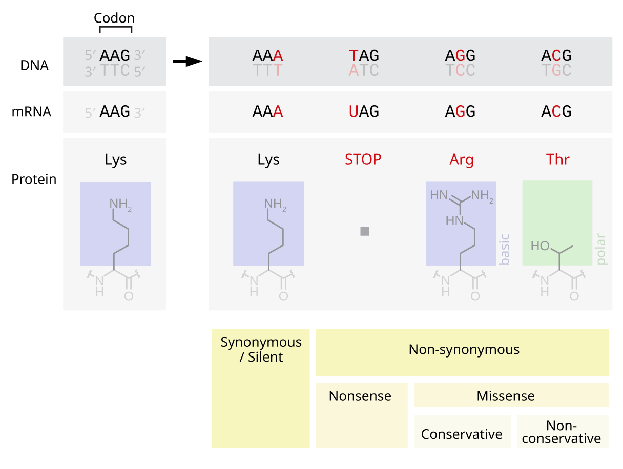
Nonsense mutations include stop-gain and start-loss. Stop-gain is a mutation that results in a premature termination codon (a stop was gained), which signals the end of translation. This interruption causes the protein to be abnormally shortened. The number of amino acids lost mediates the impact on the protein's functionality and whether it will function whatsoever.[4] Stop-loss is a mutation in the original termination codon (a stop was lost), resulting in abnormal extension of a protein's carboxyl terminus. Start-gain creates an AUG start codon upstream of the original start site. If the new AUG is near the original start site, in-frame within the processed transcript and downstream to a ribosomal binding site, it can be used to initiate translation. The likely effect is additional amino acids added to the amino terminus of the original protein. Frame-shift mutations are also possible in start-gain mutations, but typically do not affect translation of the original protein. Start-loss is a point mutation in a transcript's AUG start codon, resulting in the reduction or elimination of protein production.

Missense mutations code for a different amino acid. A missense mutation changes a codon so that a different protein is created, a non-synonymous change.[4] Conservative mutations result in an amino acid change. However, the properties of the amino acid remain the same (e.g., hydrophobic, hydrophilic, etc.). At times, a change to one amino acid in the protein is not detrimental to the organism as a whole. Most proteins can withstand one or two point mutations before their function changes. Non-conservative mutations result in an amino acid change that has different properties than the wild type. The protein may lose its function, which can result in a disease in the organism. For example, sickle-cell disease is caused by a single point mutation (a missense mutation) in the beta-hemoglobin gene that converts a GAG codon into GUG, which encodes the amino acid valine rather than glutamic acid. The protein may also exhibit a "gain of function" or become activated, such is the case with the mutation changing a valine to glutamic acid in the BRAF gene; this leads to an activation of the RAF protein which causes unlimited proliferative signalling in cancer cells.[5] These are both examples of a non-conservative (missense) mutation.

Silent mutations code for the same amino acid (a "synonymous substitution"). A silent mutation does not affect the functioning of the protein. A single nucleotide can change, but the new codon specifies the same amino acid, resulting in an unmutated protein. This type of change is called synonymous change since the old and new codon code for the same amino acid. This is possible because 64 codons specify only 20 amino acids. Different codons can lead to differential protein expression levels, however.[4]

Single base pair insertions and deletions

[edit]

Sometimes the term point mutation is used to describe insertions or deletions of a single base pair (which has more of an adverse effect on the synthesized protein due to the nucleotides' still being read in triplets, but in different frames: a mutation called a frameshift mutation).[4]

General consequences

[edit]

Point mutations that occur in non-coding sequences are most often without consequences, although there are exceptions. If the mutated base pair is in the promoter sequence of a gene, then the expression of the gene may change. Also, if the mutation occurs in the splicing site of an intron, then this may interfere with correct splicing of the transcribed pre-mRNA.

By altering just one amino acid, the entire peptide may change, thereby changing the entire protein. The new protein is called a protein variant. If the original protein functions in cellular reproduction then this single point mutation can change the entire process of cellular reproduction for this organism.

Point germline mutations can lead to beneficial as well as harmful traits or diseases. This leads to adaptations based on the environment where the organism lives. An advantageous mutation can create an advantage for that organism and lead to the trait's being passed down from generation to generation, improving and benefiting the entire population. The scientific theory of evolution is greatly dependent on point mutations in cells. The theory explains the diversity and history of living organisms on Earth. In relation to point mutations, it states that beneficial mutations allow the organism to thrive and reproduce, thereby passing its positively affected mutated genes on to the next generation. On the other hand, harmful mutations cause the organism to die or be less likely to reproduce in a phenomenon known as natural selection.

There are different short-term and long-term effects that can arise from mutations. Smaller ones would be a halting of the cell cycle at numerous points. This means that a codon coding for the amino acid glycine may be changed to a stop codon, causing the proteins that should have been produced to be deformed and unable to complete their intended tasks. Because the mutations can affect the DNA and thus the chromatin, it can prohibit mitosis from occurring due to the lack of a complete chromosome. Problems can also arise during the processes of transcription and replication of DNA. These all prohibit the cell from reproduction and thus lead to the death of the cell. Long-term effects can be a permanent changing of a chromosome, which can lead to a mutation. These mutations can be either beneficial or detrimental. Cancer is an example of how they can be detrimental.[6]

Other effects of point mutations, or single nucleotide polymorphisms in DNA, depend on the location of the mutation within the gene. For example, if the mutation occurs in the region of the gene responsible for coding, the amino acid sequence of the encoded protein may be altered, causing a change in the function, protein localization, stability of the protein or protein complex. Many methods have been proposed to predict the effects of missense mutations on proteins. Machine learning algorithms train their models to distinguish known disease-associated from neutral mutations whereas other methods do not explicitly train their models but almost all methods exploit the evolutionary conservation assuming that changes at conserved positions tend to be more deleterious. While majority of methods provide a binary classification of effects of mutations into damaging and benign, a new level of annotation is needed to offer an explanation of why and how these mutations damage proteins.[7]

Moreover, if the mutation occurs in the region of the gene where transcriptional machinery binds to the protein, the mutation can affect the binding of the transcription factors because the short nucleotide sequences recognized by the transcription factors will be altered. Mutations in this region can affect rate of efficiency of gene transcription, which in turn can alter levels of mRNA and, thus, protein levels in general.

Point mutations can have several effects on the behavior and reproduction of a protein depending on where the mutation occurs in the amino acid sequence of the protein. If the mutation occurs in the region of the gene that is responsible for coding for the protein, the amino acid may be altered. This slight change in the sequence of amino acids can cause a change in the function, activation of the protein meaning how it binds with a given enzyme, where the protein will be located within the cell, or the amount of free energy stored within the protein.

If the mutation occurs in the region of the gene where transcriptional machinery binds to the protein, the mutation can affect the way in which transcription factors bind to the protein. The mechanisms of transcription bind to a protein through recognition of short nucleotide sequences. A mutation in this region may alter these sequences and, thus, change the way the transcription factors bind to the protein. Mutations in this region can affect the efficiency of gene transcription, which controls both the levels of mRNA and overall protein levels.[8]

Specific diseases caused by point mutations

[edit]

Point mutations—single‑base changes in the DNA sequence—are one of the most common molecular causes of human disease. By altering a single nucleotide, these mutations can substitute one amino acid for another, introduce premature stop codons, or disrupt normal splicing signals. Depending on where they occur and how they affect the encoded protein, point mutations may abolish enzyme activity, destabilize structural domains, or impair regulatory interactions. In many inherited disorders, a single missense or nonsense substitution is enough to trigger a cascade of biochemical failures, leading to early‐onset or lifelong symptoms. In cancer, somatic point mutations can inactivate tumor suppressors or hyperactivate oncogenes, fueling uncontrolled cell growth.

Across the human genetic landscape, thousands of point‐mutation–driven conditions have been cataloged—from relatively common disorders like sickle‐cell anemia and cystic fibrosis to extremely rare syndromes that affect only a handful of families worldwide. Although each disease has its own pathophysiological details, they share a unifying theme: a precisely localized change in the gene sequence can compromise protein function in a way that no larger chromosomal rearrangement or copy‐number alteration could. Because point mutations are often amenable to targeted genetic testing, they also highlight how molecular diagnosis and personalized therapies (e.g., small molecules that stabilize a mutant enzyme) rely on knowing exactly which codon is altered. Although the following examples illustrate the diversity of point-mutation–mediated disorders, there are over 300,000 such mutations recorded in HGMD and over 1,000,000 variants in ClinVar, reflecting the vast spectrum of human point mutations.[9] [10] [11]

Cancer

[edit]

Point mutations in multiple tumor suppressor proteins cause cancer. For instance, point mutations in Adenomatous Polyposis Coli promote tumorigenesis.[12] A novel assay, Fast parallel proteolysis (FASTpp), might help swift screening of specific stability defects in individual cancer patients.[13]

Neurofibromatosis

[edit]

Neurofibromatosis is caused by point mutations in the Neurofibromin 1[14][15] or Neurofibromin 2 gene.[16]

Sickle-cell anemia

[edit]

Sickle-cell anemia is caused by a point mutation in the β-globin chain of hemoglobin, causing the hydrophilic amino acid glutamic acid to be replaced with the hydrophobic amino acid valine at the sixth position.

The β-globin gene is found on the short arm of chromosome 11. The association of two wild-type α-globin subunits with two mutant β-globin subunits forms hemoglobin S (HbS). Under low-oxygen conditions (being at high altitude, for example), the absence of a polar amino acid at position six of the β-globin chain promotes the non-covalent polymerisation (aggregation) of hemoglobin, which distorts red blood cells into a sickle shape and decreases their elasticity.[17]

Hemoglobin is a protein found in red blood cells, and is responsible for the transportation of oxygen through the body.[18] There are two subunits that make up the hemoglobin protein: beta-globins and alpha-globins.[19] Beta-hemoglobin is created from the genetic information on the HBB, or "hemoglobin, beta" gene found on chromosome 11p15.5.[20] A single point mutation in this polypeptide chain, which is 147 amino acids long, results in the disease known as Sickle Cell Anemia.[21] Sickle-cell anemia is an autosomal recessive disorder that affects 1 in 500 African Americans, and is one of the most common blood disorders in the United States.[20] The single replacement of the sixth amino acid in the beta-globin, glutamic acid, with valine results in deformed red blood cells. These sickle-shaped cells cannot carry nearly as much oxygen as normal red blood cells and they get caught more easily in the capillaries, cutting off blood supply to vital organs. The single nucleotide change in the beta-globin means that even the smallest of exertions on the part of the carrier results in severe pain and even heart attack. Below is a chart depicting the first thirteen amino acids in the normal and abnormal sickle cell polypeptide chain.[21]

Sequence for normal hemoglobin
AUG GUG CAC CUG ACU CCU GAG GAG AAG UCU GCC GUU ACU
START Val His Leu Thr Pro Glu Glu Lys Ser Ala Val Thr
Sequence for sickle-cell hemoglobin
AUG GUG CAC CUG ACU CCU GUG GAG AAG UCU GCC GUU ACU
START Val His Leu Thr Pro Val Glu Lys Ser Ala Val Thr

Tay–Sachs disease

[edit]

The cause of Tay–Sachs disease is a genetic defect that is passed from parent to child. This genetic defect is located in the HEXA gene, which is found on chromosome 15.

The HEXA gene makes part of an enzyme called beta-hexosaminidase A, which plays a critical role in the nervous system. This enzyme helps break down a fatty substance called GM2 ganglioside in nerve cells. Mutations in the HEXA gene disrupt the activity of beta-hexosaminidase A, preventing the breakdown of the fatty substances. As a result, the fatty substances accumulate to deadly levels in the brain and spinal cord. The buildup of GM2 ganglioside causes progressive damage to the nerve cells. This is the cause of the signs and symptoms of Tay-Sachs disease.[22]

Repeat-induced point mutation

[edit]

In molecular biology, repeat-induced point mutation or RIP is a process by which DNA accumulates G:C to A:T transition mutations. Genomic evidence indicates that RIP occurs or has occurred in a variety of fungi[23] while experimental evidence indicates that RIP is active in Neurospora crassa,[24] Podospora anserina,[25] Magnaporthe grisea,[26] Leptosphaeria maculans,[27] Gibberella zeae,[28] Nectria haematococca[29] and Paecilomyces variotii.[30] In Neurospora crassa, sequences mutated by RIP are often methylated de novo.[24]

RIP occurs during the sexual stage in haploid nuclei after fertilization but prior to meiotic DNA replication.[24] In Neurospora crassa, repeat sequences of at least 400 base pairs in length are vulnerable to RIP. Repeats with as low as 80% nucleotide identity may also be subject to RIP. Though the exact mechanism of repeat recognition and mutagenesis are poorly understood, RIP results in repeated sequences undergoing multiple transition mutations.

The RIP mutations do not seem to be limited to repeated sequences. Indeed, for example, in the phytopathogenic fungus L. maculans, RIP mutations are found in single copy regions, adjacent to the repeated elements. These regions are either non-coding regions or genes encoding small secreted proteins including avirulence genes. The degree of RIP within these single copy regions was proportional to their proximity to repetitive elements.[31]

Rep and Kistler have speculated that the presence of highly repetitive regions containing transposons, may promote mutation of resident effector genes.[32] So the presence of effector genes within such regions is suggested to promote their adaptation and diversification when exposed to strong selection pressure.[33]

As RIP mutation is traditionally observed to be restricted to repetitive regions and not single copy regions, Fudal et al.[34] suggested that leakage of RIP mutation might occur within a relatively short distance of a RIP-affected repeat. Indeed, this has been reported in N. crassa whereby leakage of RIP was detected in single copy sequences at least 930 bp from the boundary of neighbouring duplicated sequences.[35] To elucidate the mechanism of detection of repeated sequences leading to RIP may allow to understand how the flanking sequences may also be affected.

Mechanism

[edit]

RIP causes G:C to A:T transition mutations within repeats, however, the mechanism that detects the repeated sequences is unknown. RID is the only known protein essential for RIP. It is a DNA methyltransferease-like protein, that when mutated or knocked out results in loss of RIP.[36] Deletion of the rid homolog in Aspergillus nidulans, dmtA, results in loss of fertility[37] while deletion of the rid homolog in Ascobolus immersens, masc1, results in fertility defects and loss of methylation induced premeiotically (MIP).[38]

Consequences

[edit]

RIP is believed to have evolved as a defense mechanism against transposable elements, which resemble parasites by invading and multiplying within the genome. RIP creates multiple missense and nonsense mutations in the coding sequence. This hypermutation of G-C to A-T in repetitive sequences eliminates functional gene products of the sequence (if there were any to begin with). In addition, many of the C-bearing nucleotides become methylated, thus decreasing transcription.

Use in molecular biology

[edit]

Because RIP is so efficient at detecting and mutating repeats, biologists working on Neurospora crassa have used it as a tool for mutagenesis. A second copy of a single-copy gene is first transformed into the genome. The fungus must then mate and go through its sexual cycle to activate the RIP machinery. Many different mutations within the duplicated gene are obtained from even a single fertilization event so that inactivated alleles, usually due to nonsense mutations, as well as alleles containing missense mutations can be obtained.[39]

History

[edit]

The cellular reproduction process of meiosis was discovered by Oscar Hertwig in 1876. Mitosis was discovered several years later in 1882 by Walther Flemming.

Hertwig studied sea urchins, and noticed that each egg contained one nucleus prior to fertilization and two nuclei after. This discovery proved that one spermatozoon could fertilize an egg, and therefore proved the process of meiosis. Hermann Fol continued Hertwig's research by testing the effects of injecting several spermatozoa into an egg, and found that the process did not work with more than one spermatozoon.[40]

Flemming began his research of cell division starting in 1868. The study of cells was an increasingly popular topic in this time period. By 1873, Schneider had already begun to describe the steps of cell division. Flemming furthered this description in 1874 and 1875 as he explained the steps in more detail. He also argued with Schneider's findings that the nucleus separated into rod-like structures by suggesting that the nucleus actually separated into threads that in turn separated. Flemming concluded that cells replicate through cell division, to be more specific mitosis.[41]

Matthew Meselson and Franklin Stahl are credited with the discovery of DNA replication. Watson and Crick acknowledged that the structure of DNA did indicate that there is some form of replicating process. However, there was not a lot of research done on this aspect of DNA until after Watson and Crick. People considered all possible methods of determining the replication process of DNA, but none were successful until Meselson and Stahl. Meselson and Stahl introduced a heavy isotope into some DNA and traced its distribution. Through this experiment, Meselson and Stahl were able to prove that DNA reproduces semi-conservatively.[42]

See also

[edit]

References

[edit]
[edit]
Revisions and contributorsEdit on WikipediaRead on Wikipedia
from Grokipedia
A point mutation is a genetic alteration in which a single base within sequence is changed, inserted, or deleted, representing one of the smallest-scale mutations possible in the . These mutations typically arise during or due to exposure to mutagens such as ultraviolet radiation or chemicals, though cellular repair mechanisms correct many instances. While trillions occur daily across the body's cells, most point mutations are benign and have no noticeable effect on an organism's function or . Point mutations are broadly categorized into substitutions, insertions, and deletions, each with distinct molecular consequences. A substitution replaces one with another and can be further classified as silent (no change in the encoded due to codon redundancy), missense (resulting in a different that may alter or function), or nonsense (premature termination of protein synthesis by creating a ). Insertions add one or more , and deletions remove them; when not a multiple of three bases, these shift the reading frame (), often leading to a completely altered and usually nonfunctional protein downstream. The biological impact of point mutations varies widely depending on their location—such as within coding regions, regulatory sequences, or non-coding areas—and can range from neutral contributions to to pathogenic outcomes. For instance, a well-known missense substitution in the beta-hemoglobin gene causes sickle cell anemia by altering a single , leading to abnormal shape and associated health complications. In evolutionary terms, point mutations serve as a primary source of novel , driving and over generations when they confer selective advantages. Somatic point mutations in non-reproductive cells can also contribute to diseases like cancer if they activate oncogenes or inactivate tumor suppressors.

Fundamentals

Definition and Scope

A point mutation is a type of genetic mutation involving an alteration at a single position in the DNA sequence, most commonly a substitution where one nucleotide base is replaced by another. This change affects only one base pair in the double-stranded DNA molecule, which consists of four nucleotide bases: adenine (A), thymine (T), cytosine (C), and guanine (G). These bases pair specifically—A with T via two hydrogen bonds, and C with G via three hydrogen bonds—forming the rungs of the DNA double helix. While substitutions represent the typical form, the term point mutation sometimes encompasses small insertions or deletions of a single base pair, though these can shift the reading frame during protein translation. Point mutations are distinguished from larger-scale genetic alterations, such as chromosomal aberrations (e.g., deletions or duplications of extensive DNA segments) or insertions/deletions (indels) spanning multiple base pairs, which impact broader genomic regions and often lead to more severe structural changes. In contrast, point mutations are localized to one site and may result in various outcomes depending on their location and nature, including silent mutations (no change), missense mutations (altered ), nonsense mutations (premature ), or frameshift mutations (from single-base indels). These effects arise from errors in or damage but are confined without disrupting the overall chromosomal architecture. The scope of point mutations extends across all genomic contexts and organisms, occurring in both prokaryotes and eukaryotes where they serve as a primary source of genetic variation. In eukaryotic genomes, which include both coding (exons) and non-coding regions (introns, regulatory elements, and intergenic spaces), point mutations can influence protein-coding sequences or modulate if they affect non-coding functional elements. Similarly, prokaryotic genomes, lacking extensive introns, experience point mutations primarily in their compact coding and regulatory regions, contributing to adaptive in . Overall, these mutations are fundamental to , with their prevalence shaped by repair mechanisms and selective pressures in both unicellular and multicellular life forms.

Molecular Context

Point mutations occur within the context of DNA's double-helical structure, where two antiparallel strands are stabilized by hydrogen bonds between complementary base pairs: (A) with (T) via two hydrogen bonds, and (G) with (C) via three hydrogen bonds. This specific base pairing ensures the structural integrity and functional fidelity of the , as the double helix allows for accurate unwinding and separation during cellular processes. A point mutation, involving the substitution of a single , disrupts this pairing by introducing a mismatch, such as replacing an A with a G, which can lead to instability in the helix or errors in subsequent molecular interactions. During , point mutations primarily arise from errors introduced by enzymes, which synthesize new strands by adding complementary to the template. In eukaryotes, replicative polymerases such as DNA polymerase δ and ε incorporate with high selectivity, but intrinsic errors occur approximately once every 10^4 to 10^5 bases before . The overall replication fidelity is enhanced by the polymerase's 3'→5' activity and post-replicative mismatch repair, achieving an error rate of approximately 10^{-9} to 10^{-10} mutations per per . In the transcription process, point mutations within protein-coding genes alter the DNA template, leading to corresponding changes in the synthesized messenger RNA (mRNA) sequence, which can affect splicing, stability, or translation into proteins. These genomic mutations are distinct from RNA editing events, which involve post-transcriptional modifications to the mRNA itself, such as base conversions by enzymes like ADAR, without altering the underlying DNA. Point mutations can occur at various chromosomal locations within genes, including exons (coding regions that are retained in mature mRNA) and introns (non-coding intervening sequences removed during splicing), as well as in regulatory regions such as promoters upstream of the transcription start site. Mutations in exons directly impact the protein-coding sequence, while those in introns may disrupt splice sites, and alterations in promoters can impair transcription initiation by affecting binding or regulatory factor recruitment.

Causes

Spontaneous Causes

Spontaneous point mutations arise from intrinsic biochemical processes within the cell, independent of external agents, and represent a fundamental source of . These mutations occur at low but measurable rates during DNA maintenance and replication, primarily due to the chemical instability of DNA bases and the inherent limitations of enzymatic . Although most such errors are corrected by cellular repair mechanisms, those that persist can lead to base substitutions or small insertions/deletions. Key processes include hydrolytic reactions, tautomeric shifts, polymerase inaccuracies, and oxidative damage from metabolic byproducts. Depurination involves the spontaneous hydrolysis of the N-glycosidic bond linking a purine base (adenine or guanine) to the deoxyribose sugar in the DNA backbone, resulting in an apurinic (AP) site. This occurs at a rate of approximately 10,000 to 20,000 events per mammalian cell per day under physiological conditions, primarily driven by water molecules acting as nucleophiles. During subsequent DNA replication, the AP site can cause transversion mutations if the polymerase inserts an adenine opposite the vacancy, leading to a purine-to-pyrimidine substitution in the daughter strand. The chemical reaction can be represented as: \ceNglycosidic bond hydrolysis: PurineDNA+H2O>AP site+Purine\ce{N-glycosidic\ bond\ hydrolysis:\ Purine-DNA + H2O -> AP\ site + Purine} Deamination, another hydrolytic process, entails the removal of an amino group from a base, most commonly cytosine, converting it to uracil. This reaction proceeds via nucleophilic attack by water on the C4 amino group, yielding uracil and ammonia at a rate of about 100 to 500 cytosine residues per human cell per day. If unrepaired, uracil pairs with adenine during replication, resulting in a C-to-T transition mutation. The process is depicted as: \ceC+H2O>U+NH3\ce{C + H2O -> U + NH3} Less frequent deaminations affect adenine (to hypoxanthine, pairing with cytosine) and guanine (to xanthine, which still pairs with cytosine but may stall replication), but cytosine deamination predominates due to its higher susceptibility. Tautomerization refers to the reversible isomerization of DNA bases between their common keto or amino forms and rare enol or imino forms, facilitated by proton shifts under physiological conditions. This transient shift alters hydrogen-bonding patterns, promoting non-standard base pairing during DNA replication. For instance, the enol tautomer of thymine can form two hydrogen bonds with guanine instead of adenine, potentially causing a T-to-C transition if the error persists. Such events are rare, occurring at frequencies around 10^{-4} to 10^{-5} per base pair, but contribute to spontaneous mutagenesis without external triggers. Replication errors stem from the intrinsic infidelity of s, which occasionally insert incorrect due to base slippage or wobble pairing, particularly in repetitive sequences. High-fidelity polymerases like δ and ε exhibit base insertion error rates of about 10^{-5} to 10^{-7} per , exacerbated by slippage in microsatellites where the nascent strand temporarily dissociates and realigns, leading to small indels. by the polymerase's 3'→5' activity enhances fidelity by 100- to 1,000-fold, excising mismatched bases, yet deficiencies or overwhelming error loads allow some mismatches to evade correction, contributing to point mutations at an overall rate of approximately 10^{-9} to 10^{-10} per . Endogenous reactive oxygen species (ROS), generated as byproducts of cellular metabolism such as mitochondrial respiration, induce oxidative lesions in DNA that manifest as point mutations. Superoxide radicals, hydrogen peroxide, and hydroxyl radicals attack guanine preferentially, forming 8-oxoguanine (8-oxoG), one of the most abundant oxidative adducts, at rates estimated at 100 to 1,000 lesions per human cell per day. During replication, 8-oxoG mispairs with adenine, yielding G-to-T transversions if not repaired by base excision repair pathways. This process underscores how normal metabolic activity inadvertently promotes genomic instability.

Induced Causes

Induced point mutations arise from exposure to external agents that chemically or physically alter DNA bases, leading to errors during replication or repair. These mutagens include chemicals and , which can be encountered environmentally or applied deliberately in laboratory settings. Unlike spontaneous mutations from endogenous processes, induced ones often result from deliberate chemical modifications or energy deposition that targets specific DNA components. Chemical mutagens are among the most studied inducers of point mutations, primarily through or base mimicry. Alkylating agents, such as (EMS), react with bases to form O6-alkylguanine adducts, which mispair with during replication, predominantly causing G-to-A transitions. EMS is highly effective due to its ability to alkylate DNA at multiple sites, resulting in a high of up to 10^{-3} per locus in treated organisms. Another class, base analogs like 5-bromouracil (5-BU), incorporates into DNA in place of but exists in a tautomeric form that pairs with , leading to A-T to G-C transitions. The mutagenic potential of 5-BU stems from its shifted enol-keto equilibrium, increasing mispairing frequency compared to natural bases. Radiation exposure also induces point mutations by damaging DNA bases or generating reactive species. (UV) light, particularly UVB wavelengths, forms cyclobutane (CPDs) between adjacent or bases, which, if unrepaired or processed via error-prone translesion synthesis, result in C-to-T or CC-to-TT substitutions—commonly known as UV signature mutations. , such as X-rays or gamma rays, produces that cause oxidative base modifications, including , which mispairs with to yield G-to-T transversions, alongside direct strand breaks that can lead to base substitutions during repair. These effects are dose-dependent, with mutation frequencies increasing linearly with exposure levels across cell types. In experimental contexts, induced point mutations are harnessed for genetic studies using model organisms. EMS mutagenesis, for instance, is widely applied in forward and reverse genetics screens in species like Arabidopsis thaliana, Caenorhabditis elegans, and rice, where soaked seeds or larvae are treated to generate libraries of mutants with random point mutations for phenotypic analysis. This approach has facilitated the identification of thousands of genes involved in development and stress responses, with mutation rates tailored by EMS concentration (typically 0.1-1% solutions). Such techniques enable high-throughput sequencing to map induced variants, contrasting with natural mutation rates by orders of magnitude. Environmental exposures contribute to induced point mutations through chronic low-level contact with mutagens. Cigarette smoke contains polycyclic aromatic hydrocarbons and nitrosamines that act as alkylating agents, inducing G-to-T transversions in genes like TP53 and KRAS, which are hallmarks of smoking-related lung cancers. Similarly, industrial pollutants such as benzene derivatives function as alkylators, elevating point mutation rates in exposed populations via base alkylation akin to EMS. These agents often bypass standard repair mechanisms, amplifying mutation accumulation over time.

Classification

Substitution Types

Point mutations involving substitutions replace one nucleotide base with another without altering the DNA sequence length. These substitutions are categorized into two main types based on the chemical properties of the bases involved: transitions and transversions. Transitions occur when a base is substituted for another purine ( [A] to [G] or vice versa) or a base for another (cytosine [C] to [T] or vice versa). These changes involve bases with similar chemical structures and shapes, which facilitates tautomeric shifts during replication and contributes to their higher occurrence compared to other substitutions. A common example is the C-to-T transition resulting from the spontaneous of cytosine to uracil, which is often not repaired and leads to a mismatch during replication. Another frequent transition is G-to-A, particularly at hotspots in CpG dinucleotides where cytosine increases deamination rates, effectively yielding this substitution on the complementary strand. Transversions, in contrast, involve the substitution of a for a or vice versa, such as A-to-C, A-to-T, G-to-C, or G-to-T. These exchanges occur between bases with dissimilar shapes and chemical properties, making them less likely during normal replication errors and often associated with more severe DNA damage from external agents. In many genomes, including the , transitions outnumber transversions, with a typical of approximately 2:1. This arises from both mutational processes and selective pressures, influencing the overall pattern of by favoring certain synonymous changes in coding regions.

Functional Classifications

Point mutations are functionally classified based on their effects on and protein function, primarily arising from substitutions in coding or regulatory regions. This includes silent, missense, and mutations within exons, which influence the protein sequence through changes in codons, as well as regulatory mutations that alter non-coding elements like promoters and splice sites. These categories highlight how a single change can range from neutral to severely disruptive, depending on the genetic code's degeneracy and the mutation's location. Silent mutations occur when a nucleotide substitution changes a codon but does not alter the encoded amino acid, due to the degeneracy of the genetic code where multiple codons specify the same amino acid. For example, changing CGU to CGC both code for arginine, resulting in no change to the protein sequence. Such mutations are typically neutral in terms of protein function, though they may subtly affect translation efficiency in some contexts. Missense mutations involve a nucleotide change that results in a codon specifying a different , leading to a single substitution in the protein. An illustrative case is the substitution of CGU () to CAU (), which alters the protein's chemical properties. A well-known example is the GAG to GTG change in the beta-globin gene, replacing with and causing sickle cell anemia. Nonsense mutations convert a codon for an into a premature (UAA, UAG, or UGA), truncating the protein and often rendering it nonfunctional. For instance, CAG () to TAG (stop) at codon 161 in the low-density lipoprotein receptor gene leads to a shortened protein. The impact depends on the position, with early stops causing more severe loss of function. Regulatory mutations affect non-coding regions, such as promoters that influence transcription initiation or splice sites that direct pre-mRNA processing, thereby altering levels or mRNA isoform production without changing the protein sequence. Examples include mutations in splice donor sites, like c.1845+1G>A in the , which disrupts recognition and causes in type 1. Promoter variants can similarly reduce transcription rates by impairing binding. To illustrate how substitutions map to these functional classes, consider the following examples from the standard genetic code, which exhibits degeneracy primarily at the third codon position:
Original CodonMutationNew CodonAmino Acid ChangeFunctional ClassExample Amino Acid
CGUU to CCGCNoneSilentArginine to Arginine
CGUG to ACAUArg to HisMissenseArginine to Histidine
CAGC to TTAGGln to StopNonsenseGlutamine to Stop
GAG (beta-globin)A to TGTGGlu to ValMissenseGlutamic acid to Valine
This table references key positions where third-position changes often yield silent outcomes due to wobble pairing, while first- or second-position alterations typically cause missense or nonsense effects.

Small Indels as Point Mutations

Small insertions or deletions (indels) of a single nucleotide are considered a type of point mutation, distinct from larger structural variants, and they characteristically produce frameshift mutations by altering the reading frame of the downstream genetic sequence in protein-coding regions. These frameshifts occur because the genetic code is read in triplets (codons), and the addition or removal of one base disrupts this grouping, leading to a cascade of incorrect amino acid incorporations during translation. In contrast to base substitutions, which involve swapping one nucleotide for another without changing the overall length of the DNA sequence, small indels modify the sequence length itself, often resulting in a higher mutagenic potential due to the extensive alteration of the protein product. The mechanism of small indels typically involves errors during , such as polymerase slippage, where the enzyme temporarily dissociates and reassociates, adding or omitting a single base. For example, an insertion might add an extra (A) within a codon, shifting the frame so that subsequent bases are grouped differently—e.g., the sequence ATG-CGT-AAA becoming ATG-ACG-TAA-A..., producing a garbled chain and potentially introducing a premature . Deletions operate similarly by removing one base, compressing the frame and altering all downstream codons. These changes often lead to nonsense-like effects, where the protein is truncated or nonfunctional, akin to nonsense mutations but through frame disruption rather than a direct creation. Small indels frequently arise from replication slippage, particularly in repetitive sequences. Representative examples illustrate their impact: a single adenine insertion (+A) at codon 18 of the β-globin (HBB) causes a frameshift, generating a premature and resulting in β-thalassemia major due to absent functional . Similarly, a single-base deletion in the leads to a frameshift in Tay-Sachs disease, causing premature termination of the α-subunit of β-hexosaminidase A and accumulation of GM2 gangliosides. Such mutations are common in repetitive sequences like microsatellites, where they can expand or contract during replication. In terms of frequency, small s constitute the second most common form of after base substitutions across genomes, but they occur at lower rates overall—approximately 15-25% as frequent as substitutions. However, their prevalence rises significantly in microsatellites, where slippage mechanisms amplify indel rates, making them a key driver of in those loci.

Effects

Biochemical Consequences

Point mutations, which can involve a single nucleotide substitution, insertion, or deletion in the DNA sequence, often arise during replication when a polymerase incorporates an incorrect base, creating a mismatch between the template strand and the nascent strand. If not corrected by DNA mismatch repair mechanisms, this mismatch becomes permanently incorporated after the next round of replication, potentially altering the genetic code. Such mismatches can impede replication fork progression, causing transient stalling of DNA polymerase as it struggles to extend from the mismatched base, which may activate downstream repair or recombination pathways to resume synthesis. At the structural level, point mutations can influence DNA duplex stability by altering base-pairing strength; for instance, transitions from G-C to A-T pairs reduce the number of hydrogen bonds from three to two, lowering the melting temperature and overall thermodynamic stability of the . When a point mutation occurs in a , it alters the mRNA sequence during transcription, changing one or more codons and thereby affecting the read by ribosomes. For example, a might shift a codon from encoding a hydrophobic like (GUU) to a hydrophilic one like serine (UCU), disrupting the mRNA's role in specifying protein composition. Single-base insertions or deletions cause frameshift mutations, altering all downstream codons and typically leading to a garbled amino acid sequence with a high likelihood of premature termination. mutations introduce premature stop codons (e.g., UAG), leading to truncated transcripts that terminate early and produce incomplete polypeptides, while silent mutations may subtly influence mRNA secondary structure or splicing efficiency, indirectly impacting rates. At the protein level, substitutions resulting from point mutations frequently cause structural perturbations, such as misfolding due to changes in hydrophobicity; replacing a buried hydrophobic residue with a hydrophilic one can expose nonpolar cores to solvent, destabilizing the native fold and promoting aggregation. Frameshift mutations generally produce nonfunctional proteins due to the altered . These alterations often lead to loss-of-function, where the protein loses enzymatic activity or binding affinity, or gain-of-function, where novel interactions emerge, such as enhanced stability or aberrant signaling. Misfolded proteins accumulating in the trigger cellular stress responses, notably the unfolded protein response (UPR), which activates sensors like IRE1, PERK, and ATF6 to halt , upregulate chaperones, and enhance degradation pathways to restore . Persistent activation of UPR from -induced misfolding can shift from adaptive to pro-apoptotic if unresolved, though this is mitigated by repair if the is detected early. Point mutation rates in eukaryotic genomes typically range from 10^{-9} to 10^{-8} per per , reflecting the balance of replication fidelity and efficiency. In , the probability of fixation for a beneficial point mutation with small positive ss approximates 2s2s in large populations, as derived from diffusion models, while deleterious mutations have near-zero fixation probability unless under .

Phenotypic Outcomes

Point mutations can result in neutral phenotypic effects when they occur in non-coding regions or as silent mutations that do not alter the sequence of proteins, thereby contributing to without impacting organismal fitness. These neutral changes accumulate through and serve as a substrate for future evolutionary processes, maintaining polymorphism in populations. Deleterious point mutations often reduce fitness by inactivating essential enzymes or disrupting protein function, leading to phenotypes such as decreased metabolic efficiency or in homozygous individuals. For instance, a single substitution can abolish catalytic activity in enzymes critical for cellular processes, resulting in impaired growth or viability. Beneficial point mutations confer adaptive advantages, such as enhanced survival under selective pressures; a notable example is the single base change in the TEM-1 β-lactamase enzyme that increases bacterial resistance to antibiotics by improving substrate . These mutations can rapidly spread in populations exposed to antibiotics, demonstrating how point changes drive adaptive in microbes. In evolutionary terms, point mutations are primary drivers of , with the neutral theory positing that the majority are selectively neutral and fixed primarily by rather than . Proposed by Kimura, this framework explains the observed of neutral substitutions accumulating at a constant rate across lineages. Within , point mutations introduce new whose frequencies shift via drift, migration, or selection, influencing evolutionary trajectories. In cases of , such as the sickle-cell providing resistance to carriers, these mutations maintain balanced polymorphisms, stabilizing frequencies despite homozygous disadvantages.

Role in Diseases

Cancer-Associated Mutations

Point mutations play a central role in oncogenesis by altering key regulatory genes, primarily through somatic changes acquired during tumorigenesis rather than inherited variants. These mutations are detected via tumor sequencing and are distinct from mutations, which are present in all cells and contribute to hereditary cancer syndromes. In cancer genomes, somatic point mutations drive the transformation of normal cells into malignant ones by activating oncogenes or inactivating tumor suppressors, often accumulating over multiple steps in . Activating point mutations in oncogenes, such as those in the RAS family, promote uncontrolled . A prominent example is the G12D missense mutation, which substitutes with at codon 12 (changing GGT to GAT), locking the protein in an active GTP-bound state and hyperactivating downstream signaling. This mutation occurs in over 40% of pancreatic ductal adenocarcinomas (PDAC), where it initiates and sustains tumor growth. Similarly, mutations in other RAS isoforms contribute to oncogenesis across various cancers, with RAS alterations present in approximately 30% of human tumors. In tumor suppressor genes like TP53, inactivating point mutations disrupt DNA damage response and , allowing genomic instability to propagate. mutations in TP53 introduce premature stop codons, leading to truncated, nonfunctional proteins, while missense mutations at hotspots such as R175 and R248 alter the , abolishing transcriptional activity. These hotspots, including R175H and R248Q, are recurrent in diverse cancers and compromise p53's guardian function, with TP53 mutations identified in over 50% of tumors cataloged in the COSMIC database. Alterations in the RAS-MAPK signaling pathway exemplify how point mutations converge to drive multistep . Activating RAS mutations aberrantly stimulate the MAPK/ERK cascade, promoting cell survival and proliferation independent of growth factors. This pathway's deregulation, often via somatic point mutations in RAS or upstream regulators, is a hallmark of many cancers and facilitates the sequential accumulation of additional mutations required for full malignancy. According to the Catalogue of Somatic Mutations in Cancer (COSMIC), point mutations in driver genes like and TP53 are implicated in the majority of tumors, with over 90% of cancer cases harboring at least one such alteration in key pathways.

Inherited Disorders

Point mutations in the can lead to monogenic inherited disorders by altering protein function in a heritable manner, often following patterns. These mutations are transmitted from parents to offspring and manifest in affected individuals, contrasting with somatic mutations that are not inherited. In autosomal dominant disorders, a single heterozygous point mutation suffices to cause disease due to or dominant-negative effects. type 1 (NF1), for instance, arises from nonsense mutations in the NF1 gene on chromosome 17q11.2, such as the R1947X variant, which introduces a premature and produces a truncated neurofibromin protein, reducing its tumor suppressor activity. This results in , leading to symptoms like café-au-lait spots, neurofibromas, and learning disabilities; the disorder affects approximately 1 in 3,000 individuals worldwide and exhibits nearly complete but variable expressivity influenced by modifier genes. Autosomal recessive disorders require biallelic mutations, with carriers typically asymptomatic. Sickle-cell anemia exemplifies this, caused by the homozygous HBB Glu6Val missense mutation (also known as HbS) in the beta-globin gene on chromosome 11, substituting glutamic acid with valine at position 6 and promoting hemoglobin polymerization under low oxygen, leading to red blood cell sickling, vaso-occlusive crises, and chronic hemolysis. The carrier frequency reaches about 1 in 10 among African Americans, reflecting heterozygote advantage against malaria in endemic regions. Tay-Sachs disease similarly follows autosomal recessive inheritance, with point mutations in the HEXA gene on chromosome 15 causing hexosaminidase A deficiency and GM2 ganglioside accumulation in neurons; common variants include the 4-bp insertion (c.1274_1277dupTATC) in exon 11 that shifts the reading frame and the G-to-C transversion at the splice site (c.1421+1G>C). Carrier screening has reduced incidence in high-risk Ashkenazi Jewish populations, where the carrier frequency is about 1 in 27. X-linked recessive disorders predominantly affect males, with females as carriers. Hemophilia A results from point mutations in the F8 gene on , encoding coagulation factor VIII; examples include nonsense mutations like R2307X, which truncate the protein and abolish its procoagulant function, causing severe bleeding tendencies. This hotspot at CpG sites leads to a of about 1 in 5,000 males globally. Even identical point mutations can exhibit incomplete penetrance, where not all carriers develop symptoms, or variable expressivity, where phenotypes range from mild to severe due to genetic modifiers, epigenetic factors, or environmental influences. In NF1, monozygotic twins with the same NF1 mutation show differing tumor burdens and cognitive impacts, highlighting expressivity variation. Similarly, in sickle-cell , the Glu6Val mutation's severity varies with levels modulated by BCL11A variants. and carrier screening, such as newborn testing for sickle-cell trait, enable early intervention and in at-risk populations.

Repeat-Induced Point Mutation

Mechanism

Repeat-induced point mutation (RIP) is a genome defense mechanism that operates during the premeiotic stage of the sexual cycle in certain fungi, particularly , where it targets duplicated DNA sequences to introduce mutations that inactivate repetitive elements such as transposons. This process occurs in haploid nuclei shortly after fertilization but before and , specifically during or immediately following premeiotic , allowing detection of homologous sequences in the paired nuclei. RIP induces a high frequency of C-to-T (or G-to-A on the complementary strand) transitions, often resulting in the loss of up to 50% of G·C base pairs in targeted duplicates over successive generations. These mutations are densely clustered and strand-specific, effectively mutating both copies of the duplicated sequence in a bilateral manner to prevent proliferation of . The targeting of RIP is highly specific to duplicated or repetitive sequences, requiring a minimum homology length of approximately 400 base pairs with at least 80% identity, though activity can extend to shorter repeats (down to ~150 bp) at reduced efficiency. The process scans the genome for such duplications regardless of their chromosomal location—whether linked or unlinked—and applies mutagenesis symmetrically to both homologs, ensuring comprehensive inactivation without relying on recombination. This homology-dependent recognition is thought to involve direct comparison of intact double-stranded DNA molecules during the premeiotic phase, distinguishing RIP from random mutational processes. At the enzymatic level, RIP begins with the recognition and of in the duplicated regions, primarily mediated by the RID (RIP-defective) protein, a cytosine methyltransferase homologue, in conjunction with DIM-2, another methyltransferase responsible for formation. Recent studies as of have further confirmed RID's essential role in both RIP mutagenesis and associated inactivation processes. This leads to dense, asymmetric (5mC) marks on both strands of the repeats. Subsequent of these methylated to generates the characteristic C-to-T transitions, a mechanism inferred from the and supported by the near-exclusive production of A·T pairs from original G·C sites. While the specific deaminase remains unidentified, a sexual-stage-specific deaminase has been proposed based on the process. RIP exhibits strong sequence specificity, with mutations preferentially occurring at cytosines in a 5'-CpA-3' (or 5'-TA-3' on the complementary strand) dinucleotide context, accounting for the majority of changes and contributing to the AT-rich degeneration of targeted sequences. This bias amplifies the mutagenic effect, as repeated rounds of RIP can further mutate the already AT-biased products, leading to progressive silencing through associated and assembly.

Evolutionary and Biological Roles

Repeat-induced point mutation (RIP) functions primarily as a genome defense mechanism in fungi, targeting repetitive DNA sequences to suppress the proliferation of transposable elements (TEs) and prevent the spread of selfish genetic elements that could destabilize the genome. By inducing C-to-T transitions in duplicated sequences during the premeiotic stage, RIP hypermutates these elements, rendering them non-functional and limiting their accumulation, as observed in species like Neurospora crassa where it effectively curbs TE copy number increases. This process acts as a homology-dependent safeguard, diversifying repetitive sequences and reducing the risk of ectopic recombination, thereby maintaining genomic integrity against invasive DNA. In terms of evolutionary impact, RIP contributes to fungal architecture by generating AT-rich islands from mutated repeats, which are non-coding regions enriched in adenine and bases due to the bias toward A/T mutations. This mechanism promotes genome shrinkage, as RIP-active fungal lineages exhibit significantly smaller s compared to those lacking it; for instance, the loss of RIP-associated genes correlates with a 30-fold increase in genome size in certain leotiomycetes lineages approximately 120 million years ago. Furthermore, RIP influences by compartmentalizing genomic regions, altering gene density, and restricting TE-driven expansions, thereby shaping evolutionary trajectories in fungi. The biological consequences of RIP extend to nearby genes and duplicated sequences, where it can mutate loci in close proximity to repeats, potentially inactivating redundant copies and facilitating subfunctionalization—the evolutionary process by which gene duplicates partition ancestral functions. In fungal genomes, this reduces the number of paralogous genes, hindering broad gene duplication events that might otherwise promote evolutionary novelty, as evidenced by lower paralog retention rates in RIP-proficient species. Such targeted mutagenesis thus balances genome stability with adaptive evolution by selectively disabling superfluous elements. Comparatively, RIP is predominantly found in Ascomycetes, such as and Magnaporthe, with bioinformatic signatures indicating activity in some Basidiomycetes, but it is absent in other eukaryotic kingdoms like and animals, which rely on alternative defenses. This fungal-specific trait parallels other silencing mechanisms, including (RNAi), in recognizing homologous sequences and promoting formation, though RIP uniquely employs direct mutagenesis rather than RNA-guided cleavage. The enrichment of RIP-like patterns in underscores its role in phylum-specific evolution, distinct from broader eukaryotic silencing pathways. Over the long term, RIP-mutated sequences often evolve into non-functional pseudogenes, as the accumulation of mutations erodes their coding potential, contributing to a compacted, streamlined fungal with reduced genetic redundancy. This process reinforces genome defense by permanently silencing once-repetitive elements, while also influencing broader evolutionary patterns such as decreased in TE-impacted regions. In like Podospora anserina, these pseudogenes represent relics of past RIP events, highlighting the mechanism's enduring impact on fungal biology.

Laboratory Applications

In the fungus , repeat-induced point mutation (RIP) serves as a targeted tool for inactivation, where introducing a duplicate copy of a via transformation triggers extensive C-to-T during the sexual cycle, effectively knocking out the endogenous and facilitating mutant screens for functional studies. This approach, pioneered as one of the earliest methods for in Neurospora, enables high-throughput identification of with phenotypes such as altered growth or pigmentation, though it often results in multiple per , limiting precision for subtle changes.

Historical Context

Early Discoveries

The discovery of point mutations began with observations of spontaneous changes in fruit flies () in the early 1910s, which helped distinguish small-scale genetic alterations from larger chromosomal rearrangements. In 1910, identified the first sex-linked recessive mutation, the white-eyed trait, demonstrating inheritance patterns consistent with a localized gene change rather than gross chromosomal abnormalities. Calvin Bridges extended this work in the 1910s through studies of using white-eyed flies, providing cytological evidence that such mutations affected specific loci on chromosomes without disrupting their overall structure, thus laying the groundwork for separating point-like gene mutations from chromosomal mutations like deletions or duplications. A pivotal advance came in 1927 when Hermann J. Muller demonstrated that mutations could be artificially induced, proving their physical basis. By exposing sperm to X-rays, Muller observed a dramatic increase in lethal and visible mutations—about 150 times higher than spontaneous rates—many of which were heritable changes at specific gene loci, such as eye color or wing shape, without accompanying chromosomal breaks. This experiment established that mutations result from physical alterations to genes, shifting the view from vague hereditary variations to tangible, inducible events. In 1941, George W. Beadle and Edward L. Tatum built on these foundations by using X-ray-induced mutants in the fungus to propose the "one gene-one enzyme" hypothesis. They isolated auxotrophic mutants, such as those unable to synthesize (e.g., arg-1 requiring ), showing that each mutation disrupted a single enzymatic step in a biochemical pathway, linking specific genes to discrete biochemical functions. This work emphasized point mutations as precise disruptions within genes. The spontaneous nature of point mutations was further clarified in 1943 by Salvador E. Luria and through their fluctuation test in bacteria. By growing parallel cultures of and exposing them to bacteriophage T1, they found high variance in resistant mutants across cultures—indicating mutations arose randomly before selection—rather than uniformly in response to the virus, with jackpot events from early mutations amplifying clones. This confirmed pre-adaptive, spontaneous point mutations as a general mechanism across organisms.

Key Developments and Milestones

One of the earliest identifications of a point mutation at the protein level occurred in 1957, when Vernon Ingram demonstrated that sickle-cell anemia results from a single substitution in the beta-globin chain of , replacing with at position 6 due to a to GTG codon change. This discovery provided the first direct evidence of how a substitution could alter and function, marking a pivotal step in linking to . The foundational understanding of point mutations was advanced by the 1953 elucidation of DNA's double-helix structure by and , which explained the mechanisms of base substitutions during replication and their potential to cause heritable changes without disrupting the overall DNA framework. This model highlighted how a single replacement could lead to mismatched base pairing and propagate errors, laying the groundwork for subsequent research. In the 1960s, the cracking of the by Marshall Nirenberg and revealed the triplet nature of codons and demonstrated how point mutations—such as transitions or transversions—could result in missense, , or silent effects by altering specific specifications. Nirenberg's 1961 cell-free experiment using synthetic polynucleotides identified the first codon assignments, while Khorana's synthesis of repeating copolymers in the mid-1960s enabled decoding of all 64 codons, showing precisely how single-base changes disrupt . These breakthroughs, recognized with the 1968 in Physiology or Medicine, transformed the study of point mutations from phenomenological observations to predictable biochemical events. The advent of DNA sequencing in 1977 by Frederick Sanger and colleagues introduced chain-termination methods that allowed direct detection of point mutations by resolving nucleotide sequences up to several hundred bases long, revolutionizing mutation analysis beyond protein-level inferences. This technique, which earned Sanger a second Nobel Prize, enabled precise identification of base substitutions in genes, facilitating studies of mutational spectra and evolutionary rates. In the 1980s, Eric Selker's work in Neurospora crassa uncovered repeat-induced point mutation (RIP), a premeiotic process that systematically mutates duplicated DNA sequences through extensive C-to-T transitions, linking point mutations to epigenetic silencing and genome defense mechanisms. Selker's 1987 demonstration that RIP generates A/T-rich sequences prone to DNA methylation provided insights into how organisms suppress repetitive elements, influencing later research on transposable element evolution. The 2000s brought next-generation sequencing (NGS) technologies, starting with 454 in 2005, which enabled high-throughput profiling of point mutations across entire genomes at unprecedented scale and speed compared to Sanger methods. By the late 2000s, platforms like Illumina's sequencing-by-synthesis allowed simultaneous detection of millions of variants, transforming point mutation studies in cancer genomics and by revealing somatic and landscapes.

References

Add your contribution
Related Hubs
Contribute something
User Avatar
No comments yet.