Hubbry Logo
PterionPterionMain
Open search
Pterion
Community hub
Pterion
logo
8 pages, 0 posts
0 subscribers
Be the first to start a discussion here.
Pterion
Pterion
from Wikipedia

Pterion
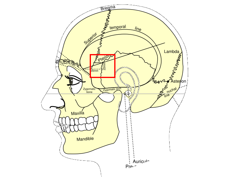
Side view of head, showing surface relations of bones (Pterion labeled at center)
Side view of the skull with arrow pointing to the Pterion
Details
Identifiers
Latinpterion
TA98A02.1.00.019
TA2421
FMA264720
Anatomical terminology
Sphenoparietal suture highlighted in red

The pterion is the region where the frontal, parietal, temporal, and sphenoid bones join.[1] It is located on the side of the skull, just behind the temple. It is also considered to be the weakest part of the skull, which makes it clinically significant, as if there is a fracture around the pterion it could be accompanied by an epidural hematoma.

Structure

[edit]

The pterion is located in the temporal fossa, approximately 2.6 cm behind and 1.3 cm above the posterolateral margin of the frontozygomatic suture.[2]

It is the junction between four bones:

These bones are typically joined by five cranial sutures:

Clinical significance

[edit]

Hematoma

[edit]

The pterion is known as the weakest part of the skull.[3] The anterior division of the middle meningeal artery runs underneath the pterion.[4] Consequently, a traumatic blow to the pterion may rupture the middle meningeal artery causing an epidural haematoma. The pterion may also be fractured indirectly by blows to the top or back of the head that place sufficient force on the skull to fracture the pterion.

Surgery

[edit]

The pterion is a structural landmark for neurosurgical approach to middle cerebral artery aneurysms.[5]

Etymology

[edit]

The pterion receives its name from the Ancient Greek root πτερόν pteron, meaning 'wing'. In Greek mythology, Hermes, messenger of the gods, was enabled to fly by winged sandals, and wings on his head, which were attached at the pterion.

References

[edit]
[edit]
Revisions and contributorsEdit on WikipediaRead on Wikipedia
from Grokipedia
The pterion is an H-shaped sutural junction on the lateral aspect of the human skull, formed by the articulation of the , , squamous portion of the , and greater of the . It is typically located approximately 3 to 4 cm superior to the midpoint of the and 2 to 3 cm posterior to the frontozygomatic suture. The term "pterion" originates from the word pteron, meaning "wing," in reference to the wing-like greater of the . Anatomically, the pterion represents the thinnest region of the , with an average bone thickness of about 3.5 mm, making it particularly vulnerable to fractures from . Beneath this site lies the anterior branch of the , and injury here can lead to laceration of the vessel, resulting in an —a rapidly expanding arterial hemorrhage in the that compresses the and may cause focal neurological deficits, , or death if not treated emergently via . This vulnerability underscores the pterion's clinical importance in and , where temporal bone fractures at this junction are common indicators of head trauma. In , the pterion serves as a critical landmark for procedures such as frontolateral craniotomies, keyhole approaches, and clipping of anterior circulation aneurysms, as well as resections involving sellar or parasellar tumors. Morphologically, it exhibits variations across populations, with the sphenoparietal type (where the sphenotemporal suture meets the parietotemporal suture) being the most prevalent at around 79%, followed by epipteric (8%) and frontotemporal (6%) forms; these differences can influence surgical planning and the risk of complications like unintended orbital penetration. Such variations also highlight the pterion's role in anthropometric studies and , reflecting genetic and developmental influences on cranial asymmetry.

Anatomy

Definition and location

The pterion is defined as the H-shaped sutural junction on the where four cranial bones converge: the , , greater wing of the , and squamous part of the . This region represents a key craniometric landmark due to its thin bony structure. It is precisely located in the on the lateral aspect of the , approximately 3.5 cm posterior to the zygomaticofrontal suture and 4 cm superior to the . Internally, the pterion overlies the anterior division of the middle meningeal artery, which courses along its undersurface, making the area particularly susceptible to vascular injury from trauma.

Bony and sutural composition

The pterion is formed by the junction of four cranial bones: the frontal bone, the parietal bone, the greater wing of the sphenoid bone, and the squamous part of the temporal bone. The frontal bone contributes to the anterosuperior aspect, meeting the parietal bone along the coronal suture. The parietal bone forms the posterosuperior portion, articulating with the frontal bone superiorly via the coronal suture and with the squamous temporal bone inferiorly via the squamous suture. The greater wing of the sphenoid bone underlies the anteroinferior region, connecting to the parietal bone through the sphenoparietal suture and to the frontal bone via the sphenofrontal suture. The squamous part of the temporal bone completes the posteroinferior boundary, joining the parietal bone along the squamous suture and the greater wing of the sphenoid via the sphenosquamous suture. Five sutures converge at the pterion, creating an H-shaped configuration that anchors these bones together. These include the between the frontal and parietal bones, the squamous suture between the parietal and temporal bones, the sphenoparietal suture between the sphenoid and parietal bones, the sphenofrontal suture between the sphenoid and frontal bones, and the sphenosquamous suture (also known as the sphenotemporal suture) between the sphenoid and temporal bones. This sutural arrangement provides structural integrity to the lateral skull wall while allowing for the flexibility needed during cranial growth. The pterion region represents the thinnest portion of the lateral skull wall in adults, with an average bone thickness of approximately 4-5 mm. This minimal thickness arises from the overlapping yet delicate layering of the contributing bones and sutures, distinguishing it from thicker areas of the cranium.

Development and variations

Embryological origins

The bones forming the pterion—frontal, parietal, greater wing of the sphenoid, and squamous part of the temporal—originate primarily from cranial neural crest-derived mesenchyme, with the parietal bone arising from paraxial mesoderm, through the process of intramembranous ossification. This contrasts with the skull base, where endochondral ossification predominates from cartilaginous precursors. Intramembranous ossification involves direct differentiation of mesenchymal cells into osteoblasts, forming bony trabeculae without an intermediate cartilage stage, which allows for the rapid expansion of the cranial vault to accommodate the growing brain. Ossification centers for these bones emerge around the 8th week of . The develops from paired centers at the frontal eminences, while the arises from paired centers at the parietal tuber. The greater wing of the sphenoid ossifies intramembranously starting from its orbital plate region, and the squamous part of the begins near the petromastoid part, both contributing to the eventual convergence at the pterion. These centers expand centrifugally, guided by underlying mesenchymal membranes, establishing the foundational architecture of the skull vault by the end of the embryonic period. The , derived from the ectomeninx, plays a crucial role in directing this by providing osteogenic inductive signals and forming membranes that outline the future sutures, including the site of the pterion. As the expands from approximately 16 weeks of , dural reflections maintain unossified zones where the ossification fronts from adjacent bones meet, ensuring proper alignment and spacing at the pterion junction.

Ossification and morphological variations

The pterion undergoes postnatal as the ossification fronts from the adjacent frontal, parietal, temporal, and sphenoid bones converge and fuse, with the area remaining partially membranous until approximately 5-7 years of age. This process begins after the closure of the around 6 months, but the sutures at the pterion continue to develop, with partial fusion of the near the pterion occurring by 2-4 years and incomplete closure extending to 6-8 years. The completion of this fusion marks the formation of the characteristic H-shaped sutural junction in the typical adult configuration. Morphological variations in the pterion include several distinct types based on the arrangement of the contributing sutures. The sphenoparietal type is the most prevalent, observed in approximately 78-80% of cases across diverse populations, where the greater wing of the sphenoid articulates directly with the , forming the central bar of the H-shape. The frontotemporal type, in which the frontal and temporal bones meet directly, occurs in about 10-15% of individuals. Less common variants include the stellate form (around 15-25%), characterized by a star-like intersection of sutures, and rare configurations such as K-shaped, T-shaped, or Y-shaped arrangements. Accessory epipteric bones, small sutural inserted between the parietal and sphenoid margins, are present in up to 15.5% of skulls and may alter the overall morphology without affecting the primary junction. Topographic variations in pterion position are well-documented and influence its clinical utility as a . The center of the pterion is typically situated 2.6-3.5 cm posterior to the frontozygomatic suture and about 3-4 cm superior to the of the . is evident, with the pterion positioned more posteriorly and superiorly in males compared to females, reflecting broader cranial differences. Ethnic variations show the pterion to be more anterior in African populations (e.g., mean distance of ~2.9 cm from the frontozygomatic suture in ) relative to Caucasians or Asians (often 3.1-3.5 cm). Laterality differences also occur, with the right pterion generally more posterior than the left by 0.1-0.5 cm on average. These population-specific patterns arise from genetic and developmental factors influencing cranial growth.

Clinical significance

Trauma and associated hematomas

The pterion represents a mechanically weak point in the due to its thin bony composition and superposition over the anterior branch of the , rendering it susceptible to fractures from lateral impacts such as temporal blows. These fractures often lacerate the underlying artery, initiating rapid hemorrhage. As noted in anatomical context, this region directly overlies the artery's course, amplifying the risk during trauma. The primary pathological outcome is an epidural (extradural) hematoma, resulting from arterial rupture that causes high-pressure bleeding between the and the inner skull table. This leads to swift blood accumulation, which strips the from the calvarium and can compress underlying tissue, potentially causing herniation if untreated. A hallmark feature is the , where the patient regains consciousness briefly after initial loss before deteriorating due to . Epidural hematomas occur in approximately 10% of traumatic injuries requiring hospitalization, most commonly implicating the . Clinically, manifestations include localized temporal swelling from the fracture and hematoma expansion, alongside neurological deficits such as ipsilateral pupil dilation (mydriasis) from oculomotor nerve compression and contralateral hemiparesis due to cerebral peduncle involvement. Diagnosis relies on non-contrast CT imaging, which typically reveals a hyperdense, biconvex (lens-shaped) collection confined by dural attachments, often associated with an overlying skull fracture. Prompt recognition is critical in emergency settings, as delays can elevate mortality rates beyond 50% in symptomatic cases.

Surgical applications

The pterional craniotomy, also known as the frontotemporosphenoidal approach, utilizes the pterion as a key anatomical landmark to provide broad surgical access to the anterior and middle cranial fossae through a keyhole incision and flap removal. This technique involves a curvilinear frontotemporal incision starting approximately 1 cm anterior to the tragus and extending posteriorly along the hairline, followed by interfascial dissection to create a flap centered at the pterion using burr holes, such as the MacCarty keyhole positioned 7 mm superior and 5 mm posterior to the frontozygomatic suture. The approach allows for wide exposure of the Sylvian fissure after dural incision and anterior reflection, facilitating access to critical neurovascular structures while preserving the frontal and temporal branches of the through subfascial techniques. Introduced by M. Gazi Yasargil in the 1970s, the pterional craniotomy revolutionized by enabling microsurgical precision for anterior circulation pathologies, replacing more invasive approaches like subfrontal or frontoparietal craniotomies. Primary indications include clipping of aneurysms in the anterior circulation, such as those of the , as well as resection of tumors like sphenoid wing meningiomas and vascular malformations including arteriovenous malformations and cavernomas in perisylvian regions. Modern modifications of the original technique incorporate endoscopic assistance for enhanced visualization in deep structures and minimally invasive variants that reduce bone removal and disruption, improving cosmetic outcomes and recovery times while maintaining efficacy for the same indications. Preoperative consideration of pterional positional variations can influence incision planning to optimize exposure and minimize complications.

History and etymology

Historical recognition

The recognition of the pterion, the H-shaped junction of cranial sutures on the lateral , traces back to ancient observations of skull , though without specific . In the 5th century BCE, described variations in cranial sutures in his treatise On Head Wounds, noting their irregular patterns and relative weaknesses as potential sites for trauma-related fractures, which implicitly encompassed regions like the pterion. Similarly, in the 2nd century CE, examined cranial injuries during dissections and trepanation studies, contributing to early understanding of skull vulnerabilities in medical texts. During the , detailed illustrations advanced the understanding of these suture confluences. , in his seminal work De Humani Corporis Fabrica, provided precise depictions of sutures, including the pterion region, highlighting its thin bony structure and variability across individuals as a point of mechanical fragility. These visualizations, based on direct cadaveric dissections, marked a shift from textual descriptions to empirical representation, influencing subsequent anatomists in identifying weak cranial points for surgical consideration. The pterion was formally identified and named as a distinct craniometric landmark in the by French anatomist (1824–1880), who coined the term in 1875 in his studies of cerebral localization and measurements, recognizing its proximity to key neurovascular structures. By the early , its clinical relevance gained prominence in , with pioneers like Fedor Krause in 1900 describing approaches to the via the pterion area, and later refinements by Walter Dandy and George Heuer in 1914 establishing it as a critical access point for intracranial procedures. This evolution underscored the pterion's role in safe planning, as detailed in early neurosurgical literature.

Linguistic origins

The term pterion derives from the word πτερόν (pterón), meaning "wing," in reference to the wing-like greater wing of the that forms part of this cranial junction. This etymological root highlights the anatomical feature's resemblance to a wing in classical descriptions of morphology. The word was first coined in French as ptérion by anatomist in 1875, drawing an analogy to inion—the established term for the —based on similar sutural formations. It entered English usage in 1878, appearing in a of anatomical texts shortly after Broca's introduction. In , the pterion carries symbolic associations with Hermes (known as Mercury in Roman tradition), the fleet-footed messenger of the gods, whose —a broad-brimmed hat adorned with wings—was depicted as attaching near this region, evoking themes of swift aerial travel. This mythological connection underscores the term's cultural resonance, linking anatomical nomenclature to ancient iconography of divine mobility.

References

Add your contribution
Related Hubs
User Avatar
No comments yet.