Hubbry Logo
Sinoatrial blockSinoatrial blockMain
Open search
Sinoatrial block
Community hub
Sinoatrial block
logo
8 pages, 0 posts
0 subscribers
Be the first to start a discussion here.
Sinoatrial block
Sinoatrial block
from Wikipedia
Sinoatrial block
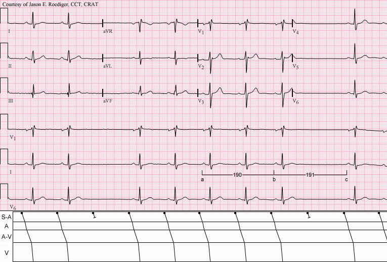
Sinus rhythm (rate = 65/min) with Type II S-A (exit) block; (see laddergram). Note that the long cycles (191) are nearly identical to twice the short cycles (190).
SpecialtyCardiology Edit this on Wikidata

A sinoatrial block (also spelled sinuatrial block)[1] is a disorder in the normal rhythm of the heart, known as a heart block, that is initiated in the sinoatrial node. The initial action impulse in a heart is usually formed in the sinoatrial node (SA node) and carried through the atria, down the internodal atrial pathways to the atrioventricular node (AV) node.[2] In normal conduction, the impulse would travel across the bundle of His (AV bundle), down the bundle branches, and into the Purkinje fibers. This would depolarize the ventricles and cause them to contract.

In an SA block, the electrical impulse is delayed or blocked on the way to the atria, thus delaying the atrial beat.[3] (An AV block, occurs in the AV node and delays ventricular depolarisation). SA blocks are categorized into three classes based on the length of the delay.

Symptoms

[edit]

Sinoatrial blocks are typically well tolerated. They are not as serious as an AV block and most often do not require treatment. In some people, they can cause fainting, altered mental status, chest pain, hypoperfusion, and signs of shock. They can also lead to cessation of the SA node and more serious dysrhythmias.

Types

[edit]

In a first degree sinoatrial block, there is a lag between the time that the SA node fires and actual depolarization of the atria. This rhythm is not easily detectable using state of the art (the highest level of scientific development at the current state in time) diagnostic equipment, thus is currently not recognizable on an ECG strip because an ECG strip does not denote when the SA node fires. It can be detected only during an electrophysiology study when a small wire is placed against the SA node from within the heart and the electrical impulses can be recorded as they leave the p-cells in the centre of the node [ see pacemaker potential ], followed by observing a delay in the onset of the p wave on the ECG.

Second degree SA blocks are broken down into two subcategories just like AV blocks are:

The first is a second degree type I, or Wenckebach block.[citation needed] This rhythm is irregular, and the R-R interval gets progressively smaller, while the P-R interval remains constant, until a QRS segment is dropped. Note that this is quite different from the Wenckebach AV block, in which the PR interval gets progressively longer, before the dropped QRS segment. The pause of a second degree type I is less than twice the shortest R-R interval and is not a multiple of the P-R interval. The cause is a gradual lengthening of conduction time from the SA node to the atria. The p-cells in the centre of the node produce the rhythm at a regular rate, but their conduction across the node to where it meets atrial tissue is where the slowing occurs.

A second degree type II, or sinus exit block, is a regular rhythm that may be normal or slow. It is followed by a pause that is a multiple of the P-P interval usually (2-4). Conduction across the SA node is normal until the time of the pause when it is blocked.

A third degree sinoatrial block looks very similar to a sinus arrest. However, a sinus arrest is caused by a failure to form impulses. A third degree block is caused by failure to conduct them. The rhythm is irregular and either normal or slow. It is followed by a long pause that is not a multiple of the P-R interval. The pause ends with a P wave, instead of a junctional escape beat the way a sinus arrest would.

Treatment

[edit]

Emergency treatment consists of administration of atropine sulfate or transcutaneous pacing.

References

[edit]
[edit]
Revisions and contributorsEdit on WikipediaRead on Wikipedia
from Grokipedia
Sinoatrial block, also known as sinoatrial exit block or SA block, is a disorder of cardiac impulse conduction in which electrical impulses generated by the sinoatrial (SA) node—the heart's primary pacemaker—fail to properly exit the node and propagate to the surrounding atrial tissue, resulting in delayed, intermittent, or absent atrial and potential pauses in heart rhythm. This condition can lead to , sinus pauses, or escape rhythms, and it is often a component of broader or sick sinus syndrome. SA block is classified into three degrees based on the extent of conduction impairment: first-degree, which is typically subclinical and not visible on standard electrocardiogram (ECG); second-degree, characterized by intermittent failure with progressive shortening of PP intervals (type 1) or abrupt dropped P waves (type 2); and third-degree, involving complete dissociation with no P waves originating from the SA node. The causes of sinoatrial block are divided into intrinsic factors affecting the SA node itself, such as (the most common , often due to age-related degeneration), ischemia from , infiltrative diseases like or , and genetic mutations (e.g., in the gene), and extrinsic factors including heightened , metabolic disturbances (e.g., ), and medications such as beta-blockers, , , or antiarrhythmics. It predominantly affects older adults; the incidence of , which includes sinoatrial block, is approximately 0.8 per 1,000 person-years in the general population, and may progress slowly over years if untreated. Symptoms of sinoatrial block vary from asymptomatic cases to manifestations of reduced , including , , , presyncope, syncope, or , particularly in the context of tachy-brady syndrome where alternates with . Diagnosis relies on ECG documentation of characteristic patterns, such as pauses exceeding 2-3 seconds without P waves, grouped beating, or failure of expected P waves following sinus activity, often confirmed with ambulatory Holter monitoring, event recorders, or electrophysiological studies to correlate findings with symptoms. Treatment focuses on addressing reversible causes (e.g., discontinuing offending drugs) and, for symptomatic patients, implanting a permanent pacemaker—typically dual-chamber (DDD) mode—to maintain appropriate heart rates, as the condition can lead to significant morbidity if causes hemodynamic instability.

Overview

Definition

Sinoatrial block, also known as sinoatrial exit block, is a cardiac conduction disorder characterized by the failure of electrical impulses generated by the sinoatrial (SA) node to propagate effectively to the surrounding atrial myocardium, resulting in delayed or blocked activation of the atria and consequent alterations in . This condition specifically involves a disruption in the conduction pathway at or near the SA node exit, while the node itself continues to generate impulses at its intrinsic rate. Unlike , which occurs lower in the conduction system, sinoatrial block affects the initiation of atrial depolarization from the heart's primary pacemaker. The physiological concept of sinoatrial block was first demonstrated in 1852 by Hermann Stannius in experiments on frog hearts. It was later characterized clinically through electrocardiographic analyses in the early , following the development of clinical by . Sinoatrial block represents a specific form of , often considered a subset of sick sinus syndrome (SSS), which encompasses broader impairments in SA node pacemaker activity and impulse conduction. However, sinoatrial block is distinguished by its focus on conduction failure rather than primary defects in impulse generation, though it frequently coexists with other SSS manifestations such as or tachyarrhythmias. A key distinction exists between sinoatrial block and sinus arrest: in sinoatrial block, the SA node generates impulses normally, but they are delayed or blocked from exiting to activate the atria, leading to pauses that are typically multiples of the underlying sinus cycle length. In contrast, sinus arrest involves a complete of the SA node to generate any impulse for a period, resulting in until an escape rhythm emerges, without the predictable timing seen in exit block. This differentiation is clinically important, as it influences diagnostic approaches and underscores that sinoatrial block primarily reflects perinodal conduction abnormalities rather than intrinsic nodal .

Normal sinoatrial node function

The sinoatrial (SA) node is anatomically positioned in the right atrium at the junction of the crista terminalis, immediately adjacent to the entrance of the superior vena cava. This location allows it to integrate with the atrial myocardium while being insulated by connective tissue to prevent premature impulse spread. The node comprises a heterogeneous cluster of specialized myocytes, primarily pacemaker cells (P cells), which are elongated and spindle-shaped with a high surface-to-volume ratio, and transitional cells (T cells), which facilitate electrical coupling to the surrounding atrial tissue. These P cells lack t-tubules and organized sarcoplasmic reticulum, distinguishing them from contractile cardiomyocytes. Physiologically, the SA node serves as the heart's primary pacemaker, initiating spontaneous s that set the rhythm for the entire . It generates these impulses at a normal resting rate of 60 to 100 beats per minute, driven by that overrides subsidiary pacemakers in the atrioventricular (AV) node or . The process begins with phase 4 depolarization, a slow, progressive rise in membrane from approximately -60 mV toward the threshold for firing, without a stable due to the absence of the inward rectifier potassium current (I_K1). Once threshold is reached, an propagates from the SA node via internodal pathways—specialized tracts of atrial myocardium—to depolarize the atria and reach the AV node, ensuring coordinated contraction. At the cellular level, SA node relies on a unique profile of ionic currents that enable phase 4 . The hyperpolarization-activated funny current (I_f), mediated by HCN4 channels, activates during to provide an inward sodium-potassium flux that initiates slow . This is amplified by voltage-gated calcium channels, including (Ca_v3.1) for early phase contributions and L-type (Ca_v1.3 and Ca_v1.2) for the upstroke of the action potential, alongside sodium-calcium exchanger activity and delayed rectifier potassium currents that shape repolarization. These mechanisms ensure reliable, rhythmic firing while allowing autonomic modulation to adjust heart rate. The SA node's rhythm integrates seamlessly with the by pacing atrial depolarization, which manifests as the on an electrocardiogram and triggers atrial to augment ventricular filling. This impulse then travels through the AV node, imposing a brief delay that optimizes atrioventricular synchrony, before exciting the ventricles for contraction. Thus, the SA node establishes the fundamental timing that coordinates the heart's pumping efficiency across the atria and ventricles.

Pathophysiology and etiology

Pathophysiology

Sinoatrial block arises from impaired conduction of electrical impulses generated by the sinoatrial (SA) node to the surrounding atrial myocardium, primarily due to disruptions in the perinodal tissue that serves as the exit pathway for these impulses. This failure often involves the transitional (T) cells in the perinodal region, where progressively infiltrates and disrupts the specialized conduction pathways, leading to delayed or blocked propagation despite normal impulse generation within the SA node itself. , resulting from or occlusion of the nodal , further compromises cellular excitability and conduction velocity in this region by reducing oxygen delivery to pacemaker and transitional cells. Autonomic imbalance, particularly enhanced parasympathetic tone, can exacerbate exit block by hyperpolarizing perinodal cells and slowing conduction, shifting the effective pacemaker site inferiorly within the SA node and hindering impulse egress. Electrophysiologically, sinoatrial block is characterized by prolongation of the sinoatrial conduction time (SACT), which measures the interval from SA node to the onset of atrial ; normal SACT ranges from 50 to 120 ms, with values exceeding 120 ms indicating delayed conduction indicative of first-degree block, though this is not visible on surface ECG. In higher-degree blocks, intermittent or complete failure of conduction manifests as absent P waves following expected sinus intervals, reflecting a breakdown in the coordinated of atrial tissue. The hemodynamic consequences of sinoatrial block include pauses in atrial and ventricular , resulting in transient or, in severe cases, short periods of that reduce and can cause cerebral hypoperfusion. These pauses typically trigger escape rhythms from subsidiary pacemakers, such as the atrioventricular (AV) junction, which generates impulses at rates of 40-60 bpm to maintain circulation until sinus activity resumes. Over time, sinoatrial blocks may progress from intermittent second-degree forms to persistent third-degree block, where no sinus impulses reach the atria, often evolving into complete sinus arrest; this chronic deterioration contributes to tachy-brady syndrome in advanced , characterized by alternating bradycardic pauses and supraventricular tachycardias due to atrial remodeling and irritability.

Causes and risk factors

Sinoatrial block can arise from intrinsic factors affecting the itself, with age-related being the most common cause, accounting for the majority of cases in older adults. This degenerative process involves progressive scarring and replacement of nodal tissue with fibrous material, leading to impaired impulse generation and conduction. Other intrinsic causes include idiopathic degeneration of the node, genetic mutations (e.g., in the gene), and infiltrative diseases such as and , which deposit abnormal substances within the nodal tissue and disrupt its function. Extrinsic causes often involve external influences that suppress or damage activity, including ischemic heart disease where compromises the blood supply to the . Medications such as beta-blockers, , and are frequent culprits, as they slow conduction through enhanced or direct nodal suppression. imbalances, particularly , and infections like can also precipitate sinoatrial block by altering membrane potentials or causing inflammation. Key risk factors for developing sinoatrial block include advanced age over 65 years, which correlates with the cumulative effects of and degeneration. Conditions such as , diabetes mellitus, , and prior further elevate risk by promoting or direct trauma to the node. Autonomic dysfunction, as seen in , may contribute through altered neural regulation of the node. Epidemiologically, the prevalence of sinoatrial block, often as part of , increases markedly with age, affecting approximately 1 in 600 cardiac patients over 65 years and rising to higher rates in those aged 70 to 89. The condition is frequently asymptomatic in its early stages, with symptomatic blocks emerging as nodal reserve diminishes. Incidence rates are estimated at 0.8 cases per 1,000 person-years in the general population, underscoring its relevance in .

Classification

First-degree sinoatrial block

First-degree sinoatrial block is defined as a prolongation of the sinoatrial conduction time (SACT > 120 ms), representing a delay in the transmission of the electrical impulse from the to the atrial myocardium without interruption of atrial . This condition maintains a normal 1:1 atrioventricular conduction pattern, with no dropped beats or pauses observable on the surface electrocardiogram (ECG). The delay occurs due to slowed impulse exit from the , often resulting from subtle impairments in nodal tissue excitability or surrounding atrial conduction pathways. Unlike higher-degree blocks, first-degree sinoatrial block produces no discernible abnormalities on standard 12-lead ECG recordings, as the atrial rhythm remains regular with consistent P-wave morphology and intervals. Diagnosis therefore requires invasive electrophysiological studies, such as intracardiac catheter mapping, to directly measure SACT. One common method involves introducing atrial premature beats and assessing the prolonged return cycle, where the interval from the premature beat to the subsequent sinus beat exceeds expectations for normal conduction (typically calculated as return cycle = sinus cycle length + 2 × SACT). Noninvasive techniques, like signal-averaged ECG or Holter monitoring, may provide indirect clues but lack the precision for definitive identification. Clinically, first-degree sinoatrial block is often subclinical and rarely causes symptoms, as the conduction delay does not significantly alter or in most cases. It holds relevance primarily as an early marker of dysfunction, with potential to progress to more advanced blocks in susceptible individuals. This form is commonly associated with age-related degenerative changes, such as mild of the and perinodal tissues, particularly in elderly patients where such structural alterations become prevalent. For instance, electrophysiological evaluations in older adults with suspected sinus node issues frequently reveal prolonged SACT as an incidental finding during assessments for broader bradyarrhythmias.

Second-degree sinoatrial block

Second-degree sinoatrial block represents an intermittent failure of sinoatrial impulses to exit the node and depolarize the atria, resulting in occasional dropped P waves on the electrocardiogram (ECG). This partial conduction disturbance differs from first-degree sinoatrial block, which involves a consistent delay without dropped beats, and from third-degree block, which is complete and persistent. It is subclassified into two types based on the pattern of conduction failure. Type I second-degree sinoatrial block, also known as Wenckebach type, features progressive shortening of the P-P intervals (reflecting sinoatrial conduction time prolongation) that culminates in a non-propagated impulse and a dropped , with the ensuing pause typically less than twice the preceding P-P interval. In contrast, type II (Mobitz type) involves abrupt failure of impulse exit without prior P-P interval changes, producing a pause approximately equal to two (or a multiple of) the basic P-P intervals. The underlying mechanism involves intermittent sinoatrial exit block, where impulses generated in the fail to propagate through perinodal transitional cells to the atrial myocardium, often due to heightened or myocardial ischemia impairing conduction. Increased vagal influence, as seen in athletes, or ischemic damage to the node or its blood supply can precipitate these episodes. Second-degree sinoatrial block is more prevalent than first-degree block, particularly in contexts like high vagal tone among athletes or medication effects (e.g., beta-blockers, calcium channel blockers), with estimates reaching 29% in patients undergoing Holter monitoring for paroxysmal arrhythmias. Diagnosis relies on identifying characteristic pauses on ECG, though differentiation from sinus arrest can be challenging due to the inability to directly observe sinoatrial node activity; type I shows grouped beating, while type II lacks progressive changes. In some cases, it may progress to third-degree sinoatrial block if conduction deteriorates further.

Third-degree sinoatrial block

Third-degree sinoatrial block represents the complete and sustained failure of impulses to propagate to the atrial myocardium, resulting in no P waves originating from the sinus node on and clinically manifesting as sinus arrest. In this condition, cardiac rhythm is sustained solely by subsidiary escape pacemakers, typically from the atrioventricular junction (rate of 40-60 bpm) or ventricles (rate of 20-40 bpm), which activate due to the absence of sinus-driven atrial . The hallmark electrocardiographic feature is prolonged asystolic pauses exceeding 3 seconds, during which no atrial or ventricular activity occurs until an escape beat emerges, often precipitating symptoms such as syncope from cerebral hypoperfusion. These pauses distinguish third-degree block from lesser degrees, as the block is total rather than partial or intermittent, and the overall escape rhythm rate commonly falls between 30 and 50 beats per minute, reflecting the slower inherent rates of backup pacemakers. As the most severe form of sinoatrial block, third-degree block is frequently linked to underlying structural heart disease, such as or ischemia of the , and carries a high risk of symptomatic or hemodynamic instability. A rare variant involves hyperkalemia-induced complete block, where elevated serum depresses and conduction, potentially resolving with prompt correction and supportive measures.

Clinical presentation

Symptoms

Sinoatrial block often presents with a spectrum of symptoms related to reduced from impaired sinus node impulse generation or conduction, with manifestations varying by the degree of block. First-degree sinoatrial block is typically and discovered incidentally during electrocardiographic evaluation, as the conduction delay does not result in dropped beats or significant pauses. In contrast, second- and third-degree blocks more commonly cause noticeable symptoms due to intermittent or complete failure of sinus impulses, leading to pauses that can provoke , , presyncope, and syncope, particularly during periods of increased demand. Associated symptoms may include palpitations, especially in cases overlapping with tachy-brady syndrome where bradycardic pauses alternate with supraventricular tachyarrhythmias. Severe bradycardia from advanced blocks can also manifest as exertional dyspnea, reflecting inadequate heart rate acceleration during physical activity. These symptoms are often intermittent and nonspecific, potentially mimicking other cardiac or neurologic conditions, and may correlate with underlying bradycardia observed on examination. The impact on is significant in symptomatic patients, as episodes of presyncope or syncope can occur unpredictably, worsening with upright posture or exercise due to the sinoatrial node's failure to provide appropriate response. In a review of 34 patients with chronic symptomatic sinoatrial block, syncope was a predominant feature, affecting cerebral and leading to recurrent events that impair daily functioning until treated.

Physical examination findings

Physical examination in sinoatrial block primarily reveals abnormalities in and rhythm assessment, reflecting impaired function. A hallmark finding is , with a often below 60 beats per minute, and in more severe cases, rates less than 50 beats per minute while awake. The pulse is frequently irregular, characterized by pauses corresponding to sinoatrial exit block or , which may last several seconds and contribute to reduced . Cardiac auscultation typically demonstrates a slow and irregular without specific murmurs unless comorbid structural heart disease is present. may vary in intensity due to the irregular , but no auscultatory features are unique to sinoatrial block. Jugular venous examination is usually unremarkable, lacking the seen in atrioventricular dissociation. General examination may show signs of hypoperfusion during symptomatic episodes, such as , , and diaphoresis, particularly in association with syncope or presyncope. In third-degree sinoatrial block, acute presentations can include hemodynamic instability with altered mental status, cool extremities, and reduced perfusion to end organs. These objective findings complement reported symptoms by indicating the degree of bradycardia-induced compromise.

Diagnosis

Electrocardiographic criteria

Sinoatrial block is challenging to diagnose on standard surface (ECG) because the sinoatrial (SA) node activity is not directly recorded, leading to indirect inference from pauses in the without visible P waves during those intervals. First-degree sinoatrial block involves a delay in conduction from the SA node to the atria but cannot be detected on a standard 12-lead ECG, as it manifests only as a normal-appearing with no overt abnormalities. Diagnosis requires advanced techniques such as intracardiac electrophysiologic study to measure sinoatrial conduction time (SACT) and identify prolonged conduction intervals. Second-degree sinoatrial block is subclassified into type I and type II based on ECG patterns. In type I (Wenckebach), there is progressive shortening of the P-P intervals preceding a pause due to incremental delays in SA conduction, followed by a pause that is less than twice the preceding P-P interval, after which the rhythm resumes with a longer initial P-P interval. In type II (Mobitz II), the P-P intervals remain constant before a sudden pause, where the duration of the pause approximates an exact multiple (typically twice) of the basic P-P interval, reflecting intermittent failure of conduction without prior prolongation. Third-degree sinoatrial block represents complete failure of SA impulses to exit the node, appearing on ECG as a prolonged pause exceeding 2.5 to 3 times the normal P-P interval, with no P waves during the pause and often an escape rhythm originating from atrial, junctional, or ventricular sites to maintain . Key diagnostic measurements on ECG include analysis of P-P intervals to identify patterns consistent with block, such as multiples or progressive changes, and the use of overdrive suppression testing during electrophysiologic studies to differentiate sinoatrial block from sinus arrest, where pauses in block are predictable multiples of the P-P interval whereas arrest pauses are irregular and unrelated to prior cycles.

Differential diagnosis and additional tests

The differential diagnosis of sinoatrial block includes conditions that present with similar bradyarrhythmias or pauses on , such as sinus arrest or pauses, atrioventricular (AV) block, variants of sick sinus syndrome, drug-induced effects, and neurocardiogenic syncope. Sinus arrest involves a transient failure of the sinus node to generate impulses, distinguishable from sinoatrial block by the irregular pause duration not being a multiple of the baseline P-P interval, whereas AV block affects conduction distal to the atria and may show progressive PR prolongation or dropped QRS complexes. Sick sinus syndrome variants often encompass sinoatrial block but can include alternating brady-tachyarrhythmias, requiring correlation of symptoms with rhythm disturbances. Drug effects from beta-blockers, , , or antiarrhythmics can mimic sinoatrial block by suppressing sinus node automaticity, necessitating a review of medication history. Neurocardiogenic syncope, triggered by vasovagal reflexes, may cause transient sinus pauses but is differentiated by situational precipitants and absence of structural sinus node disease on further testing. Laboratory evaluation plays a key role in identifying reversible causes of sinoatrial block, including assessment of (sodium, , calcium), to rule out , and serum drug levels for medications known to affect sinus node function. Abnormal levels, such as , can impair sinus node conduction and should be corrected promptly. is a metabolic cause that may contribute to and requires measurement. Additional noninvasive tests support the diagnosis when electrocardiographic findings are intermittent or equivocal. Ambulatory electrocardiographic monitoring, such as 24- to 48-hour Holter monitoring or longer-term event recorders, is essential for capturing episodic sinoatrial blocks and correlating them with symptoms like syncope or , with high specificity when spontaneous arrhythmias are observed. Exercise evaluates chronotropic incompetence by assessing the heart's rate response to exertion; failure to achieve at least 80% of the predicted maximum indicates . is recommended to identify structural cardiac abnormalities, such as ischemic heart disease, valvular disorders, or infiltrative conditions like , that may underlie or coexist with sinoatrial block. Invasive electrophysiological study (EPS) provides definitive assessment of sinus node function in symptomatic patients with nondiagnostic noninvasive tests. During EPS, sinoatrial conduction time (SACT) is measured; values exceeding 200 ms in adults suggest conduction delay. Sinus node recovery time (SNRT) is determined after overdrive atrial pacing, with prolongation defined as an uncorrected SNRT greater than 1400 ms or a corrected SNRT (CSNRT, adjusted for baseline cycle length) exceeding 525 ms indicating abnormal sinus node automaticity. These parameters help confirm sinoatrial block and guide decisions for pacemaker implantation.

Treatment

Conservative management

Conservative management of sinoatrial block primarily involves and addressing reversible factors in patients or those with mild symptoms, particularly in first-degree or second-degree cases without hemodynamic instability. For individuals with first-degree sinoatrial block or higher-degree blocks, with periodic clinical is recommended, as these often do not progress or cause significant issues. Regular follow-up includes periodic clinical , serial electrocardiograms (ECGs), and ambulatory monitoring such as Holter or event recorders to assess for symptom correlation and progression. Addressing reversible causes is a of conservative therapy to potentially resolve the block without further intervention. Common extrinsic factors include medications like beta-blockers, , or , which should be discontinued or adjusted under medical supervision. Electrolyte imbalances, such as or , require prompt correction through supplementation or dietary measures. Underlying conditions like myocardial ischemia can be managed medically with anti-ischemic therapy, such as nitrates or antiplatelet agents, while may be treated with (CPAP) to mitigate nocturnal . Infections like , if identified, warrant targeted antibiotic therapy. Lifestyle modifications play a supportive role in preventing exacerbations, particularly by avoiding triggers that enhance or . Patients are advised to maintain adequate hydration, limit alcohol and intake, and avoid strenuous activities immediately after meals to reduce vagally mediated episodes. For acute symptomatic episodes due to vagal stimulation, such as during vasovagal syncope, temporary administration of atropine (0.5 to 1 mg intravenously, repeatable up to 3 mg) can increase by blocking parasympathetic effects on the . Enhanced monitoring with implantable loop recorders is appropriate for patients with infrequent symptoms to correlate episodes with arrhythmias, enabling precise risk stratification without immediate invasive therapy. Escalation to interventional approaches is considered if symptoms persist despite these measures or if there is evidence of high-degree block with hemodynamic compromise.

Interventional therapies

For patients with symptomatic second- or third-degree sinoatrial block who do not respond adequately to conservative measures, interventional therapies are indicated to restore reliable cardiac rhythm and prevent hemodynamic compromise. Pacemaker implantation serves as the definitive treatment for persistent symptomatic sinoatrial block, particularly in cases of second- or third-degree block associated with documented pauses exceeding 3 seconds or other manifestations of . According to the 2018 ACC/AHA/HRS guidelines, permanent pacing is recommended (Class I, Level of Evidence B-R) when symptoms such as syncope, presyncope, or exertional fatigue correlate temporally with bradycardic episodes. Dual-chamber (DDD) pacemakers are preferred over single-chamber ventricular pacing to preserve atrioventricular synchrony, thereby reducing the risk of , , and (Class I, Level of Evidence B-R). In select cases of isolated sinoatrial dysfunction without atrioventricular conduction abnormalities, single-chamber atrial (AAI) pacing is recommended as a less invasive option (Class I, Level of Evidence B-R). In acute settings with hemodynamic instability due to high-degree sinoatrial block, temporary pacing via transvenous or transcutaneous approaches is indicated (Class I, Level of C-LD) to provide immediate atrioventricular support until permanent implantation can be arranged. Pharmacologic augmentation with isoproterenol infusion (typically 0.5–10 mcg/min intravenously) may be used as a bridge therapy to increase sinus node and conduction in unstable patients, particularly when pacing is not immediately available, though it carries risks of tachyarrhythmias and should be monitored closely. Adjunctive interventions target underlying etiologies or coexisting arrhythmias. For sinoatrial block occurring within tachy-brady syndrome—characterized by alternating and supraventricular tachyarrhythmias such as of the tachyarrhythmic focus (e.g., pulmonary vein isolation) can mitigate bradycardic episodes by reducing atrial remodeling and improving sinus node function, with studies showing reduced need for pacing in select patients. In cases of ischemia-related sinoatrial block, coronary via or coronary artery bypass grafting may resolve conduction disturbances by restoring perfusion to the sinus node artery, as evidenced by case reports of symptom reversal post-procedure.

Prognosis and complications

Prognosis

The prognosis of sinoatrial block varies significantly by degree of severity and underlying . First-degree sinoatrial block is typically benign and asymptomatic, often requiring no intervention and conferring a near-normal comparable to the general , as it represents only a conduction delay without clinical impact. In contrast, untreated third-degree sinoatrial block carries a poor due to the risk of prolonged pauses or , potentially leading to sudden cardiac death if escape rhythms fail to sustain adequate heart rates. Prognostic outcomes are heavily influenced by the underlying cause; reversible etiologies such as drug-induced block (e.g., from beta-blockers or calcium channel antagonists) generally yield better recovery upon discontinuation compared to irreversible intrinsic causes like degenerative or post-ischemic scarring. For patients with sick sinus syndrome encompassing sinoatrial block who undergo pacemaker implantation, 5-year survival rates exceed 70%, with one study reporting 73.1% survival, though this is lower in those with concomitant structural heart disease. As of 2025, advancements in leadless pacemaker technology have further improved outcomes for dysfunction, demonstrating reduced long-term complication rates compared to traditional transvenous systems, including lower risks and enhanced device reliability over 5 years. Overall annual complication incidence following pacemaker therapy remains below 2%, primarily driven by procedural factors rather than the block itself. Quality of life markedly improves with appropriate pacing, with symptoms resolving in approximately 90% of cases, enabling return to normal activities; however, ongoing concerns such as device battery longevity (typically 5-15 years) and rare risks persist, necessitating regular monitoring.

Potential complications

Sinoatrial block, as a manifestation of , can lead to chronic that impairs , potentially resulting in over time if untreated. Atrial stasis associated with or coexisting increases the risk of , including , with patients exhibiting a higher incidence compared to those without sinus node . Additionally, sinoatrial block may progress to atrioventricular conduction disturbances, including complete , as a pathologic in affected individuals. Pacemaker implantation, the primary treatment for symptomatic sinoatrial block, carries risks such as lead dislodgement, occurring in approximately 1-2% of cases, often requiring repositioning. Device infections affect 1-2% of patients, potentially necessitating explantation and prolonged . In patients paced in VVI mode, —characterized by symptoms from ventriculoatrial conduction—develops in approximately 20% of cases, highlighting the preference for dual-chamber pacing to mitigate this. Episodes of syncope due to sinoatrial block heighten the risk of falls and associated trauma, such as fractures or head injuries. Post-pacing, inappropriate sinus tachycardia may emerge in some patients with underlying sinus node dysfunction, contributing to palpitations and hemodynamic instability. Prevention strategies include anticoagulation for patients with coexisting atrial fibrillation to reduce thromboembolic events, alongside routine pacemaker surveillance to detect and address device-related issues early. These measures help mitigate the overall prognostic impact of complications in sinoatrial block.

References

Add your contribution
Related Hubs
User Avatar
No comments yet.