Hubbry Logo
Nasolacrimal ductNasolacrimal ductMain
Open search
Nasolacrimal duct
Community hub
Nasolacrimal duct
logo
8 pages, 0 posts
0 subscribers
Be the first to start a discussion here.
Nasolacrimal duct
Nasolacrimal duct
from Wikipedia
Nasolacrimal duct
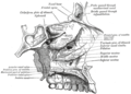
The lacrimal apparatus. Right side.
Details
Identifiers
Latinductus nasolacrimalis
MeSHD009301
TA98A15.2.07.070
TA26859
FMA9703
Anatomical terminology

The nasolacrimal duct (also called the tear duct) carries tears from the lacrimal sac of the eye into the nasal cavity.[1][2] The duct begins in the eye socket between the maxillary and lacrimal bones, from where it passes downwards and backwards. The opening of the nasolacrimal duct into the inferior nasal meatus of the nasal cavity is partially covered by a mucosal fold (valve of Hasner or plica lacrimalis).[3]

Excess tears flow through the nasolacrimal duct which drains into the inferior nasal meatus. This is the reason the nose starts to run when a person is crying or has watery eyes from an allergy, and why one can sometimes taste eye drops. This is for the same reason when applying some eye drops it is often advised to close the nasolacrimal duct by pressing it with a finger to prevent the medicine from escaping the eye and having unwanted side effects elsewhere in the body as it will proceed through the canal to the nasal cavity.

Like the lacrimal sac, the duct is lined by stratified columnar epithelium containing mucus-secreting goblet cells, and is surrounded by connective tissue.

Nasolacrimal canal

[edit]
Nasolacrimal canal
Tear system.

a = lacrimal gland
b = superior lacrimal punctum
c = superior lacrimal canal
d = lacrimal sac
e = inferior lacrimal punctum
f = inferior lacrimal canal
g = nasolacrimal canal
Details
Identifiers
Latincanalis nasolacrimalis
MeSHD009301
TA98A15.2.07.070
TA26859
FMA9703
Anatomical terms of bone

The canal containing the duct is called the nasolacrimal canal. It is formed by indentations in the inferior nasal conchae, maxilla and lacrimal bone. The canal drains into the nasal cavity through the anterior portion of the inferior meatus, which is between the inferior concha and the floor of the nasal cavity.

Clinical significance

[edit]

Obstruction of the nasolacrimal duct may occur.[4][5][6] This leads to the excess overflow of tears called epiphora (chronic low-grade nasolacrimal duct occlusion).[7] A congenital obstruction can cause cystic expansion of the duct and is called a dacryocystocele or Timo cyst. Persons with dry eye conditions can be fitted with punctal plugs that seal the ducts to limit the amount of fluid drainage and retain moisture.

During an ear infection, excess mucus may drain through the nasolacrimal duct in the opposite way tears drain. [citation needed]

In humans, the tear ducts in males tend to be larger than the ones in females.[8]

Additional image

[edit]

See also

[edit]

References

[edit]
[edit]
Revisions and contributorsEdit on WikipediaRead on Wikipedia
from Grokipedia
The nasolacrimal duct is a membranous canal approximately 12 to 18 mm in length that extends from the inferior aspect of the lacrimal sac to an opening in the inferior nasal meatus beneath the inferior turbinate, serving as the final pathway in the lacrimal drainage system to transport tears from the ocular surface into the nasal cavity. This structure, part of the broader lacrimal apparatus, connects the medial canthus of the eye to the nose via the nasolacrimal canal, which is formed by the lacrimal and maxillary bones. Its primary function is to drain excess tears and debris produced by the lacrimal glands, thereby maintaining ocular lubrication, removing irritants, and preventing overflow of tears onto the eyelids (a condition known as epiphora). Anatomically, tears enter the system through the lacrimal puncta—small openings at the medial margins of the upper and lower eyelids—before passing through the canaliculi (8 to 10 mm long) into the , which measures about 10 to 15 mm vertically and then descends through the nasolacrimal duct. The duct itself is lined with containing goblet cells that contribute to secretion, aiding in tear film stability, and it features a mucosal fold (valve of Hasner) at its distal end that helps regulate flow but can be a site of obstruction. Embryologically, the nasolacrimal duct develops from an ectodermal cord that canalizes between the 5th and 8th weeks of , achieving patency by birth in most cases, though congenital affects 5–20% of newborns. Clinically, the nasolacrimal duct plays a critical role in ocular health, as its obstruction—whether congenital, due to , , or trauma—can lead to conditions such as (inflammation of the ) or chronic tearing, often requiring interventions like probing, stenting, or . In adults, acquired blockages may arise from chronic or tumors, underscoring the duct's integration with nasal and ocular physiology for effective tear clearance and immune defense at the eye's surface.

Anatomy

Structure and histology

The nasolacrimal duct is a membranous canal that extends approximately 12 to 18 mm in length from the inferior aspect of the to the inferior , with a ranging from 3 to 4 mm. This structure facilitates the passage of tears into the while maintaining structural integrity through its specialized composition. The duct's lumen is irregular due to prominent mucosal folds that form valves, including the valve of Hasner at the distal opening, which acts as a functional barrier to prevent retrograde flow of nasal contents. Histologically, the nasolacrimal duct is lined by a pseudostratified ciliated columnar interspersed with goblet cells that secrete to lubricate the passage and protect against . The cilia on the epithelial surface beat in a coordinated manner to propel distally, enhancing drainage efficiency. Beneath the epithelium lies a consisting of helical arrangements of , elastic, and reticular fibers enclosing mixed glands and a wide luminal vascular resembling a cavernous body, which aids in tear outflow regulation. An outer fibrous layer of provides structural support and anchors the duct within the surrounding bony canal. Along its course, the nasolacrimal duct maintains a relatively consistent of approximately 2-3 mm, lined by pseudostratified ciliated columnar epithelium with regional variations in thickness. These regional differences contribute to the duct's adaptability in tear transport while minimizing resistance to flow.

Location and relations

The nasolacrimal duct originates as the inferior continuation of the , which is situated in the medial within a bony groove formed by the frontal process of the and the . It descends vertically for approximately 12-18 mm, initially traversing the intraosseous portion through the nasolacrimal canal primarily within the , before transitioning to a shorter membranous segment within the . This path directs the duct inferolaterally and slightly posteriorly, ultimately opening via a slit-like into the inferior meatus of the , directly beneath the attachment of the inferior turbinate. The is typically covered by a mucosal fold known as the valve of Hasner, which helps prevent retrograde airflow into the duct. In its orbital segment, the duct lies adjacent to the medial rectus muscle and orbital fat, with the positioned anterior (preseptal) to the , which attaches to the posterior lacrimal crest. Within the nasolacrimal canal, the duct is enclosed by the laterally and the medially, positioning it medial to the and lateral to the . Anteriorly, it relates to the middle turbinate head in about 70% of cases, while posteriorly, it is bordered by the uncinate process and bulla ethmoidalis of the , as well as a connecting to the inferior turbinate vasculature. These relations place the duct anterior to the ethmoid bone's air cells overall. On imaging, the nasolacrimal duct is identifiable on computed tomography (CT) as a vertically oriented structure within the nasolacrimal canal, measuring about 3-4 mm in diameter, with its course serving as a landmark relative to the uncinate process, which lies consistently posterior to it. In magnetic resonance imaging (MRI), the duct's membranous portion appears as a soft-tissue signal within the nasal mucosa, aiding in differentiation from adjacent turbinates and sinuses.

Nasolacrimal canal

The nasolacrimal canal is a vertical bony channel approximately 12 to 18 mm in length that provides passage for the nasolacrimal duct from the to the . It is formed by the union of the lacrimal groove on the posterior surface of the frontal process of the maxilla and the groove on the lateral surface of the , which together create the anterior and medial walls, while the posterior wall is contributed by the lacrimal process of the ; this structure opens inferiorly into the inferior of the . The canal typically measures 3 to 5 mm in width, with a mean minimum of 3.5 mm (range: 1.5–6.3 mm) in healthy individuals, showing slight where males have a larger average (3.70 mm) than females (3.35 mm). Anatomical variations in the nasolacrimal canal, particularly narrowing of its diameter, can predispose to obstruction of the enclosed nasolacrimal duct. Studies using have demonstrated that patients with primary acquired (PANDO) exhibit a significantly smaller mean minimum diameter of 3.0 mm (range: 2.0–4.2 mm) compared to controls (P = .001), with an inverse association between age and canal diameter in affected individuals (P = .007). Such constrictions may contribute to impaired tear drainage, though they are not the sole causative factor due to overlap in measurements between obstructed and normal cases. The nasolacrimal duct traverses this canal to facilitate its drainage function.

Development

Embryology

The development of the nasolacrimal duct originates from the nasolacrimal groove, which appears during the 6th to 7th week of embryonic gestation as an ectodermal thickening between the emerging lateral nasal and maxillary prominences. This groove forms through ectodermal , where surface epithelium proliferates and sinks inward, while surrounding condenses to support the structure, eventually delineating the primordium of the lacrimal drainage system. By Carnegie stage 16 (approximately 5-6 weeks, 11 mm), the groove deepens into the lacrimal lamina, an epithelial cord that detaches from the surface and embeds into the . Canalization of this solid epithelial cord initiates around the 8th to 10th week of , transforming the cord into the superiorly and the nasolacrimal duct inferiorly. Central cells within the cord undergo (), creating a lumen that extends progressively; this process is largely complete by the 12th week, though full maturation continues into the fetal period. The duct elongates and incorporates into the bony nasolacrimal canal as the and develop, ensuring alignment with the inferior meatus. The valve of Hasner, a mucosal fold at the duct's distal opening into the , forms last during late canalization and often remains imperforate at birth in up to 20% of cases, contributing to transient or persistent congenital . Genetic factors play a key role in this process, influencing mesenchymal development of the ; disruptions can lead to structural anomalies. This prenatal formation sets the stage for postnatal refinements in duct patency.

Postnatal development

At birth, the nasolacrimal duct measures approximately 8 to 10 mm in length and is often incomplete, with a thin frequently persisting at the valve of Hasner, leading to temporary obstruction in up to 20% of newborns. Full canalization typically occurs postnatally through central of the epithelial core and hydrostatic pressure from tear production, resolving most obstructions by 6 to 12 months of age. During childhood, the duct undergoes significant elongation and expansion, with the bony height increasing 1.8-fold, average diameter widening 1.4-fold, and overall volume expanding 4.6-fold from 2 weeks to 34 months, primarily in a nonlinear pattern during the first 6 months. This growth correlates closely with postnatal maxillary bone development and continues gradually thereafter, reaching adult dimensions of 12 to 18 mm by approximately 5 to 7 years, thereby reducing the risk of obstruction through improved patency and capacity. In adulthood, the ductal epithelium transitions to a stratified columnar structure with goblet cells, providing enhanced for tear lubrication, though specific thickening is observed in response to chronic exposure. With advancing age, the duct may experience or due to progressive , recurrent inflammation, and in the surrounding , contributing to primary acquired in older adults. Sex differences include a slightly wider in males, with the sectional area approximately 13% larger than in females, potentially influencing drainage efficiency.

Physiology

Tear drainage mechanism

Tears produced by the and distributed across the ocular surface enter the drainage system through the upper and lower lacrimal puncta, small openings located at the medial margins of the eyelids. From there, they travel along the canaliculi—short, approximately 10 mm long channels lined with —before converging into the , a distensible reservoir situated in the lacrimal fossa. The then funnels tears into the nasolacrimal duct, a 12-18 mm vertical passage that descends medially and opens into the inferior beneath the inferior turbinate, allowing tears to mix with nasal secretions. The primary propulsion of tears through this pathway occurs via the lacrimal pump mechanism, driven by contractions of the during , which compresses the canaliculi and while creating negative pressure to draw tears forward. This muscular action, innervated by the (cranial nerve VII), is supplemented by , which aids downward flow through the vertically oriented nasolacrimal duct, and capillary action within the narrow lumens of the canaliculi and duct, facilitating passive movement of the aqueous tear film. Valves along the system, such as the valve of Rosenmüller at the junction of the canaliculi and , and Hasner's valve at the distal nasolacrimal duct opening, promote unidirectional flow toward the by preventing reflux. Under normal conditions, the nasolacrimal system drains approximately 1 μL of per minute to match basal tear production, maintaining ocular surface by removing excess fluid and debris. During reflex tearing, such as in response to or , drainage capacity increases proportionally to handle higher volumes, preventing overflow (epiphora). The anatomical connection to the integrates tear clearance with nasal airflow, where inspired air and mucociliary transport further facilitate and removal of drained .

Regulation of flow

The flow through the nasolacrimal duct is primarily modulated by neural mechanisms that control vascular tone and mechanical propulsion, with additional influences from reflexes and hormones. Neural innervation of the nasolacrimal duct arises from both parasympathetic and sympathetic divisions of the , targeting the cavernous body—a vascular structure within the duct wall that regulates lumen diameter and tear outflow resistance. Parasympathetic fibers, conveyed via the (cranial nerve VII), release vasoactive intestinal polypeptide (VIP) to promote and glandular , thereby facilitating increased flow during heightened tear production. Sympathetic fibers originate from the and express , enabling to narrow the lumen and fine-tune drainage under varying conditions. These neural elements form a dense of myelinated and unmyelinated fibers, positive for markers such as and , which collectively adjust blood flow to optimize tear passage. Reflex mechanisms enhance duct flow through dynamic actions tied to everyday behaviors. , mediated by motor fibers from cranial nerve VII to the , generates a lacrimal pump effect: preseptal and pretarsal portions of the muscle contract to create negative intralacrimal pressure, drawing tears into the canaliculi, followed by positive pressure that propels them through the sac and duct. This process is critical for basal and reflex tear drainage, with studies demonstrating visible fluid movement and sac compression during blinks via ultrasound imaging. Sniffing or nasal inspiration complements this by producing negative pressure in the inferior meatus, aiding tear pull from the duct into the . Hormonal influences, particularly , modulate epithelial function in the lacrimal outflow system, with receptors detected in the lacrimal sac . In females, estrogen maintains epithelial integrity and permeability; declining levels during can disrupt this, contributing to altered tear dynamics and increased dry eye prevalence by affecting overall drainage efficiency. Feedback loops integrate sensory input to prevent overflow or stagnation, primarily through osmolarity detection on the ocular surface that signals the trigeminal nerve (cranial nerve V), triggering parasympathetic efferent responses via cranial nerve VII to boost tearing and subsequent duct flow as needed. This neural arc ensures adaptive regulation, with lacrimal gland secretion adjusting to maintain tear homeostasis and duct throughput.

Clinical significance

Congenital conditions

Congenital (NLDO) is the most prevalent birth-related abnormality of the nasolacrimal duct, resulting from the persistence of a membranous barrier at the valve of Hasner, the distal opening into the inferior . This condition affects 5-20% of newborns and manifests primarily as epiphora (excessive tearing) and from the affected eye, often becoming noticeable within the first few weeks of life. The obstruction arises from incomplete canalization of the nasolacrimal duct during embryonic development, a process detailed in the section. Nasolacrimal duct or represents a rarer congenital anomaly, characterized by the complete absence or failure of development of the duct, leading to severe and persistent tearing without spontaneous improvement. These defects occur in isolation or as part of genetic syndromes, such as , where up to 27% of affected children exhibit nasolacrimal drainage anomalies including , and , which is associated with lacrimal system atresias alongside branchial and renal malformations. Incidence is low, with such structural absences contributing to a small subset of the broader 1.2-30% range of congenital obstructions reported in various studies. Accessory nasolacrimal ducts or congenital lacrimal fistulas involve anomalous epithelial-lined channels connecting the lacrimal drainage system to the skin, typically originating from the common canaliculus or . These rare variants, with a prevalence of approximately 1 in 2000 live births, can cause chronic epiphora or mucoid discharge directly from the external opening, often located inferior to the medial . Overall incidence of congenital nasolacrimal duct anomalies is elevated in premature infants, with up to 16% showing some degree of blockage compared to term newborns. Additional risk factors include cesarean section delivery and genetic predispositions, though the majority of NLDO cases—around 90-96%—resolve spontaneously by 12 months of age without intervention.

Acquired disorders

Acquired disorders of the nasolacrimal duct encompass a range of non-congenital conditions that arise later in life due to , , , or medical interventions, often leading to obstruction, , or of the duct. These disorders typically manifest as epiphora (excessive tearing), recurrent s, or swelling in the medial canthal region, impairing normal tear drainage. Inflammatory conditions, particularly , represent one of the most common acquired issues, characterized by acute or chronic of the secondary to . Acute often results from bacterial pathogens such as or , causing , tenderness, and potential abscess formation over the lacrimal sac; in severe cases, it may lead to if untreated. Chronic , stemming from persistent low-grade or repeated episodes, promotes and of the duct, narrowing its lumen and perpetuating stasis of tear fluid, which fosters further bacterial overgrowth. Neoplastic disorders are rare but significant, with primary tumors such as being the most frequently reported malignancy affecting the nasolacrimal duct and . These tumors often present with insidious obstruction, leading to epiphora and medial canthal mass, and typically involve both the sac and duct at in the majority of cases; squamous cell carcinoma accounts for a substantial proportion of epithelial malignancies in this region, with aggressive local invasion contributing to delayed recognition. Traumatic injuries, particularly from midfacial fractures, can directly damage the nasolacrimal , resulting in strictures or complete obstruction of the duct. Naso-orbito-ethmoid fractures, common in high-impact trauma such as motor vehicle accidents, disrupt the bony canal housing the duct, with nasolacrimal injuries occurring in up to 15% of such cases and leading to scarring that impairs tear outflow. Iatrogenic causes arise from surgical interventions near the nasolacrimal apparatus, including endoscopic sinus surgery or orbital procedures, where inadvertent trauma induces scarring and . In , nasolacrimal duct injury or dehiscence has been reported in up to 15% of procedures, though symptomatic obstruction is less frequent at around 1-3%, often due to excessive anterior dissection or thermal damage during instrumentation. Similar risks occur in orbital surgeries, where manipulation of adjacent structures can fibrose the duct, with prevalence varying by surgical approach but highlighting the need for anatomical preservation.

Diagnosis and treatment

Diagnosis of typically begins with a clinical history of epiphora and examination, including assessment of the tear meniscus and for reflux upon pressure. The fluorescein disappearance test is a primary diagnostic tool, where 2% fluorescein is instilled into the conjunctival sac and observed for clearance over 5 minutes; retention of indicates delayed drainage and supports the of obstruction. The Jones I test further evaluates patency by instilling fluorescein into the inferior fornix and checking for its presence in the nasal inferior meatus via swab or after 5 minutes; a positive result confirms tear flow through the nasolacrimal duct, while absence suggests obstruction proximal to the duct. The Jones II test, a non-physiologic with saline through the puncta, assesses anatomical patency and localizes blockages, such as retrograde flow indicating nasolacrimal duct involvement. In children, probing of the canaliculi under topical can serve as both a diagnostic and initial therapeutic maneuver to detect and relieve membranous obstructions at the valve of Hasner. For detailed anatomical evaluation, particularly in complex or acquired cases, dacryocystography involves injecting contrast into the canaliculi to visualize filling defects or strictures via conventional . Computed tomography dacryocystography (CT-DCG) enhances this by providing multiplanar images of the lacrimal , identifying the level and nature of obstructions with high resolution and minimal invasiveness. Treatment for congenital nasolacrimal duct obstruction (NLDO) prioritizes conservative measures initially, as up to 95% of cases resolve spontaneously by 13 months of age. massage, performed by applying gentle pressure over the sac 2-3 times daily, promotes canalization and achieves success rates of 28-56% depending on age at initiation, with higher efficacy in infants under 6 months. Topical antibiotics are reserved for cases with associated infection, such as , and do not independently improve resolution rates. If symptoms persist beyond 6-12 months, office or surgical probing under is recommended, with success rates of 75-89% in children under 12 months by perforating the distal membranous obstruction. For persistent congenital NLDO after failed probing, balloon dacryoplasty offers a minimally invasive alternative, involving insertion through the canaliculi and inflation at the obstruction site to dilate the duct; success rates reach 77% in such cases, comparable to stenting without the associated complications like tube migration. In acquired NLDO, where obstructions often result from or trauma, dacryocystorhinostomy (DCR) is the definitive surgical intervention, creating a new drainage pathway from the to the ; external DCR approaches achieve anatomical and functional success rates exceeding 90%, with low complication rates of 1-6%. Endoscopic DCR variants, performed transnasally, yield similar outcomes of 89-93% success while avoiding external scars. Silicone stenting during DCR maintains ostial patency and is routinely used, with removal after 3-6 months; it contributes to sustained success in chronic cases. For enhanced endoscopic techniques, marsupialization involves everting the lacrimal sac mucosa to create a large, stable ostium, achieving primary success rates of 83% and overall 96% after revisions by reducing granulation tissue formation. Emerging options include transcanalicular laser-assisted endoscopic DCR, which uses diode or Nd:YAG lasers for precise osteotomy without incisions, offering 60-90% success rates, minimal bleeding, and faster recovery, particularly beneficial for patients with comorbidities. Recent advances as of 2025 include dacryoendoscopic-assisted laser dacryoplasty with silicone intubation, which enhances visualization and precision in probing and dilation, and sutured Ritleng tube intubation following probing, reporting success rates up to 100% in select pediatric cohorts. A 2024 clinical guideline reinforces probing as the first-line surgical treatment after conservative management failure for congenital cases. Antibiotics remain essential perioperatively for infection control in all surgical cases.

References

Add your contribution
Related Hubs
User Avatar
No comments yet.