
Community hub
Recent from talks
Contribute something
Nothing was collected or created yet.


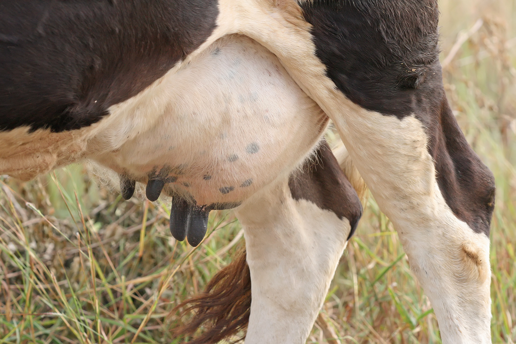
A teat is the projection from the mammary glands of mammals from which milk flows or is ejected for the purpose of feeding young.[1][2][3] In many mammals, the teat projects from the udder. The number of teats varies by mammalian species and often corresponds to the average litter size for that animal.[4][5] In some cases, the teats of female animals are milked for the purpose of human consumption.
The quality of some domesticated animals is determined by the establishment of desired characteristics, such as teat size and placement.[6][7]
Number and positioning in other animals
[edit]The number and positioning of mammary glands and teats varies widely among mammals. The protruding teats and accompanying glands can be located anywhere along the two milk lines. In general, most mammals develop mammary glands in pairs along these lines, with a number approximating the number of young typically birthed at a time. The number of teats varies from 2 (in elephants and anthropoids) to 18 (in pigs). Marsupials usually have 4 to 12 teats,[8] but the Virginia opossum has 13, one of the few mammals with an odd number.[9][10] The following table lists the number and position of teats and glands found in a range of mammals:
| Species[11] | Cranial teats (thoracic) |
Intermediate teats (abdominal) |
Caudal teats (inguinal) |
Total teats |
|---|---|---|---|---|
| Goat, sheep, horse guinea pig |
0 | 0 | 2 | 2 |
| Polar bear | 4 | 0 | 0 | 4 |
| American black bear, Asian black bear, grizzly bear | 4 | 0 | 2 | 6 |
| Camel | 0 | 0 | 4 | 4 |
| Cattle | 0 | 0 | 4 | 4 |
| Cat | 2 | 2 | 4 | 8 |
| Dog | 4 | 2 | 2 or 4 | 8 or 10 |
| Mouse | 6 | 0 | 4 | 10 |
| Rat | 6 | 2 | 4 | 12 |
| Pig | 4 | 4 | 4 | 12 |
| Elephants, Anteaters, anthropoids (including humans) | 2 | 0 | 0 | 2 |
Disease of teats
[edit]A number of diseases can affect the teats of cattle.[12]
- Pseudocowpox
- Warts caused by bovine papillomavirus
- Teat-end hyperkeratosis
- Dermatitis
- Frostbite
- Udder sores or necrotic dermatitis
Goats are also affected by diseases of the teats.[13]
Etymology
[edit]Teat is derived from the Old French or Dutch word, "tete" or the Greek word τιτθύς.[14] An alternative, but possibly not unrelated, would be the Welsh word "teth" or the Old English, "titt" which is still used as a slang term. The words "teat" and "tit" share a Germanic ancestor. The second of the two, tit, was inherited directly from Proto-Germanic, while the first entered English via Old French.[15][16]
See also
[edit]References
[edit]- ^ "teat - Wiktionary". en.wiktionary.org. Retrieved 12 August 2017.
- ^ "Definition of TEAT". www.merriam-webster.com. Retrieved 12 August 2017.
- ^ "teat - definition of teat in English - Oxford Dictionaries". Oxford Dictionaries - English. Archived from the original on August 12, 2017. Retrieved 12 August 2017.
- ^ Rowen D. Frandson; W. Lee Wilke; Anna Dee Fails (1 April 2013), Anatomy and Physiology of Farm Animals, John Wiley & Sons, pp. 449–451, ISBN 978-1-118-68601-0
- ^ "Mammalian Milk". www.earthlife.net. Retrieved 12 August 2017.
- ^ "Teat Structure Chart". American Boer Goat Association. Archived from the original on February 8, 2016. Retrieved 2017-11-22.
- ^ Blackburn, Lorrie. "More on Teats". The National Pygmy Goat Association. Archived from the original on 2017-12-07. Retrieved 2017-11-22.
- ^ Julian Lombardi (6 December 2012). Comparative Vertebrate Reproduction. Springer Science & Business Media. ISBN 978-1-4615-4937-6.
- ^ "With the Wild Things – Transcripts". Digitalcollections.fiu.edu. Archived from the original on 2013-03-23. Retrieved 2013-04-05.
- ^ Stockard, Mary (2005) Raising Orphaned Baby Opossums. Alabama Wildlife Center.
- ^ Cunningham, Merle; LaTour, Mickey A. & Acker, Duane (2005). Animal Science and Industry. Pearson Prentice Hall. ISBN 978-0-13-046256-5.
- ^ Ruegg, Pamela L. "Diseases of Bovine Teats and Skin - Reproductive System". Merck Veterinary Manual. Archived from the original on 2017-10-18. Retrieved 2017-11-22.
- ^ "Mastitis and Ketosis: Health Problems of Lactating Does". www.tennesseemeatgoats.com.
- ^ Schrevelius' Greek Lexicon, Translated Into English, with Many New Words Added, Retrieved 11 August 2018
- ^ Harper, Douglas (2001–2010). "teat". Online Etymological Dictionary. Retrieved 15 August 2011.
- ^ Harper, Douglas (2001–2010). "tit (1)". Online Etymological Dictionary. Retrieved 15 August 2011.
Definition and Overview
Biological Definition
A teat is defined as the protruding projection from the mammary gland of female therian mammals (marsupials and placentals), through which milk is ejected to nourish offspring during suckling. This structure serves as the primary outlet for lactation, enabling the transfer of nutrients, antibodies, and hydration essential for neonatal survival.[7] In mammals possessing multiple mammary glands, such as ruminants, the teats function as individual outlets within the udder, a pendulous organ that encompasses two or more glands, each terminating in a separate teat for milk discharge.[8] The evolutionary development of teats arose alongside mammary glands in the synapsid ancestors of mammals over 200 million years ago, adapting from apocrine-like cutaneous glands to support viviparous reproduction by providing post-birth nourishment that enhanced offspring viability and enabled the diversification of mammalian lineages.[7] This underscores lactation's role in mammalian evolutionary success.[9] The mammary glands, as the encompassing glandular system unique to the class Mammalia, produce the milk delivered via teats.[10]Distinction from Related Structures
The term "teat" is primarily employed in reference to the mammary projections of non-human mammals, especially quadrupeds such as cattle and goats, whereas "nipple" is the standard designation for the analogous structure in humans and certain primates.[11][12] This terminological distinction arises from contextual conventions in veterinary and medical fields, where "teat" emphasizes the functional protrusion from an udder or mammary mass in animals adapted for litter nursing.[13] Structurally, teats in many non-human mammals are elongated and pendulous, often extending downward to enable suckling by offspring positioned ventrally beneath the mother, in contrast to the shorter, more erect nipples of humans that project from the chest for direct, face-to-face nursing.[14] For instance, in cows, the teats form part of the pendulous udder containing multiple milk ducts, while human nipples are compact protrusions atop individual breast glands.[8] In veterinary literature, "teat" is the conventional term for domesticated species—such as the four teats on a bovine udder or the paired teats on a caprine udder—highlighting their role in milk delivery, whereas human medical contexts favor "nipple" to describe the same basic structure, with occasional interchangeable usage across disciplines for clarity.[11][13] This reflects broader adaptations in mammary gland positioning along the body axis, from abdominal in quadrupeds to thoracic in bipeds.[14]Anatomy and Development
Structural Components
The teat is a protruding, cylindrical structure covered by skin, extending from the mammary gland in mammals and serving as the conduit for milk ejection. Its external surface features sensory nerve endings sensitive to touch and pressure, facilitating interactions during nursing. The distal end terminates in the teat orifice, a small external opening that connects directly to the internal teat canal. The teat canal (known as the streak canal in ruminants), also referred to as the ductus papillaris in some contexts, is a narrow, keratin-lined passageway varying in length across species (approximately 1-2 cm in ruminants), acting as a primary barrier against bacterial ingress due to its stratified squamous epithelium and accumulated keratin plug. In ruminants, the proximal junction of the teat canal with the teat cistern features Furstenberg's rosette, comprising 6-10 longitudinal mucosal folds that trap pathogens and contribute to local immune defense mechanisms.[5][15] In humans and some other mammals, the nipple features multiple (typically 15-20) lactiferous ducts converging to openings on the nipple surface, differing from the single canal in many quadrupeds.[16] Internally, the teat is reinforced by dense connective tissue, including collagenous stroma that provides structural support and elasticity, varying in composition from fibrous to adipose-rich matrices across species. Bundles of smooth muscle fibers, oriented both longitudinally and circularly around the teat canal and teat wall, enable active contraction to close the orifice and prevent milk leakage between nursing sessions. A extensive vascular network supplies the teat and connects it to the alveolar structures of the mammary gland for nutrient exchange and oxygenation, with arterial supply varying by species (e.g., external pudendal in ruminants, internal thoracic in humans).[5][17][18][16] Teat size and texture exhibit interspecies variations, influenced by factors such as the density of connective tissue and the overall stromal architecture, which adapt to differing mammary gland configurations and reproductive demands.[18]Embryological Origins
The embryological development of teats in mammals originates from paired milk lines, which are ridges of thickened ectoderm that form along the ventral surface of the embryo during early embryonic development, such as approximately weeks 4 to 6 in human gestation.[19] These milk lines, also known as mammary ridges, extend bilaterally from the axillary region to the inguinal area and represent the initial site of mammary primordia formation across mammalian species.[20] The developmental process begins as the milk lines segment into a series of mammary placodes, or buds, through localized ectodermal proliferation and mesenchymal condensation.[21] In most mammals, multiple buds initially form along each ridge, but only specific ones persist to develop into functional teats, while the majority undergo programmed regression via apoptosis, ensuring species-appropriate positioning and number.[22] This selective development establishes the teat as the external outlet connected to the underlying mammary gland parenchyma.[23] Hormonal influences during fetal development contribute to sexual dimorphism in teat formation, with estrogen and progesterone playing key roles in modulating epithelial growth and mesenchymal interactions.[24] In female fetuses, these ovarian hormones promote the progression of mammary buds into more elaborate structures, whereas in males, the presence of androgens leads to inhibition and regression, resulting in rudimentary teats that remain nonfunctional postnatally.[25] This dimorphism is further supported by signaling pathways, such as parathyroid hormone-related protein (PTHrP), which is essential for maintaining bud integrity in females while allowing regression in males.[26]Physiological Role
In Lactation and Nursing
In mammals, teats serve as essential conduits for milk delivery during lactation, channeling milk synthesized in the mammary alveoli through the ductal system to the cistern and ultimately to the offspring via the teat canal. Milk production occurs in the epithelial cells of the alveoli, where it is secreted into alveolar lumens and gradually moves to the larger cistern for temporary storage, with teats facilitating this flow while maintaining structural integrity to support efficient ejection. The let-down reflex, critical for milk release, is regulated by oxytocin, which contracts myoepithelial cells surrounding the alveoli and ducts, propelling milk toward the teats.[27] During nursing, suckling by the offspring stimulates mechanoreceptors and nerve endings in the teat's skin and streak canal, sending afferent signals to the hypothalamus and triggering oxytocin release from the posterior pituitary. This hormonal surge induces the milk ejection reflex, causing coordinated contractions that force milk from the alveoli into the cistern and relax the teat sphincter muscle, allowing unimpeded flow through the teat canal. The teat sphincter, a ring of smooth muscle at the canal's base, normally remains contracted to seal the duct but relaxes under oxytocin influence during let-down, ensuring milk is available only on demand.[28][29][17] Hormonal control of lactation involves prolactin, secreted by the anterior pituitary in response to suckling stimuli, which promotes milk synthesis by stimulating alveolar epithelial cells to produce and secrete milk components like lactose, proteins, and fats. Prolactin levels remain elevated with frequent nursing, sustaining ongoing synthesis, while the teat's sphincter and the streak canal—a narrow, keratin-lined passage—prevent passive leakage of cisternal milk between feedings, preserving udder pressure and nutrient availability for the young. This dual regulation ensures teats function as both outlets and barriers in the natural nursing process.[27][17][30]Milking in Domesticated Animals
In domesticated animals such as cows, goats, and sheep, milking serves as a key agricultural practice for extracting milk intended for human consumption, distinct from natural nursing behaviors. Manual milking, often referred to as hand-stripping, involves gently squeezing the base of the teat and stripping downward to express milk into a collection vessel, typically used in small-scale operations with fewer than 15 animals. This technique requires clean, dry hands and a "strip, dip, and wipe" procedure—stripping the first few streams of milk into a separate cup to check for abnormalities, dipping the teat in a sanitizing solution like iodine, and wiping with a disposable towel—to maintain hygiene and stimulate the lactation reflex. For larger herds, machine milking predominates, utilizing portable bucket milkers or pipeline systems where milk is drawn through tubes to a central tank for cooling. These methods are adaptable across species, with adjustments for teat size in goats and sheep, which often require smaller teat cups compared to those for cows.[31][32][33] Machine milking systems replicate the natural suckling process through a combination of vacuum and pulsation mechanisms to ensure efficient and gentle extraction. A vacuum pump creates suction within teat cups, which consist of a rigid shell enclosing a flexible rubber or silicone liner that collapses and expands around the teat; this alternates between applying vacuum to draw milk and releasing to atmospheric pressure, mimicking the intermittent mouth movements of a suckling calf, kid, or lamb. The pulsator controls this rhythm, operating at approximately 60 pulses per minute for cows, 90 for goats, and 120 for sheep to match species-specific physiology and prevent tissue congestion. Proper attachment within 60-90 seconds after teat preparation optimizes milk let-down, while automated detachment sensors in modern systems halt milking when flow ceases, reducing manual intervention. These designs, refined over decades, facilitate twice-daily milking routines that align with lactation cycles of 100 days in sheep and up to 305 days in cows and goats.[34][35][36] Regarding animal welfare, milking practices must balance efficiency with minimizing stress and injury to teats, as improper techniques can lead to canal damage and compromised tissue integrity. Over-milking—continuing extraction after the udder is empty—exposes the teat canal to prolonged vacuum, causing hyperkeratosis (thickened, rough teat ends) and increasing susceptibility to bacterial entry, which heightens infection risks and discomfort during subsequent milkings. In goats and sheep, smaller teats are particularly vulnerable to bruising from ill-fitting liners or excessive pulsation, potentially leading to reluctance to enter milking parlors and reduced overall herd mobility. Modern advancements, such as softer liners and variable pulsation rates, promote better blood flow and reduce edema, enhancing comfort; for instance, attaching cups promptly after stimulation supports the natural let-down reflex without undue pressure. Welfare-focused protocols, including pre- and post-dipping, further protect teat skin and support voluntary participation in automated systems.[37][33][34] The economic significance of teat health in milking operations cannot be overstated, as it directly influences milk yield and quality across species. In dairy cows, subclinical teat issues from poor milking practices can reduce yield by 4-10% at somatic cell counts exceeding 250,000 per ml, escalating to 18% losses at higher levels, with associated costs of €150-500 per case including discarded milk and treatments. For goats and sheep, maintaining intact teats ensures consistent production—up to 1-2 liters daily per goat and 0.5-1 liter per ewe—while damage leads to elevated somatic cell counts above 1,000,000 per ml, disqualifying milk from premium markets and prompting culling. Overall, robust milking hygiene and equipment calibration can improve farm profitability through higher yields and fewer veterinary interventions, underscoring the value of welfare-integrated practices in sustainable dairy production.[38][33][36]Variations Across Mammals
Number and Positioning
The number of teats in mammals varies widely, typically ranging from 2 to 13 or more, often correlating with the species' average litter size to support multiple offspring during nursing.[39] For instance, species with small litters, such as primates and elephants, possess 2 teats, sufficient for a single young, whereas litter-bearing species like pigs have 12-14 teats to accommodate larger broods of 8-12 piglets.[40] This numerical variation reflects adaptations to reproductive strategies, where the number of functional teats ensures adequate milk distribution without excess.[41] Teats develop along paired embryonic milk lines that extend bilaterally from the thoracic region to the inguinal area, resulting in symmetrical positioning.[42] These lines give rise to glands in three primary locations: thoracic (on the chest, as in primates), abdominal (along the belly, common in carnivores and rodents), or inguinal (in the groin, typical of ungulates).[43] The milk lines originate from ectodermal thickenings during early embryogenesis, allowing potential development at multiple sites along their length. This arrangement retains an evolutionary trace of an ancestral multi-teat configuration in synapsids, where glands localized progressively along the lines for efficiency.[44] The following table illustrates representative examples of teat number and positioning across mammal species:| Species | Number of Teats | Position |
|---|---|---|
| Primates (e.g., humans) | 2 | Thoracic |
| Elephants | 2 | Thoracic |
| Horses | 2 | Inguinal |
| Dogs | 8-10 | Abdominal |
| Pigs | 12-14 | Abdominal |
| Opossums | 13 | Abdominal |
Species-Specific Adaptations
Teat morphology in mammals exhibits evolutionary adaptations that align with species-specific lifestyles and reproductive strategies, particularly in facilitating efficient nursing under diverse ecological constraints. In polytocous species, such as many rodents, which produce large litters, the presence of multiple teats—often numbering 8 to 12 or more—enables simultaneous nursing of numerous offspring, thereby maximizing survival rates in high-fecundity reproductive systems.[47] This correlation between teat number and litter size is a longstanding observation, where the average litter size approximates half the number of teats, and the maximum litter size rarely exceeds the total teat count, reflecting an evolutionary constraint that prevents overcommitment of maternal resources.[48] Conversely, in monotocous species like whales, which typically birth a single calf, only two teats are present, streamlined for focused lactation in an aquatic environment where nursing occurs underwater and calves must quickly learn to surface for air. Unique adaptations appear in basal mammals, such as monotremes (egg-laying species including the platypus and echidnas), which lack true teats altogether; instead, mammary glands secrete milk directly through specialized skin pores or patches on the abdomen, from which hatchlings lap the fluid.[49] This "pseudo-teat" mechanism represents a primitive evolutionary stage in lactation, predating the development of nipples in therian mammals (marsupials and placentals), and suits the monotremes' reptilian-like reproductive mode while still providing essential nourishment.[7] In volant mammals like bats, teat structure supports nursing amid an aerial lifestyle, with females typically possessing one or two pairs of pectoral nipples to which pups firmly attach, often remaining latched during the mother's inverted roosting or short flights.[50] This secure attachment via specialized nipple morphology ensures pup transport and feeding without detachment, an adaptation critical for species where mothers forage nocturnally and leave roosts frequently.[50]Health Issues and Management
Common Diseases
Mastitis represents one of the most prevalent diseases affecting teats in dairy mammals, particularly cattle and goats, characterized by inflammation of the mammary gland often originating from bacterial invasion through the teat canal.[51] Common pathogens include Staphylococcus aureus and various Streptococcus species, leading to symptoms such as teat swelling, pain, heat, and abnormal milk appearance including clots or discoloration.[52] In cattle, subclinical mastitis predominates, affecting up to 50% of quarters in some herds, while clinical cases involve visible inflammation and reduced milk yield; in goats, the disease similarly causes udder firmness and milk contamination, with Staphylococcus spp. as primary isolates.[53] The epidemiology highlights contagious transmission during milking and environmental exposure, with higher incidence in high-producing dairy herds.[54] Teat warts, or bovine papillomatosis, are viral lesions caused by bovine papillomavirus types 1, 2, or 6, manifesting as benign, frond-like growths on the teat skin that can interfere with milking and predispose to secondary infections.[55] These warts typically appear in young animals, spreading through direct contact or contaminated environments, and may resolve spontaneously but often persist for months, causing discomfort and economic losses from hide damage.[56] Hyperkeratosis involves abnormal thickening of the keratin layer at the teat orifice, resulting from chronic irritation and commonly observed in dairy cows subjected to mechanical milking.[55] It presents as rough, elongated teat ends that increase susceptibility to mastitis by impairing canal closure, with prevalence linked to teat shape—such as tapered or pointed teats—and milking frequency.[57] Teat atresia, a blockage of the teat orifice due to fibrosis from chronic irritation such as horn fly dermatitis, occurs sporadically in cattle, preventing milk flow from the affected quarter and requiring intervention to support nursing by neonates. This condition is associated with environmental factors like exposure to horn fly irritation in affected herds.[58] Key risk factors for these teat diseases include poor hygiene during milking, which facilitates bacterial entry via the streak canal, and physical trauma from aggressive suckling, rough handling, or malfunctioning milking equipment.[59] In goats, similar vulnerabilities stem from environmental contamination and overcrowding, exacerbating infection rates.[60]Preventive and Treatment Measures
Preventive measures for teat health in dairy cattle emphasize hygiene protocols to minimize bacterial entry during milking. Post-milking teat dips, typically containing iodophors or chlorhexidine, are applied to disinfect the teat end and form a protective barrier against pathogens, reducing new intramammary infections by up to 50% according to National Mastitis Council guidelines.[61] Premilking hygiene involves forestripping and cleaning teats with water and a mild detergent to remove dirt and bacteria before attachment of milking units.[61] Maintaining clean bedding and alleyways further limits environmental contamination of teat ends.[62] Selective breeding programs target teat morphology traits, such as teat length, diameter, and placement, to enhance resistance to injuries and infections. In dairy cattle, genetic evaluations incorporate linear scoring of udder conformation, where favorable teat traits like shorter, cylindrical teats are prioritized to improve milking efficiency and reduce hyperkeratosis.[63] Breeding indices from organizations like ABS Global include teat scores to select sires that produce daughters with robust teat structures, correlating with lower somatic cell counts.[64] Nutritional strategies support teat health by bolstering immunity through supplementation of trace minerals and vitamins. Selenium and vitamin E enhance neutrophil function, reducing susceptibility to teat canal infections, with studies showing decreased mastitis incidence in supplemented herds.[65] Zinc and copper are critical for epithelial integrity and antioxidant defense in teat tissues, as outlined in veterinary nutrition guidelines for dairy cows.[66] Treatment of teat infections primarily involves targeted antimicrobial therapy to address clinical signs such as swelling or discharge. Intramammary antibiotics like cephapirin or pirlimycin are infused directly into affected quarters during lactation, following withdrawal periods to ensure milk safety, as recommended by the American Veterinary Medical Association. For dry cows, selective dry cow therapy uses antibiotics only in infected quarters, combined with internal teat sealants to physically block pathogen ascent.[67] Topical antiseptics, including silver-based dips, are applied to superficial teat lesions to promote healing and prevent secondary infections.[68] Surgical interventions correct structural defects, such as supernumerary teats, through excision using stainless steel staples under local anesthesia, typically performed on dry cows to avoid milking interference.[69] For severe teat injuries like lacerations, debridement and suturing may be required, with postoperative antibiotic coverage to mitigate infection risk, per dairy surgery protocols.[70] Monitoring teat health relies on standardized scoring systems and imaging for early detection of abnormalities. The California Mastitis Test-adapted teat scoring evaluates hyperkeratosis and smoothness on a 0-3 scale, performed routinely in milking parlors to identify at-risk teats before infection onset.[71] Ultrasound imaging assesses teat canal integrity and keratin plug formation during the dry period, enabling non-invasive detection of subclinical issues in veterinary diagnostics.[72] Digital platforms enhance scoring consistency by analyzing teat images for automated condition assessment.[73]Etymology and Terminology
Historical Origins
The word "teat" entered Middle English around the 14th century as "tete," borrowed directly from Old French "tete" (modern French "tette"), denoting a nipple or breast. This Old French term originated from Frankish *tittā or *tittō, a diminutive form derived from Proto-Germanic *tittaz, meaning "teat" or "nipple." The Proto-Germanic root *tittaz is cognate with Old English "tit," an early term for the female breast or nipple, reflecting a shared Germanic linguistic heritage focused on suckling and nourishment. It is of expressive origin, likely imitating the sound of sucking.[74][75][11] Similarly, Egyptian iconography portrayed the goddess Hathor as a cow with prominent udders, symbolizing maternal milk flow and celestial fertility tied to the Milky Way.[76] Historically, "teat" functioned interchangeably as a synonym for "nipple" in medieval English texts, often referring to human anatomy in medical and literary descriptions of infancy and lactation. By the 18th century, as veterinary science formalized in Europe, the term shifted toward animal-specific usage in treatises on husbandry and dairy production, distinguishing bovine or ovine teats from human equivalents to address practical concerns like milking and mastitis.[12] In contemporary contexts, this evolution underscores "teat" as primarily an animal descriptor, separate from human "nipple."[12]Modern Usage and Synonyms
In contemporary scientific and agricultural terminology, "teat" serves as the primary term for the nipple-like projection of the mammary gland in mammals, particularly in veterinary and dairy contexts. Informal synonyms in farming include "dug," which refers to the teat or udder, especially when emphasizing suckling in livestock.[12] Archaic or dialectal English variants such as "pap" denote the nipple or teat, retaining usage in regional or literary expressions.[77] Regional variations in English highlight differences in preference: in British English, "tit" is employed regionally for small teats or nipples, often in informal or dialectal speech, while American English consistently favors "teat" for anatomical and practical descriptions across contexts.[12] In veterinary science, "teat" is integral to specialized procedures like teat endoscopy (theloscopy), a minimally invasive technique used to diagnose and treat milk flow obstructions in dairy cattle by visualizing the teat canal.[78] Within the dairy industry, the term features prominently in products such as internal teat sealants, non-antibiotic pastes like Orbeseal that form a physical barrier in the teat canal to prevent new intramammary infections during the dry cow period, reducing clinical mastitis risk by 29% when combined with antibiotics.[79][80] These sealants are administered via intramammary infusion and are standard in modern herd management protocols.[80] Culturally, references to the teat in literature frequently symbolize nurture and maternal sustenance, as evident in Shakespearean tragedy where lactation imagery underscores bonds of care and tyranny in maternal figures.[81] Such motifs appear in works exploring motherhood, adapting historical connotations to contemporary themes of provision and protection.References
- https://en.wiktionary.org/wiki/teat