Hubbry Logo
UdderUdderMain
Open search
Udder
Community hub
Udder
logo
8 pages, 0 posts
0 subscribers
Be the first to start a discussion here.
Udder
Udder
from Wikipedia
Udder of a cow

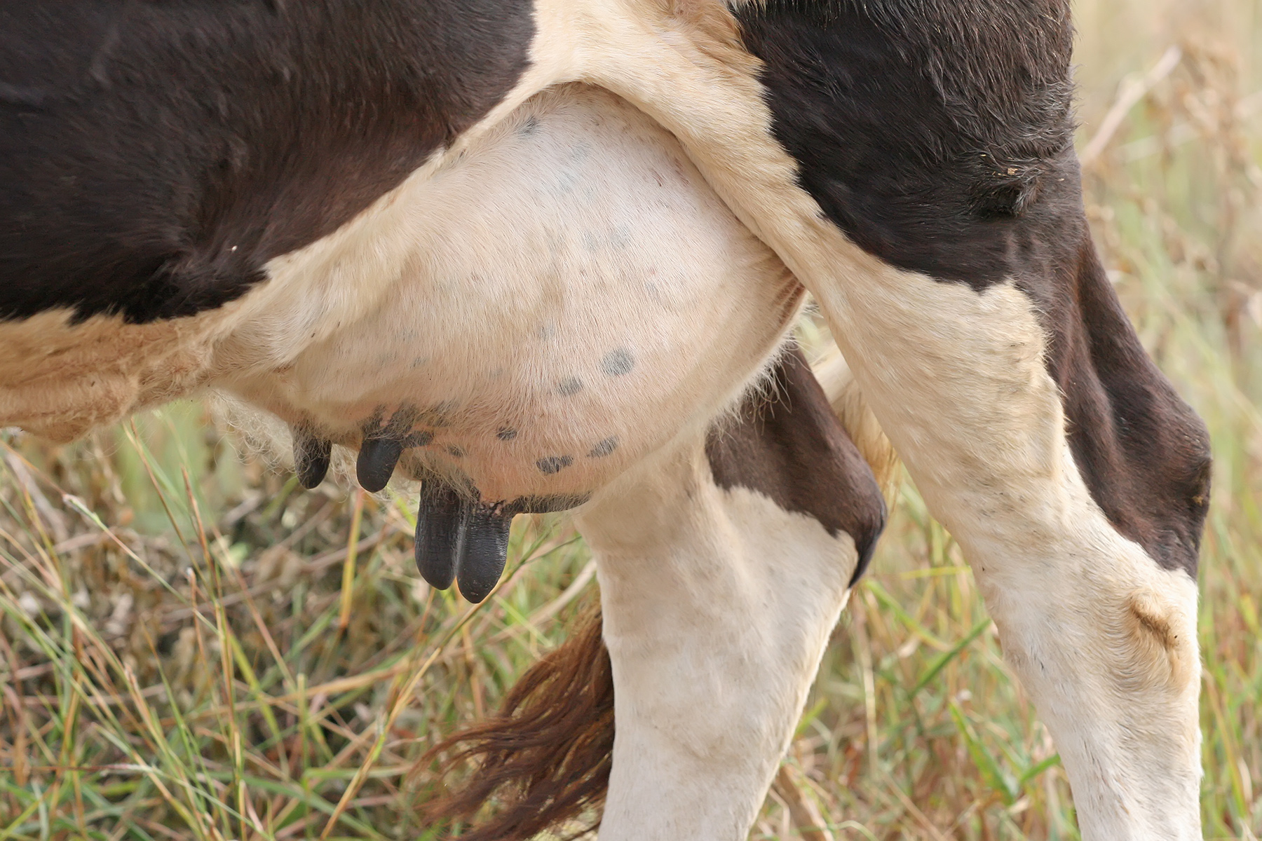
An udder is an organ formed of two or four mammary glands on the females of dairy animals and ruminants such as cattle, goats, and sheep.[1] An udder is equivalent to the breast in primates and other mammals. The udder is a single mass hanging beneath the animal, consisting of pairs of mammary glands with protruding teats. In cattle, camels and deer, there are normally two pairs, in sheep and goats, there is one pair, and in some animals, there are many pairs. In animals with udders, the mammary glands develop on the milk line near the groin. Mammary glands that develop on the chest (such as in primates and elephants) are generally referred to as breasts.[1]

Udder care and hygiene in cows is important in milking, aiding uninterrupted and untainted milk production, and preventing mastitis. Products exist to soothe the chapped skin of the udder. This helps prevent bacterial infection, and reduces irritation during milking by the cups, and so the cow is less likely to kick the cups off. It has been demonstrated that incorporating nutritional supplements into diet, including vitamin E, is an additional method of improving udder health and reducing infection.[2]

Etymology

[edit]

Udder has been attested in Middle English as udder or uddyr (also as uther, iddyr), and in Old English as ūder.[3] It was evolved from the Proto-Germanic reconstructed root *eudrą or *ūdrą, which in turn descended from Proto-Indo-European *h₁ówHdʰr̥ (“udder”). It is cognate with Saterland Frisian Jadder (“udder”), Dutch uier (“udder”), German Euter (“udder”), Swedish juver (“udder”), Icelandic júgur (“udder”), Vedic Sanskrit ऊधर् (ū́dhar), Ancient Greek οὖθαρ (oûthar), and Latin ūber.[4]

Culinary Use

[edit]

The udder, or elder in Ireland, Scotland and northern England, of a slaughtered cow was in times past prepared and consumed.[5] In other countries, like Italy, parts of Pakistan, Kenya, and some South American countries, cow udder is still consumed in dishes like the traditional teteun [it] and ubres asada.

References

[edit]
[edit]
Revisions and contributorsEdit on WikipediaRead on Wikipedia
from Grokipedia
An udder is the of female ruminants, such as , sheep, goats, and certain other mammals, consisting of multiple simple glands united into a single pendulous structure that produces and stores to nourish . In domestic , particularly cows, it typically comprises four independent quarters—two fore and two hind—each with its own vascular supply, nerve innervation, and , suspended by medial and lateral ligaments in the inguinal region. The udder's anatomy features microscopic alveoli lined with epithelial cells that synthesize from nutrients in the bloodstream, requiring approximately 500 liters of blood to produce 1 liter of . is then transported through a network of ducts to larger within each quarter for storage, before flowing into the teat and exiting via the streak , which is protected by a and lining to prevent leakage and bacterial entry. Milk ejection, or let-down, is hormonally regulated by oxytocin released in response to stimuli like calf suckling or machine milking, ensuring efficient delivery. Udder development begins in the fetal stage and continues through and pregnancies, with and enlargement enhancing milk-producing capacity over multiple lactations. In the industry, the udder is central to production, influencing yield, composition (such as and protein content varying by and diet), and overall farm profitability. However, it is highly susceptible to infections like , an inflammation often caused by bacterial entry through the canal, which leads to reduced milk output, treatment costs, discarded , and potential , making it one of the most economically damaging diseases in operations.

Etymology

Linguistic Origins

The word "udder" derives from Old English ūder, referring to the milk-secreting organ of animals, which itself stems from Proto-Germanic \ūdarą or \udr-, denoting the same glandular structure. This Proto-Germanic form traces back to the Proto-Indo-European root \h₁ówHdʰr̥ (or variant \eue-dh-r), an ancient term for "udder" that emphasized its role in milk production. This Indo-European root exhibits clear connections across ancient languages, linking to Sanskrit ū́dhar for "udder," Latin uber meaning "udder" or more broadly "fertile" and "abundant," and Greek outhár (οὖθαρ) for the mammary gland of animals. Cognates appear in other Germanic tongues, such as Old High German ūtar for "udder," reflecting shared linguistic heritage in describing bovine or ovine anatomy. By , the term evolved into forms like udder or uddyr (sometimes spelled uther or iddyr), where it solidified as a specific descriptor for the pendulous mammary glands of quadruped mammals, distinguishing it from . This period marked a stabilization in its usage within English agricultural and zoological contexts, preserving the core Proto-Indo-European sense of milk-bearing fertility.

Historical Terminology

The term "udder" first appears in Old English texts dating to around the 9th century, derived from Proto-Germanic *udr-, and was commonly used in agricultural contexts to describe the mammary glands of such as cows and . Early examples include references in Anglo-Saxon farming glossaries and charters, where it denoted the milk-producing organ in descriptions of herd management and . Regional variations of the term persisted into later centuries, particularly in dialects of Irish, Scottish, and northern English communities, where "elder" or "eldher" served as a for udder, often in culinary contexts referring to the organ as an edible item. This usage, borrowed from elder via influences, appears in 19th-century folk recipes and household accounts from and , where boiled or stewed elder (cow's udder) was valued as a cheap protein source for working-class diets, sometimes prepared with onions and herbs to mask its texture. By the mid-19th century, such dialectal terms began fading in printed cookery books as efforts in English promoted "udder" exclusively. Texts from the late 18th and 19th centuries, such as treatises on cattle breeding, emphasized udder conformation for yield optimization, using precise in works like those by the Royal Agricultural Society. This evolution aligned with mechanized innovations and improvement programs, solidifying "udder" as the dominant term in professional discourse. Notable historical mentions of udder appear in medieval European farming ordinances and rules, where it was classified as valuable for kitchen use. Such regulations underscored udder's role in sustaining large households through efficient use of remains.

Anatomy

General Structure

The udder, also known as the complex, is a pendulous organ located in the inguinal of the ventral in mammals, primarily functioning to produce and store for nourishing offspring. It consists of specialized s that develop during and expand significantly during . In ruminants, the udder typically features two or four glands; for instance, cows possess a quadrupartite udder with four distinct quarters (right fore, right hind, left fore, and left hind), while goats have a bipartite structure with two halves. The udder's key components include glandular tissue, ductal systems, connective tissues, and vascular and neural elements. Glandular tissue comprises millions of alveoli—microscopic, spherical sacs lined with secretory epithelial cells surrounded by myoepithelial cells and capillaries—where milk synthesis occurs through the uptake of nutrients from blood. Milk then flows through a branching network of ducts into larger storage areas, including the gland cistern (a central reservoir within each quarter) and the teat cistern (a smaller cavity at the base of the teat), before exiting via the teat canal. Connective tissues, such as the elastic medial suspensory ligament (dividing the udder into right and left halves) and the less elastic lateral suspensory ligaments (attaching to the abdominal wall), provide structural support to prevent sagging. The vascular supply is extensive, with arteries like the external pudic delivering blood for milk production (approximately 400–500 liters per liter of milk synthesized), and veins including the prominent subcutaneous abdominal vein (also called the milk vein) facilitating drainage along the udder's ventral surface. Innervation involves sensory nerves in the skin and teats, as well as autonomic fibers regulating glandular function. Internally, the udder is organized into independent quarters in like cows, separated externally by shallow furrows and internally by extensions of the suspensory ligaments, ensuring compartmentalization that limits spread between sections. Each quarter contains its own alveolar clusters, ductal pathways, and cisternal cavity, where 20–40% of is stored prior to or , with the remainder held in the alveoli and smaller ducts. This division allows for efficient, quarter-specific milk accumulation and release. Externally, the udder is enveloped in a thin, elastic skin that stretches during , often displaying visible vascular patterns such as the branching subcutaneous abdominal running anteriorly from the udder base. It terminates in 2–4 teats, each equipped with a streak lined by and guarded by a muscle and Fürstenberg's rosette (a series of folds at the canal's proximal end) to prevent bacterial entry. Teats vary in length and shape but generally feature a single orifice per quarter, with the skin around them highly sensitive to touch.

Variations by Species

In bovines, such as cattle, the udder consists of four distinct quarters, each served by a single teat arranged in a pendulous structure suspended between the hind legs. This configuration is supported by prominent suspensory ligaments, including medial and lateral elements composed of elastic and connective tissues that prevent sagging under the weight of the lactating gland, which can weigh around 50 kg (including milk and blood) in high-producing dairy cows. Dairy breeds exhibit larger glandular mass and overall udder size compared to beef breeds, reflecting selective breeding for enhanced milk yield. Caprine () and ovine (sheep) udders are bipartite, comprising two separate mammary glands located in the inguinal region, each with a single , resulting in a more compact and elongated overall structure than in bovines. Unlike bovines, where approximately 20-30% of is stored in the cisterns, caprine and ovine udders feature proportionally larger cisterns that hold 50-70% of total volume, facilitating efficient storage and release adapted to smaller body sizes and practices. The porcine udder is characterized by 12-14 teats arranged bilaterally along the ventral in a non-pendulous, linear fashion, allowing simultaneous by large litters of 8-12 piglets typical of the . This arrangement lacks the centralized suspension seen in ruminants, with teats embedded directly in the abdominal for accessibility to multiple offspring. In other mammals, such as equines (), the udder is bipartite with two s positioned between the hind legs, each teat typically draining two independent mammary glands for a total of four glands, supporting the needs of a single . Wild ruminants like deer exhibit a similar bipartite udder with two teats, reduced in size and glandular development compared to domesticated counterparts to suit smaller body mass and seasonal for fawns.

Physiology

Milk Production Mechanism

Milk production in the udder occurs primarily within the alveolar epithelial cells, also known as mammary epithelial cells, which line the alveoli of the mammary gland and serve as the main site for synthesizing and secreting milk components. These cells actively uptake essential nutrients from the bloodstream, including glucose via transporters such as GLUT1 and GLUT12, and amino acids through bidirectional transport across their plasma membranes to support the biosynthesis of milk constituents. Glucose is sequestered within these cells primarily through phosphorylation by hexokinase or incorporation into lactose, while amino acids are utilized for protein synthesis, with net uptake reflecting the balance of mammary gland demands. The biochemical pathways for milk synthesis in these alveolar cells involve the coordinated production of lactose, proteins, and lipids, which together determine milk composition and volume. Lactose, synthesized from glucose and UDP-galactose by the enzyme lactose synthase in a reaction unique to mammary alveolar cells, acts as the primary osmotic driver, drawing water into the alveolar lumen to form the bulk of milk volume. Milk proteins, including caseins and whey proteins such as α-lactalbumin, are assembled from amino acids within the endoplasmic reticulum and Golgi apparatus of the epithelial cells, with casein micelles forming key structural elements. Fat synthesis occurs through de novo lipogenesis, utilizing glucose-derived NADPH generated via the pentose phosphate pathway to support fatty acid chain elongation and triglyceride assembly in the epithelial cells. These processes are hormonally triggered, primarily by prolactin, to initiate and sustain synthesis during lactation. Efficient nutrient delivery to the alveolar epithelial cells relies on substantial mammary blood flow, which in lactating cows can account for 15-16% of , facilitating the transport of precursors like glucose and while also dissipating the heat generated during exothermic synthetic reactions. This high rate ensures that the receives a disproportionate share of systemic resources, with blood flow adjusting to match metabolic demands for sustained output. In the immediate , the alveolar epithelial cells initially produce , a nutrient-dense rich in immunoglobulins—particularly IgG transferred from the bloodstream across the mammary —to provide to the neonate during the first few days after birth. This transitional transitions to mature milk as the synthetic pathways shift toward higher volumes of and other components.

Lactation Cycle

The lactation cycle in the udder of mammals, particularly cows, encompasses the period from parturition to the cessation of production, characterized by distinct phases that optimize nutrient delivery to while balancing . This cycle is tightly regulated to ensure efficient synthesis and ejection, with production peaking early and gradually declining to allow for reproductive recovery. In , understanding these phases is crucial for maximizing yield and . The cycle begins with the colostrum phase, lasting the first 3-5 days postpartum, during which the udder secretes —a nutrient-dense, antibody-rich fluid essential for newborn immunity. This is followed by the transition phase (days 4-14), where milk composition shifts toward mature , with decreasing immunoglobulin levels and increasing and content. Peak occurs in weeks 4-8, marking the highest milk yield as alveolar cells reach maximum secretory capacity, often producing up to 50-60 liters per day in high-yielding breeds. Late involves a progressive decline in production, starting around day 100 and continuing through the cycle's end, as secretory tissue efficiency wanes and energy is redirected toward body maintenance. Hormonal regulation drives these stages, with playing a central role in initiating and maintaining milk synthesis by stimulating alveolar epithelial cells during and after . Oxytocin facilitates milk let-down by contracting myoepithelial cells around alveoli, enabling ejection during suckling or milking. During , elevated progesterone and promote mammary gland proliferation and ductal development, suppressing until parturition, when their decline allows dominance. In dairy cows, the full lactation cycle typically spans 305 days, influenced by breed, , and management practices, after which a dry period of about 60 days follows to facilitate udder involution, tissue regeneration, and preparation for the next . This dry period is essential for restoring mammary health and preventing metabolic disorders, with durations shorter than 40 days or longer than 70 days linked to reduced subsequent yields. Milk yield throughout the cycle is modulated by , which determine peak potential (e.g., Holsteins averaging 10,000-12,000 kg annually), to support demands during peak phases, and , where thrice-daily can boost output by 10-15% compared to twice-daily. These factors interact to shape the lactation curve, emphasizing balanced management for sustained productivity.

Health and Diseases

Common Pathologies

While the following pathologies are detailed for , similar conditions such as affect sheep and goats, often caused by comparable bacterial pathogens and managed with species-adapted hygiene and treatment protocols. is one of the most prevalent and economically significant diseases affecting the udders of , characterized by of the tissue due to microbial invasion through the canal. Common causative agents include contagious bacteria such as , which spreads between cows during milking, and environmental pathogens like , often introduced via contaminated bedding or ends. It manifests in two main subtypes: clinical mastitis, which presents with visible systemic and local signs including udder swelling, heat, pain upon , and abnormal appearance such as watery secretion, clots, or flakes; and subclinical mastitis, which lacks overt symptoms but is detected through elevated counts in , potentially leading to reduced quality and yield without immediate detection. In severe cases, clinical mastitis can cause fever, depression, and loss of appetite in affected animals, with quarter-level rates varying by but often resulting in permanent damage if untreated. Udder , a noninfectious physiologic disorder, involves excessive accumulation of in the udder's tissues, primarily occurring in the periparturient period due to hormonal changes such as elevated and progesterone levels that increase . Predisposing factors include advanced age at first calving, prolonged , , , high-salt diets prepartum, and limited exercise, which exacerbate retention. Symptoms typically include symmetrical, pitting of the udder and sometimes ventral starting shortly before or after calving, with tight, glossy skin that is usually painless unless complicated by secondary issues. If chronic, the condition can progress to , impairing udder support and milk let-down, and heightening susceptibility to by compromising end integrity. Teat injuries represent a frequent traumatic in herds, often resulting from mechanical damage during or environmental hazards, which compromises the udder's . Common causes include linear cuts or lacerations from wire, treads by herdmates, or abrasions against rough housing surfaces; cracks and fissures at the end from faulty milking machines with excessive vacuum or poor pulsation; and in cold climates leading to necrotic tissue. Symptoms vary by severity but often feature localized swelling, hemorrhage (appearing as petechial spots or hematomas), open sores with serous discharge, and pain that hinders normal movement or . These injuries significantly elevate risk by providing entry points for , potentially escalating to or formation if the canal is breached. Other notable udder pathologies include abscesses, tumors, and , each with distinct etiologies and presentations. Abscesses typically arise secondary to wounds, chronic , or infected hematomas, forming encapsulated pockets between the udder's skin and without direct involvement of milk-producing . They present as firm, fluctuating swellings with possible foul-smelling drainage upon rupture, diagnosed via needle aspiration revealing purulent material. Mammary tumors in are rare compared to other species, most commonly manifesting as benign fibropapillomas on teats induced by type 1 , appearing as wart-like growths that may bleed or ulcerate but rarely metastasize. Udder , or congenital underdevelopment of the , can result from genetic conditions such as ovarian —a recessive autosomal trait in breeds like Swedish Mountain —leading to a small, firm udder with tiny teats in heifers, often accompanied by due to associated reproductive tract anomalies like a narrow .

Prevention and Management

Maintaining udder health in cows relies heavily on rigorous practices to minimize bacterial exposure and infection risk. Post-milking dipping with antiseptics, such as solutions, forms a protective barrier on the end, significantly reducing counts (SCC) and the incidence of environmental pathogens by up to 50% in controlled studies. Dry cow therapy, involving intramammary infusion of long-acting s at the end of , eliminates existing subclinical infections and prevents new ones during the dry period, with selective application based on SCC thresholds preserving efficacy while curbing use. Additionally, providing clean, dry materials like or recycled solids lowers bacterial loads on ends, as contaminated can increase environmental exposure during the 12-14 hours cows spend lying down daily. Effective milking management is essential to prevent physical trauma and physiological stress to the udder. Optimizing settings, including levels (typically 40-42 kPa) and pulsation ratios (60:40), minimizes teat congestion and liner slip, thereby reducing the risk of trauma-induced and bacterial ingress. Frequent foremilk stripping—removing the first 2-3 streams of before attachment—stimulates oxytocin release for complete let-down, prevents udder engorgement, and allows early detection of abnormalities, contributing to lower rates when integrated into routine protocols. Overmilking should be limited to under one minute per cow to avoid teat-end , with proper cluster alignment ensuring even flow and reduced udder . Treatments for udder conditions emphasize targeted interventions to restore function swiftly. For clinical mastitis, intramammary infusions of antibiotics like cephapirin or pirlimycin are administered directly into affected quarters, achieving cure rates of 70-90% in mild cases when initiated early; however, rising antimicrobial resistance in pathogens such as Staphylococcus aureus (with penicillin resistance up to 21.8% as of 2025) underscores the need for bacterial culture and sensitivity testing to guide therapy and promote stewardship. Udder edema, characterized by fluid accumulation in periparturient cows, responds well to non-steroidal anti-inflammatory drugs (NSAIDs) such as meloxicam (0.5 mg/kg subcutaneously), which reduce swelling and pain by inhibiting prostaglandin synthesis, often resolving symptoms within 48 hours. Surgical interventions for abscesses involve lancing and drainage under local anesthesia, followed by flushing with antiseptics and systemic antibiotics, to prevent chronic suppuration and maintain milk production in the unaffected quarters. Monitoring udder health through diagnostic tools enables proactive management of subclinical issues. testing, performed via California Mastitis Test or automated analyzers on bulk tank , identifies subclinical when SCC exceeds 200,000 cells/mL, guiding selective dry cow and reducing herd-level infection prevalence by 20-30%. imaging, using 7.5-12 MHz linear transducers, visualizes structural abnormalities like parenchymal or cistern dilation noninvasively, aiding in the assessment of chronic conditions and treatment efficacy without disrupting . Regular integration of these tools into farm protocols supports sustained udder integrity and milk quality.

Uses

Culinary Applications

Udder, classified as , requires extensive preparation to make it edible due to its tough, fibrous tissue composed largely of mammary glands and connective elements. The most common methods involve prolonged or stewing, often for several hours, to tenderize the material and render it palatable; it is frequently incorporated into soups, stews, or pies as a filler or secondary ingredient. Thorough cleaning is essential prior to cooking to eliminate any residual , glandular secretions, or contaminants, preventing off-flavors and potential health risks. In various European culinary traditions, udder holds cultural significance as an economical protein source, particularly among working-class communities. Known as "elder" in Scottish, Irish, and northern , it was historically boiled and then fried or minced into variants of and other dishes. In the , particularly , uierboord (cow's udder) is a traditional dish where the organ is meticulously cleaned, braised with simple seasonings like salt and pepper, and sliced thinly for sandwiches, reflecting its roots as affordable fare for laborers before declining in popularity with the rise of cheaper meats. Across parts of Asia, including , boiled or grilled cow udder appears in street foods like sate jando, prized for its chewy texture when paired with and spices. Historically, udder consumption dates back to , where pig udders were valued in markets as premium ; the cookbook attributed to includes recipes for preparing sterile sow's vulva and udder, often boiled, stuffed, or chopped with seasonings for elite banquets. In medieval Europe, it served as sustenance for lower classes, though specific allocations in ordinances are sparsely documented. Nutritionally, cooked cow udder provides a dense source of protein (approximately 13.4 g per 100 g) and fat (15.1 g per 100 g), contributing to its role as a filling, calorie-rich in resource-scarce settings, though its high content limits digestibility without proper cooking.

Other Applications

Beyond culinary contexts, udders have found niche applications in , particularly in historical tanning practices. In ancient and early modern periods, portions of animal skins, including udder tissue, were occasionally tanned to produce small goods such as feeding teats for infants, fashioned from tanned udder-nipples or similar soft hides to mimic natural nursing. These items leveraged the udder's pliable texture for functional, albeit rudimentary, child-rearing tools, though such uses declined with the advent of synthetic alternatives. In medical and veterinary fields, preserved cow udders serve as essential educational models for teaching mammary and . Detailed anatomical replicas or preserved specimens, such as the SOMSO Udder of the Cow model, allow students to dissect and visualize the udder's internal structures, including glandular regions, ducts, arteries, veins, and lymphatic vessels, facilitating hands-on learning in veterinary programs. Similarly, functional simulators like the Bovine Milking Udder Simulator replicate udder care procedures, enabling training in detection, infection prevention, and proper techniques without relying on live animals. Udders also provide colostrum, the nutrient-rich first milk, which is harvested as a vital supplement for newborn calves lacking sufficient maternal intake. Bovine colostrum contains high levels of immunoglobulins (IgG), vitamins A, D, and E, fats, and growth factors, delivering 100–150 g of IgG per dose in replacer forms to bolster and reduce mortality rates in operations. This application supports calf health in , where timely colostrum administration—ideally 200–300 g of IgG within the first 24 hours—enhances survival and long-term productivity. Historically, udders played a pivotal role in the economic foundations of dairy farming in 18th-century Europe, where selective breeding programs targeted udder size and conformation to boost milk yields. Pioneered in regions like the Netherlands and Britain, these efforts, beginning around the early 1700s, emphasized traits such as udder capacity and attachment to improve lactation efficiency, transforming cattle from dual-purpose animals into specialized milk producers amid the Agricultural Revolution. By the late 18th century, breeds like the Holstein-Friesian emerged from such practices, with udder morphology becoming a key selection criterion for economic viability in emerging dairy economies. In folklore across various cultures, the udder symbolizes fertility and abundance, often linked to bovine deities representing nourishment and prosperity. In Irish traditions, rituals involving the cow's udder—such as burning hair from it with a blessed candle post-calving—invoked protection and bountiful milk production, underscoring the udder's emblematic role in agrarian fertility rites. Similarly, in Indo-Iranian mythology, dairy cows and their udders embody rain clouds and life's generative forces, with the udder signifying maternal wealth and cyclical renewal in ancient narratives. Contemporary leverages udder tissue for advanced research, particularly in isolation and . Bovine mammary s (MaSCs), derived from udder biopsies or milk-exfoliated epithelial cells, demonstrate regenerative potential, with studies showing their secretome promotes tissue repair and antimicrobial defense in models of treatment. These cells express markers like and CD49f, enabling expansion for therapeutic applications, such as injecting mesenchymal s to regenerate damaged udder tissue and reduce inflammation in cows. In , udder-derived cells facilitate the production of recombinant proteins; for instance, transgenic approaches have engineered cows to secrete insulin in via modifications, addressing treatment shortages. Additionally, biotech firms engineer with bovine udder genes to synthesize caseins and proteins, creating sustainable, animal-free alternatives that replicate nutritional profiles.

References

Add your contribution
Related Hubs
User Avatar
No comments yet.