

Community hub
Recent from talks
Contribute something
Nothing was collected or created yet.
Depressor anguli oris muscle
View on WikipediaThis article includes a list of references, related reading, or external links, but its sources remain unclear because it lacks inline citations. (May 2015) |
| Depressor anguli oris | |
|---|---|
 Scheme showing arrangement of fibers of Orbicularis oris (triangularis labeled at bottom right). | |
 Muscles of the head, face, and neck (labeled as triangularis near chin). | |
| Details | |
| Origin | Tubercle of mandible |
| Insertion | Modiolus of mouth |
| Artery | Facial artery |
| Nerve | Marginal mandibular branch of the facial nerve |
| Actions | Depresses angle of mouth |
| Identifiers | |
| Latin | musculus depressor anguli oris |
| TA98 | A04.1.03.026 |
| TA2 | 2076 |
| FMA | 46828 |
| Anatomical terms of muscle | |
The depressor anguli oris muscle (triangularis muscle) is a facial muscle. It originates from the mandible and inserts into the angle of the mouth. It is associated with frowning, as it depresses the corner of the mouth.
Structure
[edit]The depressor anguli oris arises from the lateral surface of the mandible.[1] Its fibers then converge. It is inserted by a narrow fasciculus into the angle of the mouth.[1] At its origin, it is continuous with the platysma muscle, and at its insertion with the orbicularis oris muscle and risorius muscle. Some of its fibers are directly continuous with those of the levator anguli oris muscle, and others are occasionally found crossing from the muscle of one side to that of the other; these latter fibers constitute the transverse muscle of the chin.
The depressor anguli oris muscle receives its blood supply from a branch of the facial artery.
Nerve supply
[edit]The depressor anguli oris muscle is supplied by the marginal mandibular branch of the facial nerve.[1]
Function
[edit]The depressor anguli oris muscle is a muscle of facial expression.[1] It depresses the corner of the mouth, which is associated with frowning.[1]
Clinical significance
[edit]Paralysis
[edit]Damage to the marginal mandibular branch of the facial nerve may cause paralysis of the depressor anguli oris muscle.[1] This may contribute to an asymmetrical smile.[1] This may be corrected by resecting (cutting and removing) the depressor labii inferioris muscle, which has a more significant impact on smiling.[1]
Hypoplasia/aplasia
[edit]Underdevelopment (hypoplasia) or complete absence (aplasia) of the depressor anguli oris can occur.[2] Similarly to paralysis, individuals with these conditions will have an asymmetric smile.[medical citation needed] These conditions are rare, and develop at or before birth (congenitally).
See also
[edit]Additional images
[edit]This gallery of anatomic features needs cleanup to abide by the medical manual of style. |
-
 Position of depressor anguli oris muscle
 -
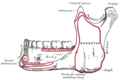 Mandible, outer surface, side view
 -
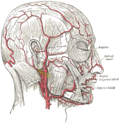 The arteries of the face and scalp


References
[edit]![]() This article incorporates text in the public domain from page 383 of the 20th edition of Gray's Anatomy (1918)
This article incorporates text in the public domain from page 383 of the 20th edition of Gray's Anatomy (1918)
- ^ a b c d e f g h Hussain, G.; Manktelow, R.T; Tomat, L.R (2004-09-01). "Depressor labii inferioris resection: an effective treatment for marginal mandibular nerve paralysis". British Journal of Plastic Surgery. 57 (6): 502–510. doi:10.1016/j.bjps.2004.04.003. ISSN 0007-1226. PMID 15308395.
- ^ "Hypoplasia: Where it can occur, causes, effects, and treatment options". 13 November 2020.