Hubbry Logo
Alpha granuleAlpha granuleMain
Open search
Alpha granule
Community hub
Alpha granule
logo
8 pages, 0 posts
0 subscribers
Be the first to start a discussion here.
Alpha granule
Alpha granule
from Wikipedia
Alpha granule
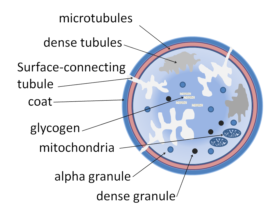
Alpha granules shown in a platelet
Details
Part ofPlatelets
Identifiers
Latingranulum alpha
THH2.00.04.1.03005
Anatomical terminology

Alpha granules, (α-granules) also known as platelet alpha-granules are a cellular component of platelets. Platelets contain different types of granules that perform different functions, and include alpha granules, dense granules, and lysosomes.[1] Of these, alpha granules are the most common,[1] making up 50% to 80% of the secretory granules.[2] Alpha granules contain several growth factors.[3]

Contents

[edit]

Contents include insulin-like growth factor 1, platelet-derived growth factors, TGF beta, platelet factor 4 (which is a heparin-binding chemokine) and other clotting proteins (such as thrombospondin, fibronectin, factor V,[4] and von Willebrand factor).[5]

The alpha granules express the adhesion molecule P-selectin[6] and CD63.[7] These are transferred to the membrane after synthesis.

The other type of granules within platelets are called dense granules.

Clinical significance

[edit]

A deficiency of alpha granules is known as gray platelet syndrome.

See also

[edit]

References

[edit]
Revisions and contributorsEdit on WikipediaRead on Wikipedia
from Grokipedia
Alpha granules are the most abundant secretory organelles within blood platelets (thrombocytes), typically numbering 50–80 per platelet and comprising approximately 10% of the platelet's volume, with diameters ranging from 200 to 500 nm. These membrane-bound structures store a diverse array of over 300 bioactive proteins, including clotting factors, molecules, growth factors, and , which are selectively released upon platelet activation to support critical physiological processes such as , , , and . Unlike other platelet granules, alpha granules exhibit heterogeneous internal organization, featuring distinct zones such as an electron-dense rich in and proteoglycans, a less dense matrix containing fibrinogen, and an electron-lucent area with tubular structures of (vWF). The contents of alpha granules are categorized into membrane-bound proteins, which integrate into the platelet surface upon release, and soluble proteins secreted into the . Key membrane proteins include P-selectin, which mediates leukocyte recruitment during inflammation, and such as αIIbβ3 that facilitate platelet aggregation. Soluble cargo encompasses hemostatic factors like fibrinogen, vWF, and coagulation factors V and XI; such as (CXCL4) and beta-thromboglobulin (CXCL7); and growth factors including (PDGF), (VEGF), and transforming growth factor-beta (TGF-β). These proteins are either synthesized endogenously by megakaryocytes (the platelet precursors) or endocytosed from plasma, enabling platelets to respond dynamically to vascular injury. Biogenesis of alpha granules occurs primarily in megakaryocytes through a complex pathway involving the trans-Golgi network, endosomal sorting, and multivesicular bodies, with maturation and cargo packaging facilitated by proteins like NBEAL2, VPS33B, and VPS16B. The granules are then trafficked along proplatelet extensions into nascent platelets, where they maintain an acidic lumen ( ≈5.2) to preserve protein stability. Upon platelet by agonists like or , alpha granules fuse with the platelet's plasma membrane or open canalicular system via SNARE-mediated , enabling graded release of contents tailored to the stimulus intensity. Functionally, alpha granules are pivotal in by delivering vWF and fibrinogen to promote platelet and clot formation at sites of vascular damage. Beyond , their secreted factors contribute to broader roles: CXCL4 and P-selectin drive inflammatory responses by attracting neutrophils and monocytes; VEGF and PDGF support and tissue repair; and like thrombocidins provide innate immune defense. Dysfunctions in alpha granule formation or release, such as those in due to NBEAL2 mutations, lead to bleeding disorders and highlight their indispensability in vascular biology.

Structure and Composition

Morphology

Alpha granules in platelets are typically ovoid or discoid organelles, ranging from 200 to 500 nm in diameter, enclosed by a single limiting , and characterized by an internal matrix that appears less electron-dense under , often surrounding a more electron-dense core or in conventional preparations. In cryosections or freeze-substituted samples, the matrix exhibits a more uniform, homogeneous appearance without prominent nucleoids, suggesting that dense cores may arise as fixation artifacts in some imaging protocols. These structures also display internal heterogeneity, such as lightly stained inclusions resembling microvesicles or exosomes, and occasional eccentric arrangements of cargo like tubules. Morphological variations exist across species; in platelets, alpha granules are generally round to ovoid and morphologically uniform, whereas in mice, they adopt a more variable, elongated rod-like shape with a consistent minor axis but differing major axis lengths. Subpopulations of alpha granules include tubular subtypes, which constitute approximately 4% of the total in resting platelets and feature extended, pipe-like projections that are detectable in about half of resting platelets but increase markedly upon . These tubular extensions, measuring 80-100 nm in width and up to 1 μm in length, often associate with the platelet's open canalicular system or plasma membrane. Visualization of alpha granule morphology relies on advanced imaging techniques, including (TEM) for basic , immunogold labeling to localize specific components within the matrix or core, and cryo-electron microscopy (cryo-EM) to preserve native states and reveal uniform matrices. Three-dimensional methods such as (STEM) tomography and serial block-face scanning electron microscopy provide insights into spatial arrangements, confirming the presence of tubular extensions and membrane interactions while quantifying volumes (approximately 0.01-0.04 μm³ per granule). Human platelets contain an average of 40 to 80 , though numbers can vary with platelet size and activation state, where granules may fuse or decondense without altering the total count. In mice, the count is lower, averaging around 19 per platelet.

Cargo Proteins

Alpha granules in platelets serve as storage organelles for a diverse array of cargo proteins essential for , , and tissue repair. These proteins are broadly classified into several categories based on their function and structure, including adhesive proteins, factors, growth factors, , and membrane-associated proteins. Adhesive proteins such as fibrinogen, (vWF), and facilitate platelet and aggregation at sites of vascular injury. factors like factor V, , factor XIII, and prothrombin support the clotting cascade upon release. Growth factors including (PDGF), (VEGF), basic fibroblast growth factor (bFGF), (EGF), hepatocyte growth factor (HGF), and (IGF) promote and . such as (PF4 or CXCL4), β-thromboglobulin (β-TG or CXCL7), (CCL5), and stromal cell-derived factor-1 () modulate leukocyte recruitment and vascular tone. Membrane proteins, notably P-selectin and like αIIbβ3 and GPIb-IX-V, are integral to the granule membrane and translocate to the platelet surface upon to mediate interactions with other cells. The origins of these cargo proteins are heterogeneous, with some synthesized endogenously in and others acquired via from plasma. Endogenously produced proteins include P-selectin, PF4, PDGF, vWF, factor V, and fibrinogen, which are generated during megakaryocyte differentiation and packaged directly into forming granules. In contrast, proteins such as additional fibrinogen (via αIIbβ3 integrin-mediated uptake), factor V, immunoglobulins, , and are endocytosed from circulating plasma, allowing platelets to incorporate extracellular components during their lifespan. This dual sourcing contributes to the functional versatility of alpha granules. Alpha granules exhibit significant heterogeneity, with distinct subpopulations containing specialized cargo subsets that enable differential release tailored to specific stimuli. For instance, procoagulant granules enriched in either fibrinogen or vWF are segregated from those harboring proinflammatory or pro-angiogenic factors like and VEGF. Other subpopulations include anti-angiogenic granules storing endostatin, separate from VEGF-containing ones, as well as granules with thrombospondin-1 or in unique compartments. This compartmentalization is evident in mature platelets, where imaging reveals non-overlapping distributions of proteins like fibrinogen and vWF. Quantitative estimates highlight the scale of cargo storage, with human platelets typically containing 50–80 alpha granules, each measuring 200–500 nm in diameter. Representative protein amounts include approximately 10 μg of fibrinogen and 20 μg of PF4 per 10^9 platelets, underscoring the granules' capacity to store bioactive molecules at concentrations sufficient for rapid deployment during activation.

Biogenesis

Formation in Megakaryocytes

Alpha granule formation begins during the early stages of differentiation in pro-megakaryoblasts, where initial protein synthesis and occur in the and Golgi apparatus. As megakaryocytes undergo polyploidization through endomitosis, driven by thrombopoietin, the number and size of alpha granules expand progressively, coinciding with increased cytoplasmic volume and production to support platelet biogenesis. This expansion phase, lasting approximately 5 days in humans, ensures sufficient granule reserves for the high output of platelets, with formation completing in mature megakaryocytes prior to . The organelles derive primarily from the trans-Golgi network via clathrin-coated vesicles, with contributions from the for nascent protein synthesis and multivesicular bodies as maturation intermediates that fuse to form electron-dense granules. Maturation of these intermediates involves fusion events facilitated by the HOPS complex components VPS33B and VPS16B, which regulate SNARE-mediated trafficking to prevent degradation. Synthetic is trafficked from the through the Golgi, while endocytic elements incorporate plasma membrane-derived components into early endosomes before convergence in multivesicular bodies. During , alpha granules are selectively inherited by nascent platelets through proplatelet formation, a process where mature extend long, branching proplatelets into sinusoids. Granules are transported along tracks at rates of 0.1–2 μm/min from the megakaryocyte cell body to proplatelet tips, ensuring even distribution to the 50–80 granules per platelet, with final platelet release occurring via shear-induced fission. Transcription factors GATA-1 and NF-E2 are essential regulators of this assembly, with GATA-1 promoting maturation and expression of genes like NBEAL2 that support granule retention and biogenesis. NF-E2, acting downstream or independently, drives late-stage differentiation and proplatelet extension necessary for granule packaging into platelets. These factors ensure coordinated initiation of alpha granule development, integrating it with overall megakaryopoiesis.

Cargo Sorting and Trafficking

Cargo sorting into alpha granules in megakaryocytes relies on specific motifs within protein sequences that direct selective packaging. Dileucine-based motifs, typically consisting of paired leucine residues flanked by acidic amino acids (e.g., [DE]XXXL[LI]), and tyrosine-based signals conforming to the YXXØ consensus (where Y is tyrosine, X is any amino acid, and Ø is a bulky hydrophobic residue) serve as primary recognition elements for cargo selection. These motifs interact with adaptor protein complexes, particularly AP-3, which binds to the cytosolic tails of transmembrane cargo proteins to facilitate their incorporation into transport vesicles. AP-3 plays a pivotal role in this process by recognizing these signals at endosomal compartments, ensuring efficient sorting of integral membrane proteins destined for alpha granules as lysosome-related organelles. Trafficking of alpha granule cargo occurs via two main routes originating in megakaryocytes. The direct biosynthetic pathway begins at the trans-Golgi network (TGN), where newly synthesized proteins are packaged into clathrin-coated vesicles mediated by AP-1 and AP-3 adaptors, followed by delivery to early endosomes for further maturation. In parallel, the endocytic recycling pathway captures extracellular proteins (e.g., fibrinogen) from the plasma membrane via AP-2-dependent , routing them through early endosomes marked by EEA1 into multivesicular bodies (MVBs) that evolve into nascent alpha granules. Both pathways converge at endosomal intermediates, where cargo is refined before fusion into mature granules, highlighting the central role of the endosomal system in alpha granule assembly. Several molecular players orchestrate these sorting and trafficking events. SNARE proteins, such as VAMP-7 (vesicle-associated membrane protein 7), mediate vesicle fusion during endosomal maturation and alpha granule formation, with VAMP-7 localizing to granule membranes to ensure proper cargo delivery. Rab GTPases, including Rab27a, regulate vesicular transport along these routes by coordinating motor protein interactions and docking at target membranes, thereby maintaining the specificity and efficiency of cargo movement to alpha granules. More recently, sorting nexin 24 (SNX24) has been identified as a critical effector in alpha-granule-specific trafficking; SNX24 associates with early endosomes and MVBs to promote cargo progression, and its depletion disrupts endosomal sorting, leading to reduced alpha granule numbers and impaired delivery of proteins like (VWF) and CD62P. Alpha granule heterogeneity arises through differential sorting mechanisms that segregate into subpopulations during trafficking. proteins like fibrinogen follow distinct endocytic itineraries involving VAMP-3-positive compartments, while growth factor-related cargos such as endostatin are directed via separate VAMP-8-enriched pathways, resulting in minimal (Pearson's coefficient ~0.21) and formation of specialized granules. This selective partitioning at early endosomes ensures functional diversity, with subpopulations retaining their distinct compositions through proplatelet extension and into mature platelets.

Function

Release Mechanisms

Platelet activation, initiated by soluble agonists such as , (ADP), and , triggers alpha granule release through binding to specific G protein-coupled and receptors. primarily acts via 1 (PAR1) and PAR4, while ADP engages P2Y1 and receptors, and binds glycoprotein VI (GPVI) to induce intracellular signaling cascades that culminate in granule . The fusion of alpha granules with the platelet plasma or the open canalicular system is mediated by the SNARE complex, comprising v-SNARE vesicle-associated 8 (VAMP-8) on the granule and t-SNAREs syntaxin-4 and SNAP-23 on the target . This core machinery is facilitated by Munc13-4, a that promotes SNARE assembly and vesicle priming in a calcium-dependent manner, ensuring efficient fusion upon elevation of intracellular Ca²⁺ levels triggered by activation signals. Alpha granule release exhibits distinct kinetics, with rapid translocation of membrane-bound cargos like P-selectin to the platelet surface occurring within seconds of to facilitate immediate leukocyte interactions, contrasted by slower secretion of soluble proteins that may extend over minutes. This temporal distinction arises from piecemeal patterns, where granules undergo partial content discharge rather than complete fusion, allowing graded responses to varying stimulus strengths. Regulation of alpha granule release involves both positive and negative modulators, with inhibitory signals such as elevated (cAMP) levels suppressing by interfering with calcium mobilization and SNARE function. Additionally, alpha granules exist as subpopulations with heterogeneous , leading to preferential release of specific proteins—such as pro-angiogenic factors from one versus anti-angiogenic from another—depending on the activating and its intensity.

Physiological Roles

Alpha granules play a central role in by releasing key factors that support platelet aggregation, fibrin formation, and clot stabilization. Upon platelet activation, they secrete fibrinogen, which binds to receptors on platelet surfaces, bridging adjacent platelets to form stable aggregates. They also release factor V, a critical cofactor in the coagulation cascade that enhances generation and promotes efficient clot formation. Additionally, alpha granule-derived factor XIII cross-links strands, reinforcing the clot structure and preventing premature dissolution to maintain vascular integrity. In and immunity, alpha granules contribute to leukocyte and defense through the release of and adhesion molecules. such as (PF4, or CXCL4) attract monocytes and neutrophils to sites of injury, facilitating immune cell infiltration and activation via pathways involving Src-kinase and Syk signaling. P-selectin, mobilized from alpha granule membranes to the platelet surface, mediates of leukocytes to by binding PSGL-1, thereby promoting rolling and firm attachment of immune cells. Certain alpha granule proteins, including thrombocidins, which are C-terminal fragments of CXC , exhibit activity against pathogens like , supporting innate immunity during infection. Alpha granules support and by delivering growth factors that stimulate tissue repair and vascular remodeling. (VEGF) released from these granules promotes endothelial , migration, and survival, driving the formation of new blood vessels essential for nutrient delivery to healing tissues. (PDGF), particularly isoforms PDGF-B and PDGF-C, recruits and cells to stabilize nascent vessels and enhances activity for deposition, accelerating formation. Recent studies (2018–2025) highlight emerging physiological roles of alpha granules in cancer and . In , alpha granule contents like P-selectin and PDGF facilitate tumor cell to and promote epithelial-mesenchymal transition, enabling cancer cell and at distant sites. In , released factors such as PF4 and PDGF contribute to plaque formation by recruiting monocytes into the vessel wall and stimulating cell proliferation, thereby promoting lesion development and stability.

Clinical Significance

Associated Disorders

Alpha granule abnormalities are implicated in various genetic and acquired disorders, leading to bleeding diatheses through defects in granule biogenesis, content degradation, or release mechanisms. (GPS) is an autosomal recessive bleeding disorder caused by biallelic mutations in the NBEAL2 gene, resulting in absent or markedly diminished alpha granules in platelets and megakaryocytes. These mutations disrupt alpha granule biogenesis by impairing cargo retention and packaging, leading to large, pale-appearing platelets on blood smears and mild to moderate mucocutaneous , often exacerbated by myelofibrosis due to ectopic release of growth factors from defective granules. Other inherited disorders feature alpha granule defects alongside broader granule pathologies. The Quebec platelet disorder, an autosomal dominant condition arising from tandem duplication of the PLAU gene, causes excessive urokinase-type accumulation in alpha granules, triggering intraplatelet generation and proteolytic degradation of multiple alpha granule proteins such as factor V, , and fibrinogen. This results in delayed postoperative or posttraumatic bleeding, ecchymoses, and , with platelets exhibiting normal morphology but reduced functional cargo. Hermansky-Pudlak syndrome (HPS), a group of autosomal recessive disorders due to mutations in s encoding biogenesis of lysosome-related organelles complexes (e.g., AP3B1 in HPS type 2), primarily impairs dense granule formation, with alpha granules generally normal in number but potential defective content release contributing to prolonged bleeding alongside oculocutaneous and ceroid lipofuscinosis. Acquired conditions can similarly disrupt alpha granule function, often through secondary effects on platelet production or activation. In myeloproliferative neoplasms such as , clonal megakaryocyte proliferation leads to abnormal alpha granule glycoprotein composition and dysregulated release, promoting both thrombotic and hemorrhagic risks despite elevated platelet counts. Diabetes mellitus is associated with platelet hyperreactivity and , which may impair overall platelet function including secretion, exacerbating prothrombotic states and vascular complications. Acquired storage pool deficiencies, observed in myelodysplastic syndromes or following , involve reduced alpha granule numbers or content, mimicking inherited forms and resulting in mild tendencies.

Diagnostic and Therapeutic Implications

Alpha granules in platelets serve as key diagnostic targets in evaluating platelet function disorders, particularly storage pool deficiencies. Electron remains the gold standard for visualizing alpha granule morphology and quantity, enabling the identification of conditions such as , where alpha granules are markedly reduced or absent. , by measuring surface expression of P-selectin (CD62P) following platelet activation, assesses alpha granule secretion and is recommended as a first-line for detecting defects in suspected inherited platelet disorders. Additionally, proteomic analyses using have mapped over 300 proteins within alpha granules, providing molecular signatures that aid in precise diagnosis of granule-related dysfunctions. Therapeutically, alpha granules are harnessed in (PRP) therapies, where activation releases growth factors like PDGF, TGF-β, and VEGF from alpha granules to promote tissue repair and regeneration. Clinical applications include accelerating healing, with studies showing improved healing rates in diabetic ulcers compared to standard care. In orthopedics, PRP injections alleviate symptoms in by modulating and cartilage repair, with meta-analyses indicating significant pain reduction and functional improvement at 6-12 months post-treatment. Emerging gene therapy approaches target alpha granules for controlled delivery of therapeutic proteins, utilizing lentiviral vectors with megakaryocyte-specific promoters to direct transgenes into granules during platelet biogenesis. For instance, fusing interferon-alpha to alpha granule sorting signals enables its storage and thrombin-triggered release, demonstrating antiviral efficacy while minimizing systemic exposure. Such strategies hold promise for hemophilia treatment by secreting clotting factors from activated platelets and for , where inhibiting alpha granule-derived P-selectin reduces tumor in preclinical models.

References

Add your contribution
Related Hubs
User Avatar
No comments yet.