Hubbry Logo
CathepsinCathepsinMain
Open search
Cathepsin
Community hub
Cathepsin
logo
8 pages, 0 posts
0 subscribers
Be the first to start a discussion here.
Cathepsin
Cathepsin
from Wikipedia
Cathepsin
Structure of Cathepsin K
Identifiers
SymbolCTP
PfamPF00112
Pfam clanCL0125
InterProIPR000668
SMARTPept_C1
PROSITEPDOC00126
MEROPSC1
SCOP21aec / SCOPe / SUPFAM
Available protein structures:
Pfam  structures / ECOD  
PDBRCSB PDB; PDBe; PDBj
PDBsumstructure summary

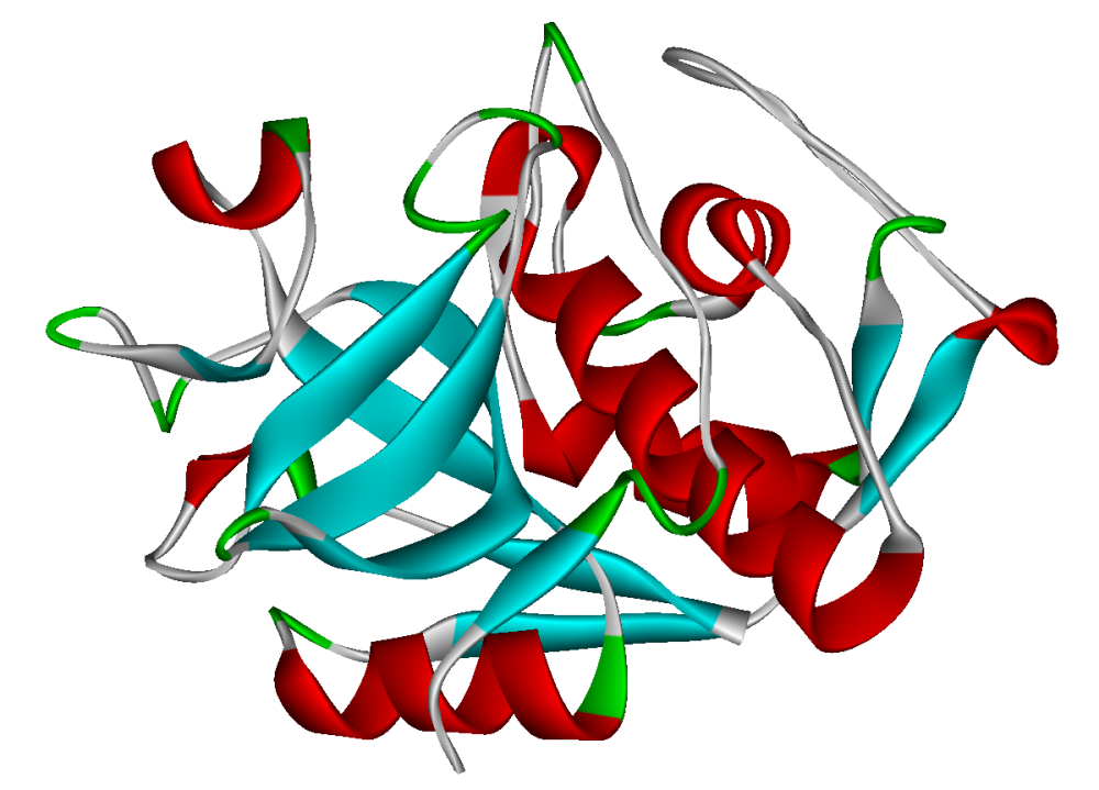
Cathepsins (Ancient Greek kata- "down" and hepsein "boil"; abbreviated CTS) are proteases (enzymes that degrade proteins) found in all animals as well as other organisms. There are approximately a dozen members of this family, which are distinguished by their structure, catalytic mechanism, and which proteins they cleave[citation needed]. Most of the members become activated at the low pH found in lysosomes. Thus, the activity of this family lies almost entirely within those organelles. There are, however, exceptions such as cathepsin K, which works extracellularly after secretion by osteoclasts in bone resorption. Cathepsins have a vital role in mammalian cellular turnover.

Classification

[edit]

Clinical significance

[edit]

Cathepsins are involved in many physiological processes and have been implicated in a number of human diseases. The cysteine cathepsins have attracted significant research effort as drug targets.[1][2]

Cathepsin A

[edit]

Deficiencies in this protein are linked to multiple forms of galactosialidosis. The cathepsin A activity in lysates of metastatic lesions of malignant melanoma is significantly higher than in primary focus lysates. Cathepsin A increased in muscles moderately affected by muscular dystrophy and denervating diseases.

Cathepsin B

[edit]

Cathepsin B may function as a beta-secretase 1, cleaving amyloid precursor protein to produce amyloid beta.[10] Overexpression of the encoded protein, which is a member of the peptidase C1 family, has been associated with esophageal adenocarcinoma and other tumors.[11] Cathepsin B has also been implicated in the progression of various human tumors[3] including ovarian cancer.

Cathepsin D

[edit]

Cathepsin D (an aspartyl protease) appears to cleave a variety of substrates such as fibronectin and laminin. Unlike some of the other cathepsins, cathepsin D has some protease activity at neutral pH.[12] High levels of this enzyme in tumor cells seems to be associated with greater invasiveness.

Cathepsin K

[edit]

Cathepsin K is the most potent mammalian collagenase. Cathepsin K is involved in osteoporosis, a disease in which a decrease in bone density causes an increased risk for fracture. Osteoclasts are the bone resorbing cells of the body, and they secrete cathepsin K in order to break down collagen, the major component of the non-mineral protein matrix of the bone.[13] Cathepsin K, among other cathepsins, plays a role in cancer metastasis through the degradation of the extracellular matrix.[14] The genetic knockout for cathepsin S and K in mice with atherosclerosis was shown to reduce the size of atherosclerotic lesions.[15] The expression of cathepsin K in cultured endothelial cells is regulated by shear stress.[16] Cathepsin K has also been shown to play a role in arthritis.[17]

Cathepsin V

[edit]

Mouse cathepsin L is homologous to human cathepsin V.[18] Mouse cathepsin L has been shown to play a role in adipogenesis and glucose intolerance in mice. Cathepsin L degrades fibronectin, insulin receptor (IR), and insulin-like growth factor 1 receptor (IGF-1R). Cathepsin L-deficient mice were shown to have less adipose tissue, lower serum glucose and insulin levels, more insulin receptor subunits, more glucose transporter (GLUT4) and more fibronectin than wild type controls.[19]

Inhibitors

[edit]

Five cyclic peptides show inhibitory activity towards human cathepsins L, B, H, and K.[20] Several inhibitors have reached clinical trials, targeting cathepsins K and S as promising therapeutics for osteoporosis, osteoarthritis, and chronic pain. Cathepsin K inhibitors, Relacatib, Balicatib, and Odanacatib, were terminated during clinical trials at phases I, II, and III, respectively, owing to adverse side effects.[21] SAR114137, a Cathepsin S inhibitor, did not progress past phase I for chronic pain. In 2022, STI-1558, a Cathepsin L inhibitor, received FDA clearance to begin phase I studies to treat COVID-19.[22]

Cathepsin zymography

[edit]

Zymography is a type of gel electrophoresis that uses a polyacrylamide gel co-polymerized with a substrate in order to detect enzyme activity. Cathepsin zymography separates different cathepsins based on their migration through a polyacrylamide gel co-polymerized with a gelatin substrate. The electrophoresis takes place in non-reducing conditions, and the enzymes are protected from denaturation using leupeptin.[23] After protein concentration is determined, equal amounts of tissue protein are loaded into a gel. The protein is then allowed to migrate through the gel. After electrophoresis, the gel is put into a renaturing buffer in order to return the cathepsins to their native conformation. The gel is then put into an activation buffer of a specific pH and left to incubate overnight at 37 °C. This activation step allows the cathepsins to degrade the gelatin substrate. When the gel is stained using a Coomassie blue stain, areas of the gel still containing gelatin appear blue. The areas of the gel where cathepsins were active appear as white bands. This cathepsin zymography protocol has been used to detect femtomole quantities of mature cathepsin K.[23] The different cathepsins can be identified based on their migration distance due to their molecular weights: cathepsin K (~37 kDa), V (~35 kDa), S (~25kDa), and L (~20 kDa). Cathepsins have specific pH levels at which they have optimum proteolytic activity. Cathepsin K is able to degrade gelatin at pH 7 and 8, but these pH levels do not allow for cathepsins L and V activity. At a pH 4 cathepsin V is active, but cathepsin K is not. Adjusting the pH of the activation buffer can allow for further identification of cathepsin types.[24]

History

[edit]

The term cathepsin was coined in 1929 by Richard Willstätter and Eugen Bamann to describe a proteolytic activity of leukocytes and tissues at slightly acidic pH (Willstätter & Bamann (1929) Hoppe-Seylers Z. Physiol. Chemie 180, 127-143). The earliest record of "cathepsin" found in the MEDLINE database (e.g., via PubMed) is from the Journal of Biological Chemistry in 1949.[25] However, references within this article indicate that cathepsins were first identified and named around the turn of the 20th century. Much of this earlier work was done in the laboratory of Max Bergmann, who spent the first several decades of the century defining these proteases.[26]

It is notable that research published in the 1930s (primarily by Bergmann) used the term "catheptic enzymes" to refer to a broad family of proteases that included papain, bromelin, and cathepsin itself.[27] Initial efforts to purify and characterize proteases using hemoglobin transpired at a time when the word "cathepsin" indicated a single enzyme;[28] the existence of multiple, distinct cathepsin family members (e.g. B, H, L) did not appear to be understood at the time. However, by 1937 Bergmann and colleagues began to differentiate cathepsins on the basis of their source in the human body (e.g. liver cathepsin, spleen cathepsin).[26]

References

[edit]
[edit]
Revisions and contributorsEdit on WikipediaRead on Wikipedia
from Grokipedia
Cathepsins are a diverse family of lysosomal proteases in humans, comprising 15 members classified into three main types based on their catalytic mechanisms: 11 proteases (B, C, F, H, K, L, O, S, V, W, X/Z), two aspartic proteases (D and E), and two serine proteases (A and G). These enzymes are primarily active in the acidic environment of lysosomes ( ~5), where they catalyze the of bonds to facilitate intracellular protein degradation and turnover. Beyond their lysosomal roles, cathepsins exhibit extracellular activities, including extracellular matrix remodeling, , and for immune responses. In physiological contexts, cathepsins are essential for processes such as , , blood coagulation, and tissue homeostasis, with tissue-specific expression patterns— for instance, cathepsin K predominates in osteoclasts for , while cathepsin S is prominent in immune cells for class II maturation. Dysregulation of cathepsin activity, often through overexpression or altered localization, contributes to numerous pathologies; elevated levels of cathepsins like B, L, and K promote tumor and in cancers, while aspartic is implicated in neurodegenerative diseases such as Alzheimer's. In inflammatory and autoimmune conditions, cathepsins modulate immune cell function and cytokine release, exacerbating diseases like and . Their therapeutic potential is highlighted by the development of selective inhibitors, such as those targeting cathepsin K for treatment, underscoring their dual roles as vital survival mechanisms and contributors to fatal disease progression.

Classification

Cysteine Cathepsins

Cysteine cathepsins represent the largest subfamily of cathepsins, comprising lysosomal proteases belonging to clan CA, family C1 ( superfamily) of peptidases, characterized by a catalytic dyad involving a residue that acts as a in . These enzymes are optimally active at acidic and are involved in intracellular , primarily within lysosomes. In humans, there are 11 members: cathepsins B, C, F, H, K, L, O, S, V, W, and X/Z. These proteases originated from the ancient superfamily, with ancestral s tracing back to the last eukaryotic common ancestor through bacterial origins in the first eukaryotic common ancestor. Their diversification arose via multiple events during , yielding eight ancestral eukaryotic C1A lineages, followed by further duplications in vertebrates that expanded functional diversity. In mammals, tandem duplications, particularly of the cathepsin L lineage, contributed to the proliferation of multigene families, enhancing specialization in processes like and immune responses. Among these, exhibits a unique hybrid activity as both an and a carboxydipeptidase, enabling it to degrade components such as IV and activate pro-urokinase-type . Cathepsin K stands out for its potent collagenolytic activity, playing a key role in degrading matrix proteins like and during . Cathepsin C functions primarily as an with dipeptidyl peptidase activity, processing N-terminal dipeptides and activating serine proteases in immune cells, such as granzymes, which is essential for immune cell function. Cathepsin L demonstrates broad specificity, capable of cleaving a wide range of substrates including histones, proteins, and the invariant chain in . The human cysteine cathepsins, their chromosomal locations, and primary substrates are summarized in the following table:
CathepsinChromosomal LocationPrimary Substrates
B8p23.1ECM components (e.g., IV), pro-uPA
C11q14.2N-terminal dipeptides, granzymes, proenzymes
F11q13Histones, invariant chain
H15q25N-terminal amino acids, peptides
K1q21.3,
L9q21.33Histones, ECM proteins, invariant chain
O4q32.1Matrix proteins
S1q21.3Invariant chain, antigens
V9q22.33, ECM proteins
W11q13.1Unknown (cytotoxic role in immune cells)
X/Z20q13.32β2 , peptides

Aspartic Cathepsins

Aspartic cathepsins constitute a small of lysosomal proteases within the broader cathepsin group, classified as pepsin-like aspartic peptidases that employ a catalytic mechanism reliant on two residues located in the cleft to facilitate through general acid-base involving a molecule as the . In humans, this is limited to two members, both endopeptidases: (CTSD, EC 3.4.23.5), which exhibits optimal activity at acidic around 3.5–4.0, and cathepsin E (CTSE, EC 3.4.23.34), which has a optimum of 3.5 but remains active up to 5.5. Unlike the more diverse cathepsin , no additional aspartic cathepsin equivalents exist in humans, reflecting their specialized roles in acidic intracellular environments. Cathepsin D plays a key role in prohormone processing, such as the cleavage of into its active 16K form and the maturation of prosaposin into sphingolipid-activating saposins, thereby contributing to hormonal regulation and . In contrast, cathepsin E is characterized by its restricted expression pattern, predominantly in immune cells including erythrocytes, dendritic cells, macrophages, lymphocytes, and , as well as in the epithelium, where it supports and local proteolytic activities without broad involvement in general . Structurally, both enzymes adopt a characteristic bilobal architecture typical of the aspartic superfamily, with the N-terminal and C-terminal lobes forming a central cleft that accommodates substrates, and the two catalytic aspartates—one protonated and one deprotonated—coordinating the hydrolytic reaction at low . Sequence conservation is high across the family, particularly in the motifs (e.g., DTG triads containing the catalytic aspartates), underscoring their phylogenetic relatedness to the family of aspartic peptidases, from which they diverged evolutionarily while retaining core catalytic and folding features adapted for lysosomal function.

Serine Cathepsins

Serine cathepsins represent the smallest and most atypical subfamily within the cathepsin protease group, functioning as serine hydrolases that employ a consisting of , , and aspartate residues to facilitate nucleophilic attack on bonds. In humans, this subfamily comprises only two members: cathepsin A (CTSA), which acts primarily as a serine carboxypeptidase involved in the sequential removal of C-terminal from and proteins, and cathepsin G (CTSG), a chymotrypsin-like capable of cleaving internal bonds with specificity for aromatic and basic residues. Cathepsin A is distinguished by its multifunctional role in the , where beyond its carboxypeptidase activity, it serves as a protective chaperone that stabilizes and activates other enzymes, such as and neuraminidase, by forming a that shields them from intralysosomal . This protective function is independent of its catalytic activity and is critical for maintaining lysosomal integrity. In contrast, cathepsin G is predominantly expressed in azurophilic granules of neutrophils, where it exerts effects by directly degrading bacterial components and factors, thereby contributing to innate immune defense. Mutations in the CTSA gene underlie the lysosomal storage disorder , highlighting its essential non-proteolytic roles. Structurally, serine cathepsins exhibit a conserved fold typical of serine proteases, with cathepsin G displaying particularly close similarity to in its two-domain architecture and geometry, despite its adaptation to the lysosomal environment. This subfamily demonstrates limited evolutionary divergence compared to the more expansive and aspartic cathepsin groups, with cathepsin G emerging as an early innovation linked to the development of adaptive immune mechanisms in vertebrates, such as enhanced neutrophil-mediated clearance.

Structure and Biochemistry

Structural Features

Cathepsins are synthesized as inactive precursors, known as preprocathepsins, consisting of a for targeting, an N-terminal propeptide domain of 20-100 residues that masks the to prevent premature activity, and the mature catalytic domain. The propeptide not only inhibits during but also facilitates proper folding and lysosomal trafficking. Upon maturation in the acidic lysosomal environment, the propeptide is proteolytically removed, yielding the active mature enzyme, which typically comprises 200-350 and exists as either a single polypeptide chain or a two-chain form linked by bonds. Mature cathepsins generally have molecular weights ranging from 20-50 , with variations depending on and chain processing. A key shared motif among cathepsins is the lysosomal targeting signal, primarily the mannose-6-phosphate (M6P) tag on N-linked glycans, which binds M6P receptors in the trans-Golgi network for directed transport to lysosomes; alternative pathways involving sortilin or may also contribute in some cases. Conserved bonds, often numbering 3-5 per molecule, stabilize the overall fold and maintain the structural integrity of the cleft. Family-specific domains distinguish the catalytic architectures: cathepsins adopt a papain-like barrel fold with left (L-) and right (R-) domains forming an interface for substrate binding; aspartic cathepsins feature a bilobal structure with N- and C-terminal lobes separated by a central cleft housing the ; serine cathepsins, such as cathepsin A, exhibit a fold homologous to carboxypeptidases with a embedded in an α/β scaffold. patterns, including high-mannose N-glycans bearing M6P, enhance stability and protect against denaturation in the lysosomal milieu. Physicochemical properties of cathepsins are adapted to the acidic lysosomal environment, with optimal enzymatic activity at 4-6, where the catalytic residues are protonated appropriately for nucleophilic attack. At neutral , most cathepsins are unstable and inactive, though exceptions like cathepsin S retain partial activity. structures of representative members reveal conserved pockets tailored for or exopeptidase functions; for instance, the structure of human cathepsin B (PDB: 1CSB) at 2.1 Å resolution shows a shallow occluding loop above the , conferring carboxydipeptidase specificity, while the bilobal arrangement in cathepsin D (PDB: 1LYA) underscores its role. These structures highlight how disulfide-stabilized domains enclose the catalytic residues, ensuring selective substrate access.

Activation and Regulation

Cathepsins are synthesized as inactive preproenzymes in the rough , consisting of a , a propeptide domain, and the mature catalytic domain. The is cleaved co-translationally upon translocation into the , yielding procathepsins that undergo N-linked to facilitate proper folding and . These proenzymes are then trafficked through the Golgi apparatus to late endosomes and lysosomes, where primarily occurs in the acidic milieu ( 4.5–5.5). involves proteolytic removal of the inhibitory propeptide, which can proceed autocatalytically for most cathepsins or be assisted by other proteases, such as for cysteine cathepsins or asparaginyl endopeptidase for some others. Glycosaminoglycans, like , can accelerate this process by promoting conformational changes that expose the cleavage site. For cysteine cathepsins, the catalytic mechanism relies on a Cys-His dyad, where the deprotonates the to generate a nucleophilic thiolate ion that initiates -dependent of bonds, including the propeptide. Regulation of cathepsin activity is multifaceted, ensuring precise control over . Endogenous protein inhibitors are central to this ; the cystatin family tightly binds the of cysteine cathepsins, with dissociation constants in the nanomolar range (e.g., Ki=0.27K_i = 0.27 nM for cystatin C against ). Serpins provide analogous inhibition for the fewer serine cathepsins, such as cathepsin G, by forming covalent complexes with the serine. pH sensitivity further restricts activity, as most cathepsins exhibit optimal function at acidic and rapid inactivation at neutral , thereby preventing deleterious extracellular unless stabilized by binding partners like glycosaminoglycans. Compartmentalization within endolysosomes maintains this acidic environment via proton pumps, isolating active enzymes from cytosolic components. Post-translational modifications contribute to regulation by influencing trafficking, stability, and activation state. N-linked , occurring in the and modified to mannose-6-phosphate in the Golgi, directs procathepsins to lysosomes via mannose-6-phosphate receptors and enhances enzymatic stability. Oxidation of the catalytic in cysteine cathepsins, often mediated by , reversibly inactivates the enzyme by forming sulfenic or derivatives, serving as a redox-sensitive switch. These mechanisms collectively ensure that cathepsin activity is confined to specific intracellular compartments, with dysregulation potentially leading to broader physiological impacts. Cathepsin-mediated substrate hydrolysis adheres to Michaelis-Menten kinetics, where the initial velocity vv is given by v=Vmax[S]Km+[S]=kcat[Et][S]Km+[S],v = \frac{V_{\max} [S]}{K_m + [S]} = \frac{k_{\text{cat}} [E_t] [S]}{K_m + [S]}, with Vmax=kcat[Et]V_{\max} = k_{\text{cat}} [E_t], kcatk_{\text{cat}} the turnover number, [Et][E_t] total enzyme concentration, [S] substrate concentration, and KmK_m the Michaelis constant reflecting substrate affinity. For cathepsin B hydrolyzing Z-Phe-Arg-AMC at pH 5.5, representative values include kcat10s1k_{\text{cat}} \approx 10 \, \text{s}^{-1} and Km0.023K_m \approx 0.023 mM, yielding a specificity constant kcat/Km4.3×105M1s1k_{\text{cat}}/K_m \approx 4.3 \times 10^5 \, \text{M}^{-1} \text{s}^{-1}. These parameters highlight the efficiency of cathepsins in lysosomal degradation while underscoring their sensitivity to environmental factors like pH.

Biological Functions

Intracellular Protein Degradation

Cathepsins serve as the primary proteases within lysosomes, where they catalyze the of proteins derived from or , thereby facilitating the breakdown of intracellular materials into for recycling. These enzymes, including , aspartic, and serine types, operate optimally in the acidic lysosomal environment ( 4.5–5.0) to ensure efficient degradation of unfolded, damaged, or obsolete proteins. This lysosomal pathway is essential for maintaining cellular , handling a significant portion of long-lived and membrane-bound proteins that are less amenable to other degradative systems. The degradation process involves cathepsins cooperating with other lysosomal hydrolases, such as glycosidases and lipases, within multivesicular bodies and mature lysosomes to sequentially dismantle complex substrates. cathepsins, in particular, exhibit broad substrate specificity, preferentially cleaving bonds after basic or hydrophobic residues, which allows for progressive starting from exposed regions of proteins. This mechanism complements the ubiquitin-proteasome by processing proteins that overflow from proteasomal degradation or require bulk handling, such as aggregated or organelle-associated materials. For instance, in hepatocytes, lysosomal contributes to a significant portion of total cellular , underscoring its role in steady-state maintenance. Cathepsins B and L play pivotal roles in bulk , acting in concert with other cathepsins to ensure comprehensive substrate clearance within lysosomes. These enzymes are particularly important for integrating with pathways, where they degrade engulfed cytoplasmic contents, including damaged organelles, to support cellular renewal and stress response. Studies depleting major cathepsins demonstrate their redundant yet essential functions in autophagy-dependent turnover, preventing accumulation of undegraded material.

Extracellular Roles

Cathepsins, primarily cysteine proteases, are secreted into the through mechanisms such as lysosomal , often triggered by inflammatory signals like cytokines (e.g., IL-1α and TNFα) or cellular stress pathways involving transcription factors such as EB or STAT signaling. This secretion allows cathepsins to function outside the , where they are stabilized at neutral pH by interactions with glycosaminoglycans (GAGs) or cell surface molecules like , preventing rapid inactivation and enabling sustained activity in the pericellular environment. In response to , lysosomal membrane permeabilization can also contribute to cathepsin release, though remains the predominant physiological route. In the extracellular milieu, cathepsins play critical roles in degrading components of the (ECM), including , , and , which facilitates tissue remodeling processes such as and . For instance, promotes keratinocyte migration during wound repair by proteolyzing ECM barriers, allowing re-epithelialization. Similarly, cathepsins S and L contribute to by cleaving ECM proteins like , releasing bioactive fragments that support vascular sprouting and endothelial cell invasion. These activities are modulated by local environmental factors, including pH gradients generated by proton pumps (e.g., V-ATPases), which create acidic microdomains in inflamed or remodeling tissues to optimize cathepsin enzymatic efficiency. Specific cathepsins exemplify these roles in specialized physiological contexts. Cathepsin K, highly expressed in osteoclasts, is essential for , where it degrades the N-terminal telopeptide and triple-helical regions of , enabling mineral release and bone matrix turnover during remodeling. Cathepsin S, meanwhile, processes such as fractalkine () extracellularly, generating soluble forms that influence leukocyte recruitment and tissue repair without direct immune activation details. These functions highlight cathepsins' adaptation to extracellular conditions, where their activity is fine-tuned by and stabilizers to support dynamic tissue .

Involvement in Immunity and Development

Cathepsins play critical roles in immune responses by facilitating , particularly through the processing of class II (MHC II) molecules. Cathepsin S, a lysosomal , is essential for cleaving the invariant chain (Ii) in antigen-presenting cells such as dendritic cells and macrophages, thereby enabling the loading of antigenic peptides onto MHC II for + T cell activation. This process is vital for initiating adaptive immune responses against pathogens. Additionally, cathepsin G, a expressed in neutrophils, contributes to innate immunity by exhibiting direct antibacterial activity against pathogens like and by processing to enhance bactericidal effects. Cathepsins also regulate processing; for instance, cathepsin C modulates -induced signaling in immune cells, influencing pro-inflammatory responses and in contexts like β-cell survival during immune challenges. In developmental processes, cathepsins support tissue remodeling and essential for embryogenesis. Cathepsin L is particularly crucial in placental development, where its expression in cells facilitates the invasion and remodeling of uterine tissues during early ; in mice, cathepsin L deficiency results in viable birth but postnatal due to multi-organ and impaired epidermal differentiation, highlighting its roles in tissue homeostasis. Furthermore, cathepsins mediate by cleaving Bid, a pro-apoptotic member, which triggers mitochondrial outer membrane permeabilization and activation; this pathway, involving cathepsins B and others released from lysosomes, is conserved in developmental events such as turnover. Cathepsins are prominently expressed in key immune cells, including dendritic cells, macrophages, and thymocytes, where they orchestrate and T cell selection. In thymic dendritic cells, cathepsin S dominates the degradation of autoantigens like myelin basic protein and proinsulin, ensuring central tolerance in adaptive immunity. This expression pattern underscores the evolutionary conservation of cathepsins in adaptive immunity across vertebrates, with homologs like cathepsin L-like genes tracing back to early jawed vertebrates and maintaining roles in MHC II-related pathways. Specific key events highlight cathepsins' regulatory functions in immunity. Cathepsin C, also known as dipeptidyl peptidase I, activates granzymes in natural killer (NK) cells by removing N-terminal pro-peptides, enabling cytotoxic granule-mediated killing of infected or transformed cells; deficiencies in cathepsin C, as seen in Papillon-Lefèvre syndrome, impair NK cell function and granzyme B maturation. Moreover, cathepsins engage in feedback loops with Toll-like receptors (TLRs); for example, cathepsins B and H cleave TLR3 in endosomes to fine-tune antiviral signaling, while lysosomal cathepsins process TLR7 and TLR9 ligands to balance innate immune activation and prevent excessive inflammation. Recent studies as of 2024 further indicate that neuronal cathepsin S exacerbates neuroinflammation in aging and Alzheimer's disease models by processing CX3CL1 via the CX3CR1 axis and JAK2-STAT3 pathway.

Clinical Significance

Roles in Cancer

Cathepsins play pivotal roles in cancer progression by facilitating tumor invasion, , and through their proteolytic activities. These lysosomal proteases, particularly cathepsins such as B, L, and S, contribute to the degradation of the (ECM) at the tumor-stroma interface, enabling migration and tissue remodeling. For instance, is upregulated in various malignancies and mediates ECM breakdown, which is essential for invasive behavior in and hepatocellular carcinomas. Additionally, cathepsins promote by processing pro-angiogenic factors like (VEGF)-C and VEGF-D; , in particular, activates these factors, enhancing vascularization in tumors such as and cancers. Cathepsin L further supports by transcriptionally upregulating VEGF-D expression in gastric cancer cells via the CDP/Cux . In , cathepsins drive podosome formation and ECM degradation, structures that facilitate pericellular and cell motility. is integral to this process, as it localizes to podosome-like invadosomes in fibroblasts and cancer cells, promoting invasive protrusions and matrix remodeling in v-Src-transformed models. Specific cathepsins exhibit distinct associations with tumor types: overexpression serves as a prognostic marker in , correlating with reduced overall survival and increased recurrence risk in node-negative patients, as evidenced by meta-analyses of clinical cohorts. Cathepsin L expression increases with progression, predominantly in tumor cells, and is linked to enhanced invasion and resistance, though it lacks direct correlation with neovascularization. In pancreatic and cancers, elevated and L levels correlate with higher tumor grades and metastatic potential, based on immunohistochemical analyses of patient samples. Experimental evidence from genetic models underscores these roles. In models of , significantly reduces burden, incidence, and invasive ductal carcinoma development compared to wild-type controls. Similarly, combined cathepsin B and S deletion in models impairs progression and by disrupting proteolytic networks. Analyses of (TCGA) datasets reveal upregulated cathepsin expression in human tumors, including higher cathepsin S in and cathepsin family members in and adenocarcinomas, associating with poor and immune infiltration patterns. Cathepsins can exhibit dual pro- and anti-tumor effects depending on cellular context and stage. While most promote progression, cathepsin X demonstrates tumor-suppressive activity in certain settings, such as by modulating myeloid-derived suppressor cell interactions and inhibiting early tumor cell in gastric and cancers; however, its overexpression later facilitates . This context-dependent functionality highlights the complexity of targeting cathepsins therapeutically.

Roles in Inflammatory Diseases

Cathepsins play critical roles in the of inflammatory diseases by facilitating maturation, processing, and immune cell recruitment, thereby exacerbating chronic inflammation and tissue damage. For instance, cathepsin S contributes to the maturation of proinflammatory cytokines such as IL-1β through its involvement in lysosomal pathways that support activation, while also processing like CXCL8 and to enhance and T-cell migration to inflamed sites. In (RA), these activities lead to synovial tissue degradation, where cathepsin S and other cysteine proteases amplify joint destruction by degrading components and promoting proliferation. In specific autoimmune and inflammatory conditions, cathepsin S has been implicated in (MS), where it is secreted by macrophages, contributes to , and may facilitate blood-brain barrier permeability, promoting immune cell infiltration into the . Mutations in the cathepsin C (CTSC) cause Papillon-Lefèvre syndrome, an autosomal recessive disorder characterized by severe periodontitis and palmoplantar due to impaired activation of serine proteases, leading to defective innate immune responses and unchecked bacterial infections in gingival tissues. Additionally, cathepsin S promotes atherosclerotic plaque instability by degradation and thinning of the fibrous cap, increasing the risk of rupture and in vulnerable lesions. Pathophysiologically, cathepsins establish feedback loops in inflamed synovium, where initial release triggers lysosomal of active cathepsins, which in turn degrade matrix proteins and perpetuate immune cell influx, sustaining chronic in . They also link to activation, with cathepsins B, L, and S released from damaged lysosomes acting as danger signals that promote caspase-1-mediated IL-1β and IL-18 processing, thereby amplifying and storms in autoimmune settings. Clinically, elevated cathepsin levels in correlate with disease activity in patients; for example, cathepsins B and S are markedly increased in RA compared to , reflecting active and inflammation severity. Genetic variants in cathepsin genes, such as loss-of-function mutations in CTSC for Papillon-Lefèvre syndrome and polymorphisms in CTSB associated with RA susceptibility, further underscore their role in disease predisposition by altering activity and immune regulation.

Roles in Bone and Tissue Disorders

Cathepsin K plays a central role in by degrading within the of , a process mediated by that becomes dysregulated in various skeletal disorders. In , particularly postmenopausal , hyperactivity of leads to excessive Cathepsin K activity, resulting in accelerated and reduced bone mineral density. This mechanism contributes to the pathophysiology of the disease, where deficiency post-menopause enhances differentiation and function, amplifying Cathepsin K-mediated degradation of the bone matrix. Genetic mutations in the CTSK gene, which encodes Cathepsin K, underlie , a rare autosomal recessive disorder characterized by deficient , leading to , , and increased bone density due to impaired activity. In osteoarthritis, Cathepsin K is upregulated in articular cartilage and synovial tissues, where it cleaves type II collagen, promoting cartilage breakdown and contributing to joint degeneration. This proteolytic activity exacerbates the loss of extracellular matrix integrity in affected joints, distinguishing it from normal remodeling processes. Similarly, in abdominal aortic aneurysm, Cathepsin K facilitates elastin degradation in the vascular wall, driven by inflammatory infiltrates and smooth muscle cell apoptosis, which weakens the aortic structure and promotes aneurysmal expansion. Studies in animal models demonstrate that Cathepsin K deficiency attenuates elastin breakdown and reduces aneurysm formation, highlighting its pathological contribution. Diagnostically, elevated serum levels of Cathepsin K serve as a for increased bone turnover in , correlating with disease severity and fracture risk in postmenopausal women. These levels are higher in patients with compared to healthy controls and decrease with antiresorptive therapies, providing a measure of treatment efficacy. In bone disorders involving lytic lesions, such as those seen in hyperresorptive states, imaging techniques like MRI or CT can correlate with Cathepsin K activity, revealing areas of excessive matrix degradation.

Therapeutic Inhibitors

Therapeutic inhibitors of cathepsins encompass a range of pharmacological agents designed to modulate their proteolytic activity, primarily targeting or aspartic subtypes based on their catalytic mechanisms. For cathepsins such as K, S, and L, covalent traps like -based compounds have been developed; odanacatib, a selective inhibitor of cathepsin K, binds irreversibly to the residue, preventing substrate . Aspartic cathepsins like D are targeted by transition-state mimics, such as statine-containing peptides exemplified by pepstatin A, which occupy the by mimicking the tetrahedral intermediate of cleavage. Additionally, epoxysuccinyl derivatives like E-64 serve as covalent inhibitors for multiple cathepsins (B, H, L), forming a thioether bond with the catalytic , though they exhibit broader reactivity. Development of cathepsin inhibitors has advanced through preclinical and clinical stages, with notable examples in and autoimmune disorders. Odanacatib progressed to phase III trials in postmenopausal women, demonstrating significant reductions in markers and vertebral fracture risk over five years, but development was halted in 2016 due to an imbalance in events compared to . For cathepsin S, implicated in , selective inhibitors like RO5459072 (a reversible ) have entered phase II trials for primary Sjögren's syndrome, reducing disease activity scores, with preclinical evidence supporting potential efficacy in by attenuating T-cell responses. As of 2025, BI 1291583, a selective cathepsin C inhibitor, is in phase II clinical trials (e.g., AIRTIVITY study, NCT06872892) for , aiming to reduce neutrophil activity and exacerbation rates. Key challenges in cathepsin inhibitor development include achieving isoform selectivity to minimize off-target effects on related proteases, as non-selective inhibition can disrupt lysosomal function and lead to accumulation of undigested substrates. Delivery remains problematic, particularly for lysosomal-targeted agents, where poor compartmental access or rapid clearance limits efficacy in extracellular disease contexts like cancer or . Furthermore, identifying reliable biomarkers—such as specific proteolytic fragments or activity assays—is essential for monitoring therapeutic response, yet current options like urinary peptides for cathepsin show variability across patient cohorts. Emerging strategies aim to overcome these hurdles through innovative modalities, including PROTACs designed for ubiquitin-mediated degradation of cathepsins, with cathepsin B-responsive nano-PROTACs enabling tumor-specific and enhanced intracellular degradation of disease-associated . Dual inhibitors targeting cathepsins B and L have shown promise in cancer models, where combined genetic or pharmacological blockade yields synergistic antitumor effects by impairing and more effectively than single-target approaches.

Research Methods

Zymography

Zymography is a -based assay designed to detect and characterize the proteolytic activity of cathepsins by incorporating protein substrates directly into the matrix. During the procedure, cathepsin-containing samples are separated by sodium dodecyl sulfate- (SDS-PAGE) under non-reducing conditions, which preserves structure and allows differentiation based on molecular weight. Post-electrophoresis, the SDS is removed to renature the enzymes, enabling them to digest the embedded substrate during incubation at an acidic optimal for cathepsin activity, typically around 5.5–6.0. Substrate degradation manifests as clear bands upon , providing a visual readout of active localization and intensity. Commonly, is copolymerized into the resolving at concentrations of 0.1–1 mg/mL to serve as a substrate, making it particularly effective for assessing cathepsins such as B, , L, S, and V, which exhibit gelatinolytic activity. For enhanced specificity, adaptations include using alternative substrates like for cathepsin or manipulating incubation to selectively amplify or suppress activities—e.g., pH 7 favors cathepsin while pH 4 enhances cathepsins L and V. These modifications exploit differences in pH optima and substrate preferences among cathepsins, allowing multiplex detection in a single where enzymes migrate to distinct positions (e.g., cathepsin at ~37 , S at ~25 ). The protocol begins with sample preparation: tissues or cell lysates are homogenized in a non-denaturing buffer (e.g., 20 mM Tris-HCl, pH 7.5, with protease inhibitors but without reducing agents), centrifuged to clarify, and normalized to 1–10 µg protein per lane using non-reducing loading buffer. Electrophoresis is performed on 10–12.5% polyacrylamide mini-gels at 4°C or room temperature (e.g., 200 V for 10–90 min), followed by renaturation through washes in 2.5% Triton X-100 or 20% glycerol buffer (3 × 10–15 min). The gel is then equilibrated in activation buffer (e.g., 0.1 M sodium phosphate, pH 6.0, containing 2 mM dithiothreitol and 1 mM EDTA) for 30 min, and incubated at 37°C for 4 hours to overnight to permit proteolysis. Finally, the gel is stained with 0.1–0.5% Coomassie Brilliant Blue R-250 for 1 hour and destained in methanol-acetic acid-water until clear bands appear. Optimized protocols reduce run and incubation times for faster throughput while maintaining detection limits in the subnanomolar range. This technique finds applications in evaluating cathepsin activity within complex tissue extracts, such as those from tumors or inflamed sites, where it distinguishes active mature forms from inactive proenzymes based on migration shifts and band intensity. Quantification is achieved via software (e.g., ), correlating band area or optical density to enzyme amounts, often using recombinant standards for calibration. It is particularly valuable for profiling multiple cathepsins simultaneously in pathological samples, aiding studies on disease progression without the need for purification. Zymography offers high sensitivity for low-abundance active cathepsins, detecting as little as 0.05 ng for cathepsins K, S, and V, and is advantageous for its antibody-free, species-independent nature, cost-effectiveness, and ability to confirm activity visually through band position and intensity. However, limitations include variable detection thresholds (e.g., 10 ng for cathepsin L), potential artifacts from non-specific by contaminating proteases, and diffusion during renaturation that can blur bands, particularly for low-activity samples. Time optimizations, such as shortened incubations, may compromise sensitivity in certain tissues, and post-translational modifications like can alter migration patterns.

Biochemical and Imaging Techniques

Biochemical assays for cathepsins primarily rely on fluorogenic substrates that release fluorescent products upon enzymatic cleavage, enabling sensitive detection of activity in solution-phase kinetics. A widely used example is Z-Arg-Arg-AMC for cathepsin B, where cleavage liberates 7-amino-4-methylcoumarin (AMC), detectable at excitation 380 nm and emission 460 nm, allowing real-time monitoring of activity with detection limits in the nanomolar range. These assays are performed in buffers mimicking lysosomal conditions (pH 5.5–6.5) and have been validated across multiple cathepsin family members, such as Z-Phe-Arg-AMC for cathepsins L and S. HPLC-based methods complement fluorogenic assays by quantifying cleavage products through separation and UV or fluorescence detection, providing structural confirmation of substrate hydrolysis without interference from off-target activities. For instance, reverse-phase HPLC analysis of peptide fragments from cathepsin B digestion of synthetic substrates like Z-Arg-Arg-pNA yields precise product yields, useful for verifying specificity in complex lysates. Inhibitor screening often employs these fluorogenic assays to determine IC50 values, where dose-response curves measure residual activity; for example, the cysteine protease inhibitor E-64 exhibits IC50 values in the low nanomolar range against cathepsin B under standard conditions. Imaging techniques utilize activity-based probes (ABPs) conjugated to fluorescent tags for visualizing cathepsin activity in live cells via . These irreversible inhibitors, such as BODIPY-labeled epoxysuccinyl derivatives, covalently bind active-site cysteines, enabling of cathepsin localization and dynamics in endolysosomal compartments with minimal background . For in vivo applications, (PET) tracers like 18F-labeled nitrile-based inhibitors target cathepsin K in tumors, accumulating in osteoclast-rich sites and providing quantitative uptake data in preclinical models. These methods support kinetic studies, including pH dependence curves that reveal optimal activity around 5–6 for most cathepsins, with activity dropping sharply above pH 7 due to protonation changes at the . In mouse models, ABPs and PET tracers have mapped distribution, showing elevated cathepsin S activity in tumor microenvironments of xenografts. High-throughput screening adaptations of fluorogenic assays facilitate discovery of novel inhibitors, processing thousands of compounds to identify leads with sub-micromolar potency. Specificity constants (k_cat/K_m) for efficient substrates reach up to 10^6 M^{-1} s^{-1}, underscoring the high catalytic proficiency of cathepsins like toward optimal peptide sequences.

History

Early Discoveries

isolated from gastric juice in 1836, identifying it as the first recognized and laying foundational groundwork for understanding proteolytic activity in animal tissues. In 1930, John Howard Northrop crystallized , confirming its protein nature and advancing the biochemical characterization of proteases through purification techniques. Early investigations into intracellular focused on acid-dependent enzymes in organs like the and liver. In 1903, Sven Gustaf Hedin reported two distinct proteolytic activities in minced bovine tissue: one optimal at neutral (later associated with cathepsin G, a ) and another active under acidic conditions. These findings highlighted the presence of multiple spleen-derived enzymes capable of protein breakdown, predating formal lysosomal concepts. The term "cathepsin," derived from katahepsein meaning "to boil down" or "digest," was coined in by Richard Willstätter and Eugen Bamann to describe an acidic activity observed in leukocyte and tissue extracts, particularly from . This , initially isolated from spleen autolysates, was later identified as , an aspartyl , with purification achieved in the late through improved extraction methods involving acidic conditions and sulfhydryl agents. In the 1940s, subcellular fractionation studies by Albert Claude on rat liver homogenates revealed that acid hydrolases, including phosphatases and proteases, sedimented in granular fractions distinct from mitochondria and microsomes, suggesting compartmentalized intracellular digestion. Similar observations in spleen tissue supported the idea of sedimentable acid enzymes involved in autolysis. In 1948, A.B. Gutman and J.S. Fruton described cathepsin C as a dipeptidyl aminopeptidase from beef spleen and liver, active on dipeptide substrates under acidic conditions. These isolations marked key milestones in identifying specific cathepsin family members before the 1955 discovery of lysosomes by Christian de Duve.

Modern Characterization

The modern understanding of cathepsins advanced following Christian de Duve's 1955 identification of lysosomes as organelles housing acid hydrolases, including cathepsins, which earned him the in 1974. Purification of additional cathepsins progressed in the mid-20th century; cathepsin B, a , was first isolated from bovine in 1970 by Keilová and fully purified by Barrett in 1973. The 1980s saw the of cathepsin genes, beginning with cDNA from rat liver in 1984, enabling studies on expression and regulation. Structural biology breakthroughs in the 1990s revealed the papain-like fold of cysteine cathepsins; for example, the of human procathepsin B was determined at 2.5 Å resolution in 1997, elucidating the and activation. Genomic analyses confirmed 15 human cathepsins, with 11 types (B, C, F, H, , L, O, S, V, X, W). Research in the highlighted extracellular roles and associations, such as cathepsin in , leading to inhibitor development; odanacatib, a cathepsin inhibitor, reached phase III trials for by 2013 but was discontinued in 2016 due to risk. As of 2025, ongoing studies explore cathepsin inhibitors for cancer and neurodegeneration, with activity-based probes aiding imaging.

References

Add your contribution
Related Hubs
User Avatar
No comments yet.