Hubbry Logo
HamstringHamstringMain
Open search
Hamstring
Community hub
Hamstring
logo
8 pages, 0 posts
0 subscribers
Be the first to start a discussion here.
Contribute something
Hamstring
Hamstring
from Wikipedia
Hamstring
Rotating view of the hamstring muscles
Details
Origintuberosity of the ischium, linea aspera
Insertiontibia, fibula
Arteryinferior gluteal artery, profunda femoris artery
Nervesciatic nerve (tibial nerve and common fibular nerve)[1][2]
Actionsflexion of knee, extension of hip
AntagonistRectus femoris muscle
Identifiers
MeSHD000070633
Anatomical terms of muscle

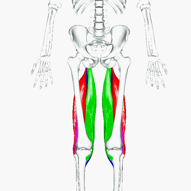
A hamstring (/ˈhæmstrɪŋ/) is any one of the three posterior thigh muscles in human anatomy between the hip and the knee: from medial to lateral, the semimembranosus, semitendinosus and biceps femoris.[3][4]

Etymology

[edit]

The word "ham" is derived from the Old English “ham” or “hom” meaning the hollow or bend of the knee, from a Germanic base where it meant "crooked". It gained the meaning of the leg of an animal around the 15th century.[5] String refers to tendons, and thus the hamstrings' string-like tendons felt on either side of the back of the knee.[6]

Criteria

[edit]

The common criteria of any hamstring muscles are:

  1. Muscles should originate from ischial tuberosity.
  2. Muscles should be inserted over the knee joint, in the tibia or in the fibula.
  3. Muscles will be innervated by the tibial branch of the sciatic nerve.
  4. Muscle will participate in flexion of the knee joint and extension of the hip joint.

Those muscles which fulfill all of the four criteria are called true hamstrings.
The adductor magnus reaches only up to the adductor tubercle of the femur, but it is included amongst the hamstrings because the tibial collateral ligament of the knee joint morphologically is the degenerated tendon of this muscle. The ligament is attached to the medial epicondyle, two millimeters from the adductor tubercle.

Structure

[edit]

The three muscles of the posterior thigh (semitendinosus, semimembranosus, biceps femoris) flex (bend) the knee, while all but the biceps femoris extend (straighten) the hip. The three 'true' hamstrings cross both the hip and the knee joint and are therefore involved in knee flexion and hip extension. The short head of the biceps femoris crosses only one joint (knee) and is therefore not involved in hip extension. With its divergent origin and innervation, it is sometimes excluded from the 'hamstring' characterization.[7]

Muscle Origin Insertion Nerve
semitendinosus ischial tuberosity medial surface of tibia tibial part of sciatic
semimembranosus ischial tuberosity medial tibial condyle tibial part of sciatic
biceps femoris - long head ischial tuberosity lateral side of the head of the fibula tibial part of sciatic
biceps femoris - short head linea aspera and lateral supracondylar line of femur lateral side of the head of the fibula (common tendon with the long head) common peroneal

A portion of the adductor magnus is sometimes considered a part of the hamstrings.[7]

Biceps femoris, semitendinosus and semimembranosus muscles of the right leg

Function

[edit]

The hamstrings cross and act upon two joints – the hip and the knee – and as such they are termed biarticular muscles. The hamstrings contract when the knee is bent, and lengthen when the knee is extended, and when the hips are extended.

Semitendinosus and semimembranosus extend the hip when the trunk is fixed; they also flex the knee and medially (inwardly) rotate the lower leg when the knee is bent.

The long head of the biceps femoris extends the hip, as when beginning to walk; both short and long heads flex the knee and laterally (outwardly) rotate the lower leg when the knee is bent.

The hamstrings play a crucial role in many daily activities such as walking, running, jumping, and controlling some movement in the gluteus. In walking, they are most important as an antagonist to the quadriceps in the deceleration of knee extension.

Clinical significance

[edit]

Sports running injuries

[edit]

A common running injury in several sports, excessive stretch of a hamstring results from extensive hip flexion while the knee is extended.[4][8] During sprinting, a hamstring injury may occur from excessive muscle strain during eccentric contraction late in the leg swing phase.[4][8] The overall incidence of a hamstring injury in sports and professional dancers is about two per 1000 hours of performance.[4] In some sports, a hamstring injury occurs at the incidence of 19% of all sports injuries, and results in an average time loss from competition of 24  days.[4]

Imaging

[edit]
Tear of the hamstrings muscles at the ischial tuberosity seen on MRI (visible on a coronal STIR MRI sequence). The arrowheads indicate the tuber[which?] and the retracted tendon stump.[which?] Significant bleeding around and into the muscles.
Picture of pulled hamstring showing location of hamstring

Imaging the hamstring muscles is usually performed with an ultrasound and/or MRI.[9]  The biceps femoris is most commonly injured, followed by semitendinosus. Semimembranosus injury is rare. Imaging is useful in differentiating the grade of strain, especially if the muscle is completely torn.[10] In this setting, the level and degree of retraction can be determined, serving as a useful roadmap prior to any surgery. Those with a hamstring strain of greater than 60 mm (2.4 in) in length have a greater risk of recurrence.[11]

Use in surgery

[edit]

The distal semitendinosus tendon is one of the tendons that can be used in the surgical procedure ACL reconstruction. In this procedure, a piece of it is used to replace the anterior cruciate ligament (ACL). The ACL is one of the four major ligaments in the knee, which also include the posterior cruciate ligament (PCL), medial collateral ligament (MCL), and lateral collateral ligament (LCL).

See also

[edit]

References

[edit]
[edit]
Revisions and contributorsEdit on WikipediaRead on Wikipedia
from Grokipedia
The hamstrings, also known as the hamstring muscles, are a group of three muscles located in the posterior compartment of the that primarily function to extend the joint and flex the joint. These muscles consist of the femoris (long head), semitendinosus, and semimembranosus, all of which originate from the of the and insert into structures on the proximal and . The femoris short head, while sometimes included in broader discussions of the posterior , originates from the and is considered a separate monoarticular muscle that only flexes the . Innervated primarily by branches of the —with the semitendinosus, semimembranosus, and biceps femoris long head supplied by the tibial division, and the biceps femoris short head by the common fibular division—the hamstrings play a crucial role in locomotion, including walking, running, and squatting, by facilitating pelvic tilting and tibial rotation during knee flexion. Their biarticular nature, crossing both the and joints (except the short head), allows coordinated movement but also predisposes them to strain injuries, which are among the most common musculoskeletal issues in sports, particularly sprinting and kicking activities. Such injuries often affect the biceps femoris long head and can range from mild strains to complete ruptures, with recurrence rates as high as 33% due to factors like and eccentric loading during activity.

Etymology and Definition

Etymology

The term "hamstring" originates from the combination of two words: "," meaning the bend or hollow behind the , derived from Proto-Germanic *hamma-, and "," referring to a due to its fibrous, cord-like appearance. The noun form "hamstring" first appeared in English in the mid-16th century, specifically around 1565, as recorded in a translation by Golding, denoting the tendons at the back of the knee. This usage reflected the visible, string-like tendons in the posterior , which were prominent in both and animal . By the , the term evolved in English literature and early medical descriptions, with the verb form emerging around 1641 in the writings of , meaning to disable by cutting these tendons—a practice historically employed to lame or enemies without killing them. In modern anatomical contexts, "hamstring" has become the standard term for the group of muscles and tendons in the posterior , retaining its descriptive roots while entering formal medical nomenclature by the 18th and 19th centuries in texts like those of anatomists describing lower limb structures.

Anatomical Criteria

The hamstring muscles are defined as the three superficial muscles located in the posterior compartment of the : the biceps femoris (long head), semitendinosus, and semimembranosus. These muscles are distinguished by their shared anatomical features that enable coordinated movement across the and joints. Key criteria for classifying a muscle as part of the hamstring group include a proximal origin from the of the —forming a common tendon in some cases, often described as bipartite due to the medial and lateral attachments—distal insertion on the proximal portions of the or , innervation primarily by the tibial division of the (L5-S2), and primary actions of flexion and extension. The short head of the biceps femoris represents a partial exception, originating from the of the and innervated by the common fibular division of the ; due to these differences, it is not classified as a hamstring muscle. These characteristics ensure the hamstrings act as a biarticular muscle group, spanning both the and . Other posterior thigh muscles, such as the adductor magnus, are excluded from the hamstring group despite a partial "hamstring portion" originating from the and sharing innervation and hip extension function; this portion is classified within the medial compartment due to its primary adductor role. This delineation maintains the hamstrings as a distinct superficial posterior unit.

Anatomy

Muscle Composition

The hamstring muscle group comprises three primary muscles in the posterior compartment: the semimembranosus, semitendinosus, and femoris, all of which are biarticular structures crossing the and joints. The semimembranosus is the deepest and largest of the medial hamstrings, featuring a broad, flat, and membranous shape with a muscle belly and extensive aponeurotic expansions. Its (PCSA) measures approximately 15 cm², reflecting its substantial size relative to the other hamstrings, and it exhibits unipennate proximally transitioning to bipennate distally. Positioned superficial to the semimembranosus on the medial side, the semitendinosus is a slender, strap-like muscle distinguished by its long, cord-like tendinous portions that dominate its , with a belly spanning about 30 cm. It has a PCSA of around 8 cm² and includes a tendinous inscription separating superior and inferior regions. Laterally, the biceps femoris differs from the medial pair by consisting of two heads: the long head, a slender, , bipennate muscle with a PCSA of about 10 cm² and a of roughly 42 cm, and the short head, a thinner, broader approximately 30 cm long with a PCSA of 3 cm². The long head shares an with the short head, and it is the only hamstring with a fibular insertion. In terms of layering, the medial hamstrings form a superficial-to-deep arrangement with the semitendinosus overlying the semimembranosus, while the biceps femoris occupies a distinct lateral position; overall, the semimembranosus contributes the largest volume (up to 324 cm³ in healthy adults), underscoring its dominant role in the group's mass.

Origins, Insertions, and Relations

The hamstring muscles, comprising the semitendinosus, semimembranosus, and biceps femoris, exhibit distinct origins that anchor them primarily to the pelvis and femur. The long heads originate from the ischial tuberosity, with the semitendinosus from the inferomedial impression and the long head of biceps femoris sharing a conjoined tendon, while the semimembranosus originates separately from the superolateral aspect; the short head of the biceps femoris arises from the lateral lip of the linea aspera on the posterior femur, positioning it more distally along the thigh. Regarding insertions, the semitendinosus and semimembranosus converge medially at the , with the semitendinosus inserting via a long onto the medial surface of the proximal as part of the pes anserinus, alongside the sartorius and gracilis tendons. The semimembranosus inserts primarily on the posterior medial tibial condyle, with expansions forming the and attaching to the and . The biceps femoris, both heads uniting distally, inserts on the head of the and the lateral tibial condyle, enabling lateral stabilization. Anatomically, the hamstrings occupy the posterior compartment of the , with key relations to surrounding structures that influence their . Proximally, their origins at the lie adjacent to the and , while distally, the semitendinosus and semimembranosus form the superomedial border of the , and the biceps femoris defines its superolateral boundary, enclosing the fossa alongside the gastrocnemius medially and laterally. The courses posteriorly along the , lying deep to the long head of the biceps femoris and entering the between the hamstring tendons. In terms of muscular interactions, the hamstrings antagonize the femoris across the for balanced flexion-extension, while proximally, they interface with the adductor magnus, whose vertical fibers blend with the hamstring origins at the , contributing to shared hip adduction and extension dynamics.

Innervation and Vascular Supply

The hamstring muscles are primarily innervated by branches of the , which divides into the tibial and common peroneal (fibular) nerves within the posterior . The semimembranosus, semitendinosus, and long head of the femoris receive motor innervation from the tibial division of the (L5-S2 spinal segments). In contrast, the short head of the femoris is innervated by the common peroneal division of the (L5-S1 spinal segments). The vascular supply to the hamstring muscles arises mainly from the profunda femoris artery (), with contributions from its perforating branches that supply the mid-thigh portions of the muscle group. Proximal aspects of the hamstrings receive blood flow from the , a branch of the profunda femoris, along with minor input from the . These vessels form an anastomotic network that ensures robust perfusion during muscle activity. Due to the close anatomical proximity, the lies anterior to the hamstring muscles in the proximal and middle thirds of the , increasing the risk of nerve compression or injury from hamstring-related trauma or .

Function and Biomechanics

Primary Muscle Actions

The hamstring muscles, comprising the femoris (long and short heads), semitendinosus, and semimembranosus, primarily function as biarticular muscles that cross both the and joints, except for the biceps femoris short head, which acts solely at the . This biarticular configuration allows them to generate unique force vectors by influencing motion at two joints simultaneously, facilitating coordinated lower limb movements. At the knee joint, the primary action of all hamstring components is flexion, achieved through their insertions on the and , which pull these bones posteriorly relative to the . This flexion is particularly critical during eccentric contractions, where the muscles lengthen under tension to control deceleration of knee extension, such as in the late swing phase of to prevent excessive forward momentum of the shank. The semitendinosus and semimembranosus, as medial hamstrings, also contribute secondary internal (medial) of the during knee flexion, while the biceps femoris induces external (lateral) . At the hip joint, the long heads of the biceps femoris, semitendinosus, and semimembranosus primarily extend the by drawing the posteriorly from their common origin on the , aiding in propulsion during activities like walking or running. This extension action complements their knee flexion role, with the biarticular nature enabling efficient energy transfer between joints, though it can impose length-tension constraints based on combined and angles.

Role in Locomotion and Daily Activities

The hamstrings are integral to the cycle, facilitating smooth transitions between phases through coordinated contractions. In the late swing phase (approximately 50% to 90% of the cycle), the hamstrings undergo eccentric lengthening under load to decelerate the forward swing of the , absorbing and controlling flexion and extension to prepare for ground contact. This eccentric action peaks in the femoris, with negative work increasing with speed, such as 0.46 J/kg at maximum speed. Transitioning to the early stance phase (0% to 50% of the cycle), the hamstrings shift to concentric shortening to extend the , contributing to forward propulsion while the foot is fixed on the ground. Positive work during this phase also escalates with speed, reaching 0.43 J/kg for the femoris. These dynamics ensure efficient energy transfer and joint stability throughout walking. In sports activities, the hamstrings support explosive movements by enhancing and control. During sprint , they generate substantial extensor torques, with eccentric knee flexor peak torque averaging 2.29 Nm/kg and correlating positively with horizontal ground reaction production (P = 0.04). This activation, particularly in the biceps femoris during late swing, facilitates the "pawing" action that boosts initial speed. In , the hamstrings provide stabilization during landing by modulating and eccentric control, which helps dissipate impact and maintain knee alignment through negative mechanical work at the (averaging 11.03% body weight × height in high absorbers). For daily activities, the hamstrings enable essential lower-body functions through their dual actions of extension and flexion. In , they activate to extend the hip and control descent, working in tandem with other muscles to generate the necessary power for ascent. During , the hamstrings assist in knee flexion and hip stabilization, allowing controlled lowering and rising from a seated position. In maintaining posture during walking, they contribute to the antagonist relationship with the , where the hamstring-to-quadriceps thickness ratio influences flexion moments (r = 0.373, P = 0.042), promoting balanced joint kinetics and upright stability. Additionally, their eccentric role in energy absorption during activities like landing from a step underscores their importance in routine impact moderation.

Clinical Significance

Common Injuries and Risk Factors

Hamstring tightness is a common condition distinct from acute injuries such as strains or tears. Signs and symptoms of tight hamstrings typically include a sensation of tightness or stiffness in the back of the thigh, reduced flexibility (e.g., difficulty fully extending the leg or bending forward), trouble transitioning from sitting to standing, and challenges with activities like picking things up off the floor. Pain is not always present but may occur in the lower back, hips, or knees due to compensatory strain. Severe pain, swelling, bruising, or sudden sharp pain often indicates a strain or injury rather than simple tightness. Hamstring injuries primarily manifest as strains, which are classified into three grades based on severity: grade 1 involves mild damage with minimal fiber disruption and no loss of strength; grade 2 represents a moderate partial with noticeable and ; and grade 3 indicates a severe or complete leading to significant functional impairment. can occur at the muscle- junction or within the itself, while tendinopathies, often proximal, involve chronic degeneration and of the . Among the hamstring muscles, the femoris is the most frequently affected, accounting for a substantial portion of injuries due to its role in rapid deceleration. Epidemiologically, hamstring strains constitute 12-24% of all injuries in professional soccer players, with recent data as of the 2021/22 season showing an increase to 24%, and incidence rates ranging from 0.4 to 0.5 injuries per 1,000 training hours and up to 4.99 per 1,000 match hours. In sprinters and track athletes, recurrence rates can reach 5-60%, often within months of the initial injury, highlighting a high reinjury burden. Data from 2020-2025 indicate an overall incidence of approximately 0.5-1.0 injuries per 1,000 exposure hours across various sports, with soccer and sprinting showing elevated rates compared to other disciplines. Key risk factors include a history of prior hamstring injury, which increases susceptibility by up to twofold, and older age, with peak incidence in athletes aged 20-30 years due to cumulative wear. Muscle imbalances, such as quadriceps dominance where the overpower the hamstrings, contribute to vulnerability, alongside from prolonged activity and poor flexibility in the . These factors are modifiable through targeted , though neuromuscular deficiencies and excessive loads exacerbate the risk in high-intensity . Injuries typically arise from eccentric overload, where the hamstrings lengthen under tension during the late swing phase of sprinting or the follow-through of kicking, leading to excessive strain at speeds exceeding 85% of maximum. This mechanism is compounded by the biarticular nature of the hamstrings, spanning both and joints, which predisposes them to high mechanical stress during rapid hip flexion and knee extension. Such overload is prevalent in sports involving explosive movements, accounting for over 70% of cases in soccer.

Diagnosis and Imaging

Diagnosis of hamstring injuries typically begins with a detailed clinical history, where patients report sudden onset of in the posterior , often accompanied by a popping or tearing sensation during activities such as sprinting or kicking. Physical examination involves along the posterior to identify tenderness, swelling, or ecchymosis, followed by assessment of strength through resisted knee flexion and extension, as well as evaluation of for deficits in extension or flexion. These clinical findings help differentiate hamstring strains from other posterior pathologies, such as irritation or referred . Imaging modalities are employed to confirm the , assess severity, and guide when clinical suggests significant involvement. X-rays are primarily used to rule out avulsion fractures, particularly at the proximal hamstring origins on the in adolescents or during high-force injuries. serves as an accessible initial imaging tool for acute strains, enabling dynamic assessment of muscle tears and integrity with high sensitivity (approximately 85%) for detecting abnormalities in the early post-injury phase. (MRI) is considered the gold standard for detailed , utilizing T2-weighted or STIR sequences to visualize , hemorrhage, and fiber disruption, while proton density sequences help delineate partial versus full-thickness tears at the musculotendinous junction. Coronal T2 fat-saturated images provide an overview of extent, measuring length and retraction to inform . Hamstring injuries are commonly graded on a three-tier system based on the degree of muscle disruption observed clinically or via . Grade 1 injuries represent mild strains with less than 10% involvement, manifesting as minimal and no significant loss of strength or function. Grade 2 injuries involve partial tears with 10-50% disruption, leading to moderate , swelling, and noticeable deficits in strength and . Grade 3 injuries indicate complete ruptures with greater than 50% disruption, often resulting in severe , a palpable gap, and substantial functional impairment. Recent advances since 2020 have incorporated artificial intelligence to enhance MRI interpretation for hamstring injuries, improving diagnostic accuracy and prognostic assessment. Deep learning algorithms now enable automatic 3D segmentation and quantification of hamstring muscle edema on MRI, facilitating precise measurement of injury volume and aiding in the prediction of recovery timelines. Additionally, the Proximal Hamstring Objective Magnetic Resonance Imaging Score (PHOMRIS) provides a reliable, MRI-based grading tool for proximal injuries, correlating imaging features like tendon retraction and edema with clinical outcomes in surgical candidates.

Treatment and Rehabilitation

The initial treatment of acute hamstring injuries follows the protocol, which involves rest to avoid further strain, ice application to reduce swelling, compression to minimize hemorrhage, and elevation to promote fluid drainage. Nonsteroidal anti-inflammatory drugs (NSAIDs) are commonly used for and to control , typically introduced after the first 48 hours to avoid interfering with early processes. Rehabilitation for hamstring injuries is structured in progressive phases aligned with tissue healing timelines. The protection phase, spanning 0-2 weeks post-injury, focuses on minimizing load through relative rest and, if necessary for severe strains, brief immobilization to protect the damaged tissue while initiating gentle isometric exercises at shortened muscle lengths. The repair phase, from 2-6 weeks, emphasizes controlled mobility with gentle stretching and submaximal strengthening to support alignment and early load tolerance. In the remodeling phase, beginning around 6 weeks and extending to full recovery, eccentric strengthening exercises—such as Nordic hamstring curls—are introduced to enhance muscle length-tension properties and resilience, allowing gradual return to dynamic activities. Recent evidence-based guidelines from 2023 highlight the importance of individualized, criteria-based progressive loading in rehabilitation protocols to optimize tissue and minimize reinjury risk. These approaches, incorporating -specific demands and monitoring symptoms alongside strength metrics, have demonstrated reductions in recurrence rates when including progressive , trunk stabilization, and eccentric loading. Typical outcomes for mild to moderate (grade 1-2) include an average return to within 11-25 days, depending on and athlete response, while grade 3 often require surgical intervention for complete tears to restore function.

Surgical Uses and Prevention Strategies

Hamstring tendons, particularly the semitendinosus and gracilis, are commonly harvested as autografts for (ACL) reconstruction due to their suitable length, diameter, and biomechanical properties, allowing for quadrupled or five-strand configurations to enhance graft strength. This approach provides comparable clinical outcomes to other autografts, with low failure rates and good stability reported in systematic reviews. For proximal hamstring avulsions, surgical repair typically involves suture anchors placed at the to reattach the tendons, often using 2-5 anchors in configurations like suture bridge or all-suture constructs to achieve secure fixation and minimize displacement under load. Biomechanical studies confirm that all-suture anchors offer superior load-to-failure resistance compared to traditional anchors, supporting their use in acute repairs to restore function and prevent chronic weakness. Prevention strategies for hamstring injuries emphasize eccentric strengthening programs, such as the Nordic hamstring exercise (NHE), which has been shown in meta-analyses to reduce injury incidence by up to 51% through improved eccentric strength and muscle fascicle length. Dynamic warm-ups incorporating progressive sprinting and , combined with flexibility routines targeting hamstring extensibility, further mitigate by enhancing neuromuscular control and reducing strain during high-speed activities. monitoring, including tracking acute-to-chronic ratios and high-speed running volumes, is essential for athletes, as spikes in demands correlate with elevated injury , allowing coaches to adjust loads proactively. Following hamstring , rehabilitation protocols integrate early —typically starting within days of repair—to promote alignment, prevent adhesions, and avoid stiffness while protecting the repair site through controlled range-of-motion exercises. Emerging trends in 2025 include biomechanical screening tools, such as field-based protocols using video analysis, to assess sprint mechanics and identify at-risk athletes. These tools enable personalized prevention by quantifying risk factors like sprint mechanics asymmetries, with ongoing validation in elite sports settings.

References

Add your contribution
Related Hubs
Contribute something
User Avatar
No comments yet.