Hubbry Logo
Tinea crurisTinea crurisMain
Open search
Tinea cruris
Community hub
Tinea cruris
logo
8 pages, 0 posts
0 subscribers
Be the first to start a discussion here.
Tinea cruris
Tinea cruris
from Wikipedia
Tinea cruris
Other namesEczema marginatum, crotch itch, crotch rot, dhobi itch, gym itch, jock itch, jock rot, scrot rot[1][2]: 303 
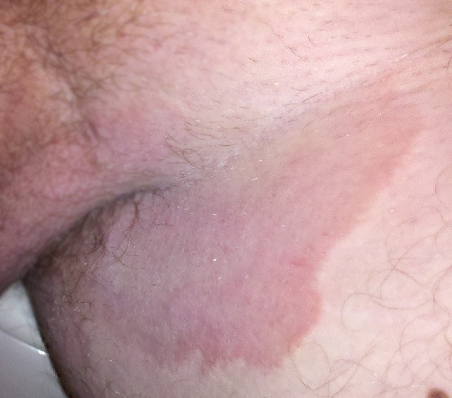
Tinea cruris on the groin of a man
SpecialtyDermatology
SymptomsItch, rash in groin
Risk factors
  • Excessive sweating
  • Diabetes
  • Obesity
Diagnostic methodMicroscopy and culture of skin scrapings
Differential diagnosis
Prevention
  • Treat any fungal infections of feet or nails
  • Keep groin region dry
  • Avoid tight clothing
  • Losing weight if obese
MedicationTopical antifungal medications

Tinea cruris (TC), also known as jock itch, is a common type of contagious, superficial fungal infection of the groin and buttocks region, which occurs predominantly but not exclusively in men and in hot-humid climates.[3][4]

Typically, over the upper inner thighs, there is an intensely itchy red raised rash with a scaly well-defined curved border.[3][4] It is often associated with athlete's foot and fungal nail infections, excessive sweating, and sharing of infected towels or sports clothing.[4][5][6] It is uncommon in children.[4]

Its appearance may be similar to some other rashes that occur in skin folds including candidal intertrigo, erythrasma, inverse psoriasis and seborrhoeic dermatitis. Tests may include microscopy and culture of skin scrapings.[7]

Treatment is with topical antifungal medications and is particularly effective if symptoms have recent onset.[5][6] Prevention of recurrences include treating concurrent fungal infections and taking measures to avoid moisture build-up including keeping the groin region dry, avoiding tight clothing and losing weight if obese.[8]

Names

[edit]

Other names include "jock rot",[9] "dhobi itch",[10] "crotch itch",[11] "scrot rot",[12] "gym itch", "ringworm of groin" and "eczema marginatum".[13]

Signs and symptoms

[edit]

Typically, over the upper inner thighs, there is a red raised rash with a scaly well-defined border. There may be some blistering and weeping, and the rash can reach near to the anus.[3] The distribution is usually on both sides of the groin and the center may be lighter in colour.[8] The rash may appear reddish, tan, or brown, with flaking, rippling, peeling, iridescence, or cracking skin.[14]

If the person is hairy, hair follicles can become inflamed resulting in some bumps (papules, nodules and pustules) within the plaque. The plaque may reach the scrotum in men and the labia majora and mons pubis in women. The penis is usually unaffected unless there is immunodeficiency or there has been use of steroids.[4]

Affected people usually experience intense itching in the groin which can extend to the anus.[3][4]

Causes

[edit]
Macroconidia from Epidermophyton floccosum

Tinea cruris is often associated with athlete's foot and fungal nail infections.[4][5] Rubbing from clothing, excessive sweating, diabetes and obesity are risk factors.[6][8] It is contagious and can be transmitted person-to-person by skin-to-skin contact or by contact with contaminated sports clothing and sharing towels.[3][5]

The type of fungus involved may vary in different parts of the world; for example, Trichophyton rubrum and Epidermophyton floccosum are common in New Zealand.[7] Less commonly Trichophyton mentagrophytes and Trichophyton verrucosum are involved.[8] Trichophyton interdigitale has also been implicated.[5]

Diagnosis

[edit]

Tests are usually not needed to make a diagnosis, but if required, may include microscopy and culture of skin scrapings, a KOH examination to check for fungus, or skin biopsy.[3][7]

Differential diagnosis

[edit]

The symptoms of tinea cruris may be similar to other causes of itch in the groin.[3] Its appearance may be similar to some other rashes that occur in skin folds including candidal intertrigo, erythrasma, inverse psoriasis and seborrhoeic dermatitis.[7]

Prevention

[edit]

To prevent recurrences of tinea cruris, concurrent fungal infections such as athlete's foot need to be treated. Also advised are measures to avoid moisture build-up including keeping the groin region dry, avoiding tight clothing, and losing weight if obese.[8] People with athlete's foot or tinea cruris can prevent spread by not lending their towels to others.[5]

Treatment

[edit]

Tinea cruris is treated by applying antifungal medications of the allylamine or azole type to the groin region. Studies suggest that allylamines (naftifine and terbinafine) are a quicker but more expensive form of treatment compared to azoles (clotrimazole, econazole, ketoconazole, oxiconazole, sulconazole).[6] If the symptoms have been present for long or the condition worsens despite applying creams, terbinafine or itraconazole can be given by mouth.[5]

The benefits of the use of topical steroids in addition to an antifungal are unclear.[15] There might be a greater cure rate but no guidelines currently recommend its addition.[15] The effect of Whitfield's ointment is also unclear,[15] but when given, it is prescribed at half strength.[5]

Wearing cotton underwear and socks, in addition to keeping the groin dry and using antifungal powders, is helpful.[16]

Prognosis

[edit]

Tinea cruris is not life-threatening and treatment is effective, particularly if the symptoms have not been present for long.[5] However, recurrence may occur. The intense itch may lead to lichenification and secondary bacterial infection. Irritant and allergic contact dermatitis may be caused by applied medications.[8]

Epidemiology

[edit]

Tinea cruris is common in hot-humid climates, and is the second most common clinical presentation for dermatophytosis.[8] It is uncommon in children.[4]

References

[edit]
[edit]
Revisions and contributorsEdit on WikipediaRead on Wikipedia
from Grokipedia
Tinea cruris, commonly known as jock itch, is a superficial that primarily affects the of the , inner thighs, pubic region, , and perianal area. Caused by pathogenic fungi such as , , , and less commonly species of , it manifests as a pruritic, erythematous, scaly with annular plaques featuring raised borders and central clearing. This condition is favored by warm, moist environments and is highly contagious through direct contact, shared towels or clothing, or autoinoculation from other s like tinea pedis (). The infection typically presents with intense itching, redness, scaling (which may appear as flaking, peeling, white flakes, or crusty white/yellow patches), and sometimes blistering or cracking in the affected areas, typically sparing the scrotum in males. Tinea cruris rarely affects the scrotum directly, and symptoms under the scrotum, such as white flakes, may indicate other conditions such as candidal intertrigo. Symptoms may worsen with sweating, from tight clothing, or in individuals with risk factors such as , , , or excessive perspiration. It is more common in adolescent and adult males and is a infection with global prevalence estimated at 20-25%, particularly in tropical climates and among athletes due to occlusive sportswear and communal showers. Diagnosis is primarily clinical based on the characteristic appearance, though confirmed by microscopic examination with (KOH) preparation, fungal culture, or if atypical features are present. Treatment typically involves topical antifungal agents such as terbinafine (often applied for 1-2 weeks) or azoles like clotrimazole (typically applied for 2-4 weeks), achieving cure rates of 80-90%; severe or recurrent cases may require oral antifungals such as . Prevention strategies include maintaining dryness, wearing loose-fitting , promptly treating concurrent fungal infections, and avoiding shared personal items to minimize recurrence, which can occur due to reinfection or antifungal resistance. Complications are rare but may include secondary bacterial infections from scratching or permanent discoloration.

Nomenclature and Overview

Other Names

Tinea cruris is widely known in colloquial terms as jock itch, a name originating from its high incidence among male athletes, where "jock" refers to athletic supporters or jockstraps that create moist conditions conducive to the infection. Other informal names include crotch rot and jock rot, terms emphasizing the affected area and the discomfort associated with moisture and friction. Gym itch is another variant, highlighting the role of gym environments in promoting the condition through sweat and shared facilities. Medically, synonyms include epidermophytosis cruris, an older term reflecting the involvement of Epidermophyton species as a primary causative fungus, and ringworm of the groin, as "tinea" denotes superficial dermatophyte infections resembling ringworm.

Definition and Classification

Tinea cruris is a superficial dermatophytosis characterized by a fungal infection primarily affecting the groin, inner thighs, pubic region, and buttocks, caused by dermatophyte fungi that invade the stratum corneum and keratinized tissues. This condition manifests as an inflammatory response in the intertriginous areas, leading to pruritic, erythematous lesions confined to the superficial layers of the skin. Commonly referred to as jock itch, it represents a localized form of ringworm infection. Within dermatological and mycological classifications, tinea cruris is categorized as a type of tinea , where "tinea" denotes superficial ringworm-like dermatophytoses, and "cruris" specifies the involvement. It falls under the broader umbrella of superficial mycoses, which are cutaneous fungal limited to the , , and without systemic or deep tissue dissemination. This distinguishes it from deeper fungal infections, such as subcutaneous or systemic mycoses, which involve dermal or subcutaneous layers and often require more invasive diagnostic and therapeutic approaches. Furthermore, tinea cruris is differentiated from non-fungal inguinal rashes, like or , by its specific fungal etiology and characteristic annular morphology, confirmed through mycological examination.

Clinical Features

Signs and Symptoms

Tinea cruris typically presents with initial symptoms of intense pruritus, often accompanied by a burning or stinging sensation in the groin area. These sensations arise in the inguinal folds and may extend to the inner thighs, prompting patients to scratch, which can exacerbate irritation. The hallmark physical sign is an erythematous, scaly rash forming annular plaques with well-defined, raised borders and central clearing, resembling a ring. The rash commonly exhibits scaling, flaking, peeling, or cracking of the skin, which may appear as white flakes or crusty white/yellow patches. The rash is often red to brown, depending on skin tone, and may include small blisters or vesicles along the advancing edge; it commonly affects both groins but spares the scrotum and penis. The condition rarely affects the scrotum directly, and symptoms such as white flakes under the scrotum more typically indicate other conditions, such as candidal intertrigo. As the condition progresses, the rash can spread to the upper thighs, buttocks, or perianal region, potentially developing satellite lesions or fissuring in the skin folds. Secondary bacterial infection may occur, leading to increased inflammation, malodorous exudate, or pustules, particularly in areas of maceration. Variations in presentation include milder symptoms in dry climates, where scaling predominates without significant inflammation, contrasted by more severe, weeping lesions in moist, humid environments that promote maceration and discomfort.

Risk Factors

Tinea cruris is more prevalent among adult males, with a reported male-to-female ratio of approximately 4:1, attributed to anatomical factors such as the apposition of the and thighs that promote moisture retention. Adolescents and young adults also face elevated risk due to hormonal changes and increased leading to . further heightens susceptibility by creating that trap heat and moisture, fostering an environment conducive to infection. Behavioral factors significantly contribute to the development of tinea cruris, including the use of tight-fitting or occlusive that restricts and promotes sweating in the area. Excessive , often from strenuous exercise or hot weather, exacerbates this by maintaining a damp environment, while sharing towels, , or using communal facilities like locker rooms increases exposure to potential contaminants. Poor personal hygiene, such as infrequent changing of undergarments or reusing sweaty attire, compounds these risks. Certain medical conditions predispose individuals to tinea cruris, notably diabetes mellitus, which impairs immune responses and healing in moist areas. , as seen in conditions like or those requiring therapy, similarly elevates vulnerability by reducing the body's ability to combat fungal overgrowth. Concurrent infections such as tinea pedis serve as reservoirs, allowing spread to the groin through autoinoculation. Environmental and occupational exposures play a key role, particularly in warm, humid climates where ambient hinders drying of the skin. Professions involving prolonged physical labor in humid settings, such as farming or , heighten risk due to persistent sweating and limited opportunities for . Athletes, especially those in contact sports or training in enclosed, steamy environments, are particularly affected owing to frequent and shared equipment.

Etiology and Pathogenesis

Causative Agents

Tinea cruris is primarily caused by fungi from the genera Trichophyton and Epidermophyton. Among these, is the most prevalent causative agent, accounting for approximately 70-90% of cases depending on the geographic region and study population. Other primary include and , which together represent the majority of remaining infections, though T. mentagrophytes has shown increasing prevalence in recent decades. Dermatophytes responsible for tinea cruris are classified based on their host preferences: anthropophilic species, which are adapted to humans and cause chronic, less inflammatory infections, predominate over zoophilic species derived from animals that typically produce more acute, inflammatory responses. T. rubrum is a classic anthropophilic dermatophyte, thriving in human skin environments and contributing to its high incidence in groin infections. In contrast, zoophilic strains like certain variants of T. mentagrophytes are less common in tinea cruris but may occur through animal contact. Less commonly, Candida species, particularly Candida albicans, can mimic tinea cruris in intertrigo-like presentations within moist skin folds, but these are non-dermatophyte yeasts distinguished by their opportunistic nature and different microscopic features, such as pseudohyphae. The prevalence of T. rubrum in tinea cruris has notably increased since the late 1980s, reflecting broader global trends in dermatophytoses likely influenced by factors such as population mobility and urbanization. An emerging causative agent is Trichophyton indotineae, a terbinafine-resistant first described in 2014, which often causes extensive, inflammatory, and pruritic tinea cruris, corporis, and faciei. Previously misidentified as T. mentagrophytes, it has spread rapidly from to other regions, with the first confirmed cases reported in 2023 and increasing global prevalence as of 2025, contributing to recalcitrant infections.

Transmission and Pathophysiology

Tinea cruris is primarily transmitted through direct skin-to-skin contact with an infected individual, particularly in shared environments such as locker rooms or during close physical activities. The infection can also spread via fomites, including contaminated towels, clothing, or athletic equipment, which harbor fungal spores in warm, humid conditions. Autoinoculation is a common route, where the fungus spreads from an existing infection site like tinea pedis on the feet to the area through , hand contact, or contaminated . In the pathophysiology of tinea cruris, fungi, such as , invade the by producing keratinases and other proteinases that degrade , the primary structural protein in the outer skin layer. This enzymatic activity allows fungal hyphae to penetrate and colonize the skin, while arthroconidia and hyphae proliferate in the warm, moist areas of the , where occlusion and perspiration create an alkaline pH conducive to conidia germination and growth. The infection elicits a localized inflammatory response, characterized by pruritus and , driven by the host's recognition of fungal antigens. The host's plays a crucial role in containing the infection to the superficial layers of the skin, preventing deeper or in immunocompetent individuals. However, trapped moisture in the can prolong viability and facilitate ongoing , as the stratum corneum's barrier function is compromised by maceration. Systemic spread is rare in healthy hosts due to effective immune surveillance, though moisture retention exacerbates local persistence. Recent studies since 2015 have highlighted the role of formation by dermatophytes in enhancing persistence, where fungal communities embedded in an resist agents and host defenses, contributing to chronic or recurrent tinea cruris cases. For instance, ex vivo models have demonstrated species-specific development by isolates, increasing tolerance to treatments and underscoring the need for targeted antibiofilm strategies.

Diagnosis

Diagnostic Methods

Diagnosis of tinea cruris typically begins with a clinical evaluation based on patient history and , which often reveals a characteristic pruritic, erythematous, scaly annular plaque with a raised and central clearing in the , upper thighs, , or perianal region. This presentation is usually sufficient for presumptive diagnosis in straightforward cases, particularly in individuals with risk factors such as or . A Wood's lamp examination may be employed to assess for , which can occur with certain strains like species, though it is uncommon in tinea cruris predominantly caused by or Epidermophyton; non-fluorescence helps differentiate from conditions like . Laboratory confirmation is recommended for atypical, recurrent, or treatment-resistant cases to verify the fungal . using (KOH) preparation is a rapid, inexpensive first-line test; scrapings from the active border of the are treated with 10-20% KOH to dissolve , revealing branching septate hyphae or arthroconidia under light , with a reported false-negative rate of about 15%. The cellophane tape method can enhance sample collection by preserving the specimen for better visualization. Fungal culture provides definitive species identification when microscopy is inconclusive, involving inoculation of scrapings onto Sabouraud dextrose agar (SDA), often supplemented with antibiotics to inhibit ; results typically take 7-14 days to show growth and microscopic morphology. For atypical presentations, a may be performed, with histologic examination using periodic acid-Schiff () stain to highlight fungal elements in the , such as the "sandwich sign" where hyphae sandwich neutrophils. Emerging , particularly (PCR) assays, offer rapid detection since around 2020, especially valuable in outbreaks or for identifying drug-resistant strains; multiplex PCR targeting DNA from skin scrapings achieves sensitivities of 80-100% and specificities around 80-90% compared to , with turnaround times reduced to hours rather than weeks. These methods, including real-time PCR and arrays, are increasingly adopted in clinical practice for precise identification, such as , the most common cause.

Differential Diagnosis

Tinea cruris must be differentiated from other conditions affecting the inguinal folds, as misdiagnosis can lead to ineffective treatment and prolonged symptoms. Common mimics include intertrigo, which may be bacterial or candidal in origin, presenting with erythematous, macerated plaques but lacking the annular morphology and central clearing typical of tinea; instead, it often shows satellite pustules and involves the scrotum more prominently. Contact dermatitis, triggered by irritants or allergens such as soaps or fabrics, manifests as diffuse erythema without scaling borders or fungal elements, and patient history typically reveals exposure to a potential irritant. Psoriasis inversus appears as well-demarcated, shiny red plaques with uniform scaling across the groin and other flexural areas, but lacks the raised advancing edge and responds negatively to (KOH) examination for hyphae. Seborrheic features greasy, yellowish scales on an erythematous base, often extending to the trunk or face, and does not demonstrate central clearing or hyphae on microscopy. , caused by , presents as fine, scaly brown patches without an active border; it is distinguished by coral-red fluorescence under Wood's lamp and absence of fungal elements on KOH preparation. Rare mimics include , resulting from chronic scratching and showing thickened, lichenified skin with exaggerated skin markings but no hyphae or annular pattern. Fixed drug eruption may present as recurrent, round erythematous patches in the following medication intake, resolving with but lacking fungal confirmation on testing. Clinicians should suspect a non-fungal if lesions fail to respond to topical antifungals after two to four weeks or if initial KOH microscopy is negative for hyphae. In such cases, further evaluation with or may be warranted to identify alternative causes.

Management

Treatment Options

The primary treatment for tinea cruris involves agents, which are considered first-line therapy for localized infections. Allylamines such as terbinafine cream or butenafine cream, applied once or twice daily for 2 to 4 weeks, are highly effective due to their broad-spectrum activity against dermatophytes and lower relapse rates compared to other classes. Azoles, including clotrimazole or miconazole creams, are suitable for milder cases and are typically applied twice daily for the same duration, offering good efficacy with minimal side effects. In Canada, as of 2025-2026, terbinafine cream (e.g., Lamisil or generic equivalents) is considered the best over-the-counter (OTC) treatment for tinea cruris (jock itch). It is fungicidal, often more effective, and typically requires a shorter treatment course of 1-2 weeks compared to clotrimazole (e.g., Canesten) or miconazole. Apply twice daily to clean, dry skin and continue for 1-2 weeks after symptoms resolve. Consult a pharmacist or doctor if there is no improvement in 2 weeks or if the condition is severe. For extensive, refractory, or disseminated infections, oral antifungals are recommended to ensure systemic coverage. Terbinafine at a dose of 250 mg daily for 2 weeks is the preferred option, demonstrating high mycological cure rates in clinical studies. Alternatively, or can be used, with administered as 150 mg weekly for 2 to 4 weeks in responsive cases. Non-pharmacological adjunct measures support resolution by addressing environmental factors that promote fungal growth. Patients should maintain the affected area clean and dry, wear loose-fitting underwear to reduce moisture and , and avoid sharing personal items to prevent reinfection. In cases of significant , short-term use of low-potency topical corticosteroids like (1%) may alleviate symptoms, but prolonged application must be avoided to prevent tinea incognito, a masked that complicates . Recent advancements include the topical luliconazole 1% cream, approved in regions like the post-2013 for tinea cruris, which allows once-daily application for just 1 week and achieves faster clinical cure rates than traditional . Increasing terbinafine resistance in , the primary causative agent, has been reported globally since 2020, often linked to squalene epoxidase gene mutations, necessitating susceptibility testing via culture for refractory cases and consideration of alternative agents like .

Prevention Strategies

Maintaining personal is essential to prevent tinea cruris by reducing and fungal growth in the area. Individuals should take daily showers using mild and thoroughly dry the and inner thighs with a clean , ensuring the feet are dried last to avoid transferring fungi from . Applying absorbent powders, such as those containing miconazole or , after bathing can further help keep the area dry, particularly for those prone to sweating. Choosing appropriate clothing and adopting certain habits can minimize and retention that favor fungal proliferation. Wearing loose-fitting underwear promotes airflow and reduces chafing, while changing underwear and workout clothes immediately after sweating or exercising prevents buildup of dampness. It is also advisable to avoid sharing personal items like towels, clothing, or athletic supporters, as these can serve as fomites for fungal transmission. Environmental controls are particularly important in shared facilities to curb indirect spread. In gyms, locker rooms, or public showers, disinfecting surfaces regularly and wearing protective footwear like flip-flops can limit exposure to contaminated areas. Concurrent infections, such as tinea pedis, should be treated promptly to prevent autoinoculation to the , with recommendations to don socks before underwear during this period. For at-risk groups, such as those in humid climates or with recurrent episodes, additional measures include education on consistent practices to mitigate environmental factors. In cases of frequent recurrence, prophylactic application of topical antifungals like terbinafine or once weekly after initial cure can reduce reinfection rates, though the optimal duration may vary.

Prognosis and Epidemiology

Clinical Outcomes

With appropriate antifungal treatment, tinea cruris has an excellent , achieving cure rates of 80-90% in most patients. Resolution of symptoms and lesions typically occurs within 2-4 weeks of initiating . However, recurrence is common, with rates varying from 15-65% in studies without preventive measures such as maintaining dryness in the area. Complications arise infrequently but can include secondary bacterial infections due to scratching-induced skin breakdown. Chronicity is more likely in patients with diabetes mellitus, where persistent impairs and healing. Misuse of topical corticosteroids can lead to tinea incognito, masking the infection and promoting spread, or cause atrophy. Factors influencing clinical outcomes include early diagnosis to prevent progression, adherence to the full treatment course, and underlying immune status. In immunocompromised individuals, such as those with , infections can be more extensive and severe. Long-term effects are minimal, with scarring being rare even in untreated or recurrent cases. Nonetheless, chronic itching can impose a psychological burden, contributing to distress and reduced in affected individuals.

Global Distribution

Tinea cruris exhibits a worldwide distribution, with cutaneous fungal infections, including this condition, affecting approximately 20-25% of the global population. The lifetime risk of acquiring a infection like tinea cruris is estimated at 10-20%, predominantly among adolescent and adult males. Prevalence varies significantly by region, with higher rates reported in tropical and subtropical climates due to favorable conditions of heat and humidity that promote fungal growth. In developing countries, particularly in and the , prevalence can exceed 20%, while global ranges span from 0.3% to 53% depending on environmental and host factors. In tropical regions such as and , tinea cruris accounts for a substantial portion of dermatophytoses, comprising up to 25% of all cases in and up to 80% of chronic or recurrent infections. Demographic patterns reveal a peak incidence in the 20-40 age group, aligning with increased and , and a male-to-female ratio of approximately 3:1, though cases in females may be underreported due to and less frequent seeking of care. In the United States, superficial fungal infections, including tinea cruris, led to approximately 51 million physician visits from 1995 to 2004 (about 5 million annually), with an estimated 29.4 million cases as of 2004. Trends indicate a rising incidence in urban settings worldwide, driven by factors such as , which increases and moisture retention, and the proliferation of gym culture, where shared equipment facilitates transmission. In contrast, remains relatively stable in developed nations with access to resources and treatments. Socioeconomic disparities exacerbate distribution, with higher rates in low-income communities characterized by poor , , and limited ; for instance, screening in varying socioeconomic groups revealed culture-positive rates up to 70.7%. Outbreaks are notably common in sports communities, particularly contact sports like wrestling and football, where close physical interaction and communal facilities heighten risk. Post-2020 data highlight the influence of on spread, as rising global temperatures and increased humidity—projected to intensify in tropical regions—create optimal environments for proliferation, potentially elevating infection rates in affected areas. Recent emergence of resistant strains like Trichophyton indotineae has increased chronic and recurrent cases in regions like and . and events, more frequent due to climate shifts, further contribute by promoting dissemination through flooding and disrupted . These environmental pressures, combined with , underscore the need for enhanced in vulnerable populations.

References

Add your contribution
Related Hubs
User Avatar
No comments yet.