Hubbry Logo
Palmoplantar keratodermaPalmoplantar keratodermaMain
Open search
Palmoplantar keratoderma
Community hub
Palmoplantar keratoderma
logo
8 pages, 0 posts
0 subscribers
Be the first to start a discussion here.
Palmoplantar keratoderma
from Wikipedia
Palmoplantar keratoderma
Other namesVohwinkel's syndrome
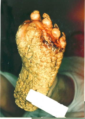
Patient with severe plantar keratosis
SpecialtyDermatology Edit this on Wikidata

Palmoplantar keratodermas are a heterogeneous group of skin disorders characterized by abnormal thickening (scleroderma) of the stratum corneum of the palms and soles.

Autosomal recessive, dominant, X-linked, and acquired forms have all been described in medical literature.[1]: 505 [2]: 211 [3]

Types

[edit]

Clinically, three distinct patterns of palmoplantar keratoderma may be identified: diffuse, focal, and punctate.[1]: 505 

Diffuse

[edit]
Diffuse palmoplantar keratoderma

Diffuse palmoplantar keratoderma is a type of palmoplantar keratoderma that is characterized by an even, thick, symmetric hyperkeratosis over the whole of the palm and sole, usually evident at birth or in the first few months of life.[1]: 505  Restated, diffuse palmoplantar keratoderma is an autosomal dominant disorder in which hyperkeratosis is confined to the palms and soles.[4] The two major types can have a similar clinical appearance:[4]

  • Diffuse epidermolytic palmoplantar keratoderma (also known as "Palmoplantar keratoderma cum degeneratione granulosa Vörner", "Vörner's epidermolytic palmoplantar keratoderma", and "Vörner keratoderma"[4]) is one of the most common patterns of palmoplantar keratoderma, an autosomal dominant condition that presents within the first few months of life, characterized by a well-demarcated, symmetric thickening of palms and soles, often with a "dirty" snakeskin appearance due to underlying epidermolysis.[1]: 506 
  • Diffuse nonepidermolytic palmoplantar keratoderma (also known as "Diffuse orthohyperkeratotic keratoderma", "Hereditary palmoplantar keratoderma", "Keratosis extremitatum progrediens", "Keratosis palmoplantaris diffusa circumscripta", "Tylosis", "Unna–Thost disease", and "Unna–Thost keratoderma"[4]) is inherited as an autosomal dominant condition and is present from infancy, characterized by a well-demarcated, symmetric, often "waxy" keratoderma involving the whole of the palms and soles.[1]: 506–8 [2]: 213 

Focal

[edit]

Focal palmoplantar keratoderma, a type of palmoplantar keratoderma in which large, compact masses of keratin develop at sites of recurrent friction, principally on the feet, although also on the palms and other sites, a pattern of calluses that may be discoid (nummular) or linear.

  • Focal palmoplantar keratoderma with oral mucosal hyperkeratosis (also known as "Focal epidermolytic palmoplantar keratoderma",[4] "Hereditary painful callosities",[4][5] "Hereditary painful callosity syndrome",[1] "Keratosis follicularis",[1] "Keratosis palmoplantaris nummularis",[1] and "Nummular epidermolytic palmoplantar keratoderma"[4]) is an autosomal dominant keratoderma that represents a clinical overlap syndrome with pachyonychia congenita type I but without the classic nail involvement.[1]: 510 

Punctate

[edit]

Punctate palmoplantar keratoderma is a form of palmoplantar keratoderma in which many tiny "raindrop" keratoses involve the palmoplantar surface, skin lesions which may involve the whole of the palmoplantar surface, or may be more restricted in their distribution.[1]: 505 [4]

  • Type 1: Keratosis punctata palmaris et plantaris (also known as "Autosomal-dominant hereditary punctate keratoderma associated with malignancy", "Buschke–Fischer–Brauer disease", "Davis Colley disease", "Keratoderma disseminatum palmaris et plantaris", "Keratosis papulosa", "Keratoderma punctatum", "Keratodermia punctata", "Keratoma hereditarium dissipatum palmare et plantare", "Palmar and plantar seed dermatoses", "Palmar keratoses", "Papulotranslucent acrokeratoderma", "Punctate keratoderma", "Punctate keratoses of the palms and soles", and "Maculosa disseminata") is a skin condition, an autosomal dominant palmoplantar keratoderma with variable penetrance, characterized clinically by multiple, tiny, punctate keratoses over the entire palmoplantar surfaces, beginning over the lateral edge of the digits.[1]: 509 [2]: 212–213  It has been linked to 15q22-q24.[6]
  • Type 2: Spiny keratoderma (also known as "Porokeratosis punctata palmaris et plantaris", "Punctate keratoderma", and "Punctate porokeratosis of the palms and soles") is an autosomal dominant keratoderma of late onset that develops in patients aged 12 to 50, characterized by multiple tiny keratotic plugs, resembling the spines on a music box, involving the entire palmoplantar surfaces.[1]: 509 [4]
  • Type 3: Focal acral hyperkeratosis (also known as "Acrokeratoelastoidosis lichenoides", and "Degenerative collagenous plaques of the hand") is a late-onset keratoderma, inherited as an autosomal dominant condition, characterized by oval or polygonal crateriform papules developing along the border of the hands, feet, and wrists.[1]: 509  It is considered similar to Costa acrokeratoelastoidosis.[7]

Ungrouped

[edit]
  • Palmoplantar keratoderma and spastic paraplegia (also known as "Charcot–Marie–Tooth disease with palmoplantar keratoderma and nail dystrophy"[1]) is an autosomal dominant or x-linked dominant condition that begins in early childhood with thick focal keratoderma over the soles and, to a lesser extent, the palms.[1]: 513 
  • Palmoplantar keratoderma of Sybert (also known as "Greither palmoplantar keratoderma",[1] "Greither syndrome",[4] "Keratosis extremitatum hereditaria progrediens",[1] "Keratosis palmoplantaris transgrediens et progrediens"[1] "Sybert keratoderma",[4] and "Transgrediens and progrediens palmoplantar keratoderma"[4]) is an extremely rare autosomal dominant[8] keratoderma (a skin condition involving horn-like growths) with symmetric severe involvement of the whole palmoplantar surface in a glove-and-stocking distribution.[1]: 509  It was characterized by Aloys Greither in 1952.[9][10][11] It was characterized by Virginia Sybert in 1988.[12] An autosomal recessive form which is known as Mal de Meleda has been described.[13] This is associated with mutations in the Secreted Ly-6/uPAR-related protein 1 (SLURP1) gene.
  • Striate palmoplantar keratoderma (also known as "Acral keratoderma",[1] "Brünauer-Fuhs-Siemens type of palmoplantar keratoderma",[1] "Focal non-epidermolytic palmoplantar keratoderma",[4] "Keratosis palmoplantaris varians",[1] "Palmoplantar keratoderma areata",[4] "Palmoplantar keratoderma striata",[4] "Wachter keratoderma",[4]: 778, 785  and "Wachters palmoplantar keratoderma"[1]) is a cutaneous condition, an autosomal dominant keratoderma principally involving the soles with onset in infancy or the first few years of life.[1]: 509 
  • Carvajal syndrome (also known as "Striate palmoplantar keratoderma with woolly hair and cardiomyopathy"[4] and "Striate palmoplantar keratoderma with woolly hair and left ventricular dilated cardiomyopathy",[1]) is a cutaneous condition inherited in an autosomal recessive fashion, and due to a defect in desmoplakin.[4]: 811  Striate palmoplantar keratoderma, woolly hair, and left ventricular dilated cardiomyopathy has been described in both autosomal dominant and autosomal recessive forms, but only the recessive forms have a clear association with dilated cardiomyopathy.[1]: 513  The skin disease presents as a striate palmoplantar keratoderma with some nonvolar involvement, particularly at sites of pressure or abrasion.[1]: 513 
  • Scleroatrophic syndrome of Huriez (also known as "Huriez syndrome", "Palmoplantar keratoderma with scleroatrophy",[4] "Palmoplantar keratoderma with sclerodactyly", "Scleroatrophic and keratotic dermatosis of the limbs", and "Sclerotylosis") is an autosomal dominant keratoderma with sclerodactyly present at birth with a diffuse symmetric keratoderma of the palms and soles.[1]: 513 [2]: 576  An association with 4q23 has been described.[14] It was characterized in 1968.[15]
  • Vohwinkel syndrome (also known as "Keratoderma hereditaria mutilans",[4] "Keratoma hereditaria mutilans",[4] "Mutilating keratoderma of Vohwinkel",[2]: 213  "Mutilating palmoplantar keratoderma"[4]) is a diffuse autosomal dominant keratoderma with onset in early infancy characterized by a honeycombed keratoderma involving the palmoplantar surfaces.[1]: 512  Mild to moderate sensorineural hearing loss is often associated.[1] It has been associated with GJB2.[16] It was characterized in 1929.[17]
  • Olmsted syndrome (also known as "Mutilating palmoplantar keratoderma with periorificial keratotic plaques", "Mutilating palmoplantar keratoderma with periorificial plaques"[4] and "Polykeratosis of Touraine") is a keratoderma of the palms and soles, with flexion deformity of the digits, that begins in infancy.[1]: 510 [2]: 214 [4][18] Treatment with retinoids has been described.[19] It has been associated with mutations in TRPV3.[20]
  • Aquagenic keratoderma, also known as acquired aquagenic palmoplantar keratoderma,[4]: 788  transient reactive papulotranslucent acrokeratoderma,[4] aquagenic syringeal acrokeratoderma,[4] and aquagenic wrinkling of the palms,[2] is a skin condition characterized by the development of white papules on the palms after water exposure.[2]: 215  The condition causes irritation of the palms when touching certain materials after being wet, e.g., paper, cloth. An association with cystic fibrosis has been suggested.[21] The association with cystic fibrosis suggests an increased salt content in the skin.[22]

Genetics

[edit]

Epidermolytic palmoplantar keratoderma has been associated with keratin 9 and keratin 16.[23]

Nonepidermolytic palmoplantar keratoderma has been associated with keratin 1 and keratin 16.[24]

See also

[edit]

References

[edit]
[edit]
Revisions and contributorsEdit on WikipediaRead on Wikipedia
from Grokipedia
Palmoplantar keratoderma (PPK), also known as keratosis palmaris et plantaris, encompasses a diverse group of disorders marked by persistent hyperkeratosis leading to abnormal thickening of the epidermal layer on the palms and soles. PPK is rare, with prevalence varying by subtype but generally estimated at about 1-4 per 100,000 population. These conditions can manifest as diffuse, focal/striate, or punctate patterns of skin involvement, often causing pain, fissuring, and impaired function due to the rigid, scaly plaques that form. PPK may occur in isolation or as part of syndromic features, such as hearing loss or alopecia, and affects individuals across all ages, with varying severity that can significantly impact quality of life. PPK is broadly classified into inherited (epidermolytic or nonepidermolytic) and acquired forms. Inherited PPK arises from monogenic mutations, predominantly autosomal dominant, in genes encoding keratins (e.g., KRT9 for epidermolytic PPK), connexins (e.g., GJB2 in Vohwinkel syndrome), or other structural proteins like desmosomes, leading to early-onset often present at birth or in infancy. Notable subtypes include diffuse nonepidermolytic PPK (Unna-Thost disease), striate PPK, and punctate PPK (e.g., due to AAGAB mutations), with some associated with extracutaneous risks like esophageal in Howel-Evans syndrome. In contrast, acquired PPK develops later in life without and is often secondary to underlying factors. Causes of acquired PPK include chronic friction or , systemic diseases (e.g., in ), malignancies (e.g., paraneoplastic associations with , , or ), infections (e.g., human papillomavirus), nutritional deficiencies, or medication side effects (e.g., exposure). In diffuse forms, clinical features typically involve yellowish, hyperkeratotic plaques covering more than 50% of the palmar or plantar surfaces, sometimes with , , or secondary bacterial infections; in inflammatory variants, blistering or erosions may occur. Diagnosis relies on clinical examination, family history, and histopathological findings such as orthokeratotic or depending on the subtype, with (e.g., next-generation sequencing) essential for inherited cases to identify specific mutations. Management focuses on symptom relief and addressing underlying etiologies, as no curative therapy exists for inherited forms. Topical keratolytics such as , , or creams soften plaques and reduce thickness, while emollients prevent fissuring; oral retinoids (e.g., ) offer benefit for severe or widespread disease but carry risks like . For acquired PPK, treating the primary cause—such as resection or eradication—can lead to resolution, supplemented by mechanical or phototherapy in cases. Emerging approaches, including gene-targeted therapies like , show promise in preclinical studies for monogenic subtypes as of 2025. Long-term monitoring is crucial, particularly for syndromic PPK with risks.

Overview

Definition and characteristics

Palmoplantar keratoderma (PPK) is a heterogeneous group of disorders characterized by abnormal thickening, or , of the on the palms and soles, resulting in that can become fragile, rigid, or prone to fissuring. This leads to a rough, hardened texture on the affected areas, often impairing mobility and causing discomfort due to pressure or friction. The condition manifests primarily on the palmar surfaces of the hands and the plantar surfaces of the feet, though in some cases it may extend to adjacent areas such as , knuckles, elbows, or dorsa of the hands and feet. Onset typically occurs congenitally or during early childhood, with many forms presenting within the first year of life, although acquired variants can develop later in adulthood. Progression generally worsens with age, exacerbated by mechanical friction or trauma, leading to more pronounced thickening and potential complications like painful cracks or secondary infections, though the course can vary from stable to relentlessly advancing. Histologically, PPK features orthokeratotic or parakeratotic , often accompanied by acanthosis and , but without significant underlying inflammation. PPK is not a single disease entity but rather a clinical shared across multiple etiologies, including hereditary and acquired forms, and is broadly classified into diffuse, focal, and punctate types based on the pattern of involvement. This distinction helps differentiate it from other hyperkeratotic conditions, such as calluses from mechanical stress or variants, emphasizing its role as a descriptive term for a of palm and sole-specific dermatoses.

Epidemiology

Palmoplantar keratoderma (PPK) encompasses a heterogeneous group of rare dermatological disorders, with an overall global estimated at less than 1 in 100,000 individuals, though this varies significantly by subtype and population. Specific inherited forms, such as epidermolytic PPK (Vörner type), have a reported of at least 4.4 per 100,000 in , while the Bothnian type (Unna-Thost) reaches 0.3-0.55% in isolated northern Swedish communities. In , the of hereditary PPK has been documented at 5.2 per 10,000, highlighting higher rates in certain regional or ethnic groups due to founder effects. Acquired forms are generally less common than inherited ones but can occur secondary to underlying conditions, contributing to the overall rarity of the disorder. Demographically, hereditary PPK typically manifests in infancy or , with autosomal dominant forms like Unna-Thost and Vörner showing onset by age 3-4 years, while recessive variants such as Mal de Meleda present in the first months of life. Autosomal recessive subtypes are more prevalent in consanguineous populations, where increases homozygosity for causative mutations, as observed in Algerian and Slovenian families with high rates of first-degree cousin marriages. There is no strong overall bias, though some acquired PPK cases show a slight male predominance, potentially linked to occupational exposures. Punctate forms appear more frequently among Europeans compared to diffuse types in other groups. Geographically, PPK exhibits notable variations, with higher incidence in specific regions tied to genetic isolates. Mal de Meleda, an autosomal recessive form, has an estimated worldwide prevalence of 1 in 100,000 but is more common in the Mediterranean basin, including the (originally described on the Adriatic of ), , and the due to historical . In , Nagashima-type PPK shows elevated rates of 1.2 per 10,000 in and 3.1 per 10,000 in , reflecting founder mutations in SERPINB7. Risk factors for inherited PPK include family history and , while acquired variants are associated with environmental triggers like chronic friction or heat exposure. Recent genetic studies from 2024-2025 have enhanced recognition of PPK through expanded testing, potentially increasing reported incidence in diverse populations due to migration and improved diagnostics. For instance, novel in genes like SERPINA12 have been identified in European cases, previously thought limited to Asian cohorts, underscoring emerging patterns in multicultural settings. These advancements highlight the disorder's impact in isolated communities while emphasizing the need for targeted screening in high-risk groups.

Classification

Diffuse forms

Diffuse palmoplantar keratoderma is characterized by symmetrical, uniform that covers most or all of the palms and soles, resulting from excessive epidermal proliferation and altered cornification. This form typically manifests as thick, yellowish plaques with sharply demarcated erythematous borders, often beginning in infancy or and reaching full expression by age 3–4 years. Subtypes of diffuse palmoplantar keratoderma are broadly classified into epidermolytic and nonepidermolytic variants based on histopathological features. The epidermolytic subtype, exemplified by the Vörner type, presents with severe blistering and a characteristic "dirty snakeskin" appearance due to and epidermolysis; it is associated with mutations in the KRT9 gene, which encodes a palmoplantar-specific . In contrast, the nonepidermolytic subtype, such as the Unna-Thost type, features smoother, waxy yellow plaques with minimal or absent blistering and is linked to mutations in the KRT1 gene, providing mechanical stability to . Clinically, diffuse forms often initiate with localized redness and fine scaling on weight-bearing areas, progressing to rigid, diffuse plaques that impair mobility and function. High-friction sites are particularly susceptible to fissuring, secondary infections, , and excessive sweating or , with a onset age of around 5 years and symptoms worsening in about 23% of cases over time. Inheritance is primarily autosomal dominant, facilitating within families, though rare autosomal recessive cases occur, particularly in consanguineous populations. These genetic patterns underscore the role of filament disruptions in the palmoplantar .

Focal forms

Focal palmoplantar keratoderma refers to a subtype of hereditary palmoplantar keratodermas characterized by discrete areas of thickening confined to specific sites of mechanical stress on the palms and soles, such as the balls of the feet, heels, and of the hands, while sparing the central portions of the palms and soles. These lesions typically manifest as yellow-brown, callus-like plaques that can be painful and are often induced by or . Clinical features may also include associated blistering, minor nail abnormalities, and follicular in some cases, with the plaques appearing nummular-shaped and primarily affecting the plantar skin. The condition usually presents with onset in childhood or , though it can vary by subtype, and is less common than diffuse forms of palmoplantar keratoderma. Inheritance is typically autosomal dominant, with affected individuals having a 50% chance of passing the trait to . Genetic mutations associated with focal forms include those in genes such as KRT6A, KRT16, and desmosomal genes like DSG1, though detailed molecular mechanisms are beyond the scope here. Progression of focal palmoplantar keratoderma often worsens with repeated trauma or mechanical stress, leading to deeper fissures that increase the risk of secondary bacterial infections. Unlike more widespread variants, these localized lesions do not typically lead to spontaneous complications like autoamputation but can significantly impair mobility due to pain. Representative examples include Jadassohn-Lewandowsky syndrome, also known as pachyonychia congenita type 1, which features focal hyperkeratotic plaques on weight-bearing areas of the soles, accompanied by nail dystrophy affecting over 90% of toenails and oral leukokeratosis, with onset often before age 5 upon weight-bearing. Another example is Richner-Hanhart syndrome, a type II-related disorder presenting with painful focal keratotic lesions on palms and soles starting in infancy or , though it is metabolic in nature rather than purely structural. Isolated non-syndromic focal forms, such as striate palmoplantar keratoderma, show linear hyperkeratotic bands on palms and discrete plantar plaques, emphasizing the localized pattern at friction sites.

Punctate forms

Punctate palmoplantar keratoderma is a rare subtype of hereditary palmoplantar keratoderma characterized by multiple discrete, small hyperkeratotic papules, typically measuring 1-5 mm in diameter, resembling raindrops or seeds, primarily affecting the palms and soles and occasionally extending to the digits. These lesions are usually and non-painful in their initial presentation, though they may cause mild discomfort with prolonged pressure or friction. The condition manifests as scattered, firm keratotic plugs that do not involve the entire palmoplantar surface, distinguishing it from more generalized forms. Several subtypes of punctate palmoplantar keratoderma have been identified based on clinical presentation, age of onset, and genetic associations. Type 1, also known as the Buschke-Fischer-Brauer type, is the most common and features an autosomal dominant inheritance pattern with early onset during late childhood or ; it is linked to the 15q22.33-q23 locus and mutations in the AAGAB gene, which encodes a protein involved in recycling. Type 2, often described as the spiny or porokeratotic variant, typically presents with late onset in early adulthood and is characterized by small, spiny keratotic projections or plugs that may resemble porokeratoses, also following autosomal dominant inheritance, though specific genetic loci remain less well-defined in some cases. Type 3, referred to as acral hyperkeratosis or the acrokeratoelastoidosis type, predominantly involves the margins of the hands and feet, including the toes, with keratotic papules appearing in or adulthood and autosomal dominant transmission. The inheritance of punctate palmoplantar keratoderma is predominantly autosomal dominant, with affected individuals having a 50% chance of passing the to , though rare autosomal recessive forms exist in specific variants. Clinically, the lesions may progress over time by increasing in number and size, potentially coalescing into larger plaques, particularly in areas subject to mechanical stress; exacerbation is commonly noted with exposure to moisture, such as water immersion, or occupational , leading to worsening in manual laborers. The Buschke-Fischer-Brauer type exemplifies this progression, with lesions often becoming more prominent with age. While the global prevalence is estimated at approximately 1 in individuals, higher rates have been observed in certain ethnic groups, including those of European, African, and Asian descent, reflecting varied genetic across populations.

Other variants

Striate palmoplantar keratoderma, also known as the Brunauer-Fohs-Siemens type, is an autosomal dominant disorder characterized by linear or striate primarily along the volar aspects of the fingers and toes, often extending to the palms and soles without significant involvement elsewhere. This variant typically manifests in infancy or with longitudinal ridges of thickened that may cause discomfort but rarely leads to . It is caused by mutations in the desmoglein 1 (DSG1) gene, which encodes a desmosomal protein essential for epidermal adhesion. Mutilating variants, such as Vohwinkel syndrome, represent severe progressive forms featuring diffuse honeycombed palmoplantar accompanied by constricting fibrous bands that can result in autoamputation of digits. Distinctive starfish-shaped keratotic plaques often appear on the dorsal aspects of hands and feet, with onset in early childhood and autosomal dominant inheritance due to mutations in the GJB2 gene encoding connexin 26, a protein. These bands, known as pseudoainhum, arise from circumferential constriction and may necessitate surgical intervention to prevent tissue loss. Loricrin keratoderma serves as a syndromic overlap, presenting with honeycomb-like palmoplantar , mild erythroderma, and pseudoainhum, often resembling a progressive symmetric erythrokeratoderma variant but with predominant acral involvement. This autosomal dominant condition stems from frameshift mutations in the LORICRIN gene, leading to truncated loricrin protein accumulation that disrupts the cornified envelope. Non-classic features may include subtle ichthyosiform scaling on the limbs, distinguishing it from purely mutilating types. Among rare forms, Nagashima-type palmoplantar keratoderma is an autosomal recessive disorder prevalent in East Asian populations, marked by well-demarcated, transgressive of palms and soles that extends to the wrists and ankles, often with periorificial around the eyes, nose, and mouth. It arises from biallelic mutations in the SERPINB7 gene, which encodes a inhibitor crucial for epidermal , and features a nonprogressive course without epidermolysis. In contrast, aquagenic palmoplantar keratoderma is an acquired or transient variant triggered by water exposure, causing rapid edematous wrinkling, whitish papules, and burning pain on the palms within minutes of immersion, sometimes linked to underlying conditions like . This form lacks persistent and resolves spontaneously, highlighting its distinct environmental provocation. Recent genetic studies from 2024 and 2025 have identified biallelic DSG1 splice-site variants causing mixed phenotypes, including non-syndromic striate palmoplantar keratoderma with transgressive features and variable severity across families. These reports expand the spectrum of DSG1-related disorders, previously associated mainly with milder dominant forms, to include recessive presentations with hyperkeratotic plaques and occasional nail dystrophy. A 2024 of 76 families confirmed DSG1 variants in multiple mixed phenotypes, underscoring the gene's role in diverse morphological expressions.

Etiology

Genetic causes

Palmoplantar keratoderma (PPK) encompasses a heterogeneous group of hereditary disorders primarily caused by mutations in genes encoding structural proteins essential for epidermal integrity, such as keratins, desmosomal components, and proteins. These mutations lead to abnormal keratinization and hyperproliferation of in the palmoplantar . Most hereditary forms follow an autosomal dominant inheritance pattern, though autosomal recessive and rare X-linked modes also occur. Mutations in genes represent a major genetic cause, particularly for nonepidermolytic and epidermolytic diffuse PPK variants. For instance, heterozygous mutations in KRT1 (OMIM 139350) are associated with nonepidermolytic diffuse PPK, where missense variants in the rod domain disrupt keratin filament assembly, causing cytoskeletal instability and compensatory epidermal thickening. Similarly, mutations in KRT9 (OMIM 607606), specific to palmoplantar , underlie epidermolytic palmoplantar keratoderma (EPPK1; OMIM 144200), with over 30 reported variants, many in the helix initiation motif, leading to clumping of keratin intermediate filaments. Overlap with pachyonychia congenita occurs in cases of KRT16 (OMIM 148067) mutations, where focal palmoplantar keratoderma accompanies nail due to impaired keratin 6/16 heterodimer formation. Other non-keratin genes contribute to syndromic and nonsyndromic PPK through diverse molecular mechanisms. Biallelic mutations in GJB2 (OMIM 121011), encoding connexin 26, cause Vohwinkel syndrome (OMIM 124300), an autosomal dominant disorder with mutilating keratoderma and , resulting from impaired communication and cellular . Autosomal recessive Mal de Meleda (OMIM 248300) arises from SLURP1 (OMIM 606398) variants, which encode a secreted protein modulating signaling and . TRPV3 (OMIM 601627) gain-of-function mutations lead to Olmsted syndrome (OMIM 614594), featuring severe mutilating PPK and periorificial keratoses due to dysregulated calcium influx and . Rare X-linked forms, such as Olmsted syndrome variants, involve MBTPS2 (OMIM 300294) mutations affecting proteolytic processing of epidermal transcription factors. Autosomal recessive inheritance predominates in certain subtypes, exemplified by Nagashima-type PPK (OMIM 615598), caused by SERPINB7 (OMIM 608955) mutations that inhibit activity, leading to unchecked desmosomal degradation and transgressive . Recent genetic studies have expanded the spectrum, including 2024 findings of 8 DSG1 (OMIM 125670) variants, including 5 novel alleles such as splice variants, in diverse nonpunctate PPK phenotypes across families, emphasizing variable expressivity and the need for targeted sequencing. These mutations in desmoglein 1, a desmosomal , compromise and intercellular cohesion in the suprabasal . Overall, mutations disrupt networks or cell-cell junctions, precipitating cytoskeletal instability and aberrant differentiation.

Acquired causes

Acquired palmoplantar keratoderma (PPK) refers to non-hereditary forms of excessive epidermal thickening on the palms and soles, often triggered by external environmental exposures, underlying diseases, or secondary systemic conditions, in contrast to genetic forms that typically present with early onset and familial patterns. These cases usually develop later in life and may resolve upon addressing the underlying cause. Reactive forms of acquired PPK arise from chronic mechanical irritation, thermal exposure, or chemical contact, commonly observed in occupational settings such as farmers exposed to and or athletes subjected to repeated on the feet. For instance, prolonged immersion in water can lead to aquagenic PPK, characterized by transient wrinkling and keratotic plaques shortly after exposure. Chemical agents like or chloracnegens have also been implicated in inducing through direct toxic effects on the skin. Paraneoplastic PPK manifests as a cutaneous sign of internal malignancies, most frequently associated with bronchial carcinoma, , or breast tumors, where the hyperkeratosis often regresses following tumor treatment. Specific variants include acrokeratosis paraneoplastica (Bazex syndrome) and tripe palms, presenting with psoriasiform or velvety thickening. Recent case reports from 2024 document resolution of diffuse PPK after surgical excision of a mixed serous of the , highlighting the reversible nature when the malignancy is addressed. A reported instance linked PPK to , emphasizing the need to screen for occult cancers in atypical presentations. Endocrine disorders, such as leading to , can also cause acquired PPK. Inflammatory dermatoses frequently cause secondary resembling PPK, with leading to diffuse or central palmoplantar involvement marked by scaly plaques, while eczema and variants produce localized thickening due to chronic inflammation. can induce yellow-orange on the palms and soles as part of its erythrodermic features. These conditions differ from primary PPK by their pruritic or erythematous components and response to anti-inflammatory therapies. Infectious etiologies are rare but include human papillomavirus (HPV) inducing punctate lesions, fungal overgrowth such as tinea pedis causing hyperkeratotic borders, and other pathogens like , , or mycobacterial infections leading to reactive epidermal proliferation in predisposed individuals. For example, has been reported to cause focal PPK through granulomatous inflammation. Drug-induced PPK occurs as an adverse reaction to various medications, with commonly implicated in producing psoriasiform that may persist during therapy but improves upon discontinuation. Other agents include drugs like and hydroxyurea, as well as and verapamil, where cases resolve within 4-5 weeks after withdrawal. Nutritional deficiencies, particularly scarcity in , can precipitate acquired PPK alongside gingival changes and ecchymoses, as evidenced by a 2022 case where severe palmoplantar thickening resolved after four weeks of supplementation and dietary correction. Protein and other deficits, often seen in states, similarly contribute to epidermal fragility and . Emerging reports from 2023-2025 associate post- inflammatory triggers with new-onset PPK, including cases of palmoplantar following vaccination, potentially mediated by immune dysregulation. A 2023 case described diffuse triggered by COVID-19 vaccination, while aquagenic variants have been noted post-infection, underscoring viral-induced as a factor. Additionally, a 2025 report linked severe PPK to poorly controlled , resolving with topical keratolytics and glycemic management.

Pathophysiology

Molecular mechanisms

Palmoplantar keratoderma (PPK) arises from disruptions in keratinocyte function, primarily driven by genetic mutations that lead to abnormal keratin aggregation and compromise desmosomal integrity, thereby weakening the epidermal barrier. In hereditary forms, mutations in keratin genes such as KRT1 and KRT9 alter the structure of intermediate filaments, causing to aggregate improperly and fail to maintain stable cell-cell adhesions via desmosomes. This dysfunction manifests as hyperproliferation and incomplete differentiation of in the palmoplantar , contributing to the characteristic thickening. In epidermolytic variants of PPK, tonofilament disruption is a central mechanism, where mutations in suprabasal keratins like KRT1 or KRT9 induce clumping of keratin filaments within , leading to and localized blistering beneath the hyperkeratotic areas. These aggregates destabilize the cytoskeletal network, impairing mechanical resilience and promoting compensatory epidermal as the skin attempts to reinforce its barrier. Signaling pathways in PPK involve dysregulated expression of cornified envelope components, including upregulation of involucrin and in the , which enhances cross-linking and rigidifies the envelope but exacerbates . In acquired forms, inflammatory mediators activate pathways such as , amplifying proliferation through signaling and further disrupting epidermal . Epidermal turnover is impaired in PPK due to alterations in cornified envelope proteins, notably loricrin mutations in variants like Vohwinkel syndrome, which reduce by forming defective, insoluble envelopes that resist shedding and perpetuate accumulation of corneocytes. This leads to a thickened, adherent with diminished barrier renewal. Recent insights from 2024 studies highlight the role of TRPV3 mutations in erythrokeratoderma-associated PPK, such as in Olmsted syndrome, where gain-of-function variants cause constitutive activation of calcium channels, resulting in excessive Ca²⁺ influx that triggers EGFR/PI3K/ signaling, , and hyperproliferative responses culminating in severe palmoplantar .

Histopathological features

Histopathological examination of palmoplantar keratoderma (PPK) lesions reveals characteristic epidermal changes, primarily involving thickening and altered differentiation of the and underlying layers. The most consistent feature across subtypes is , often compact and orthokeratotic in nonepidermolytic forms, manifesting as marked thickening of the without parakeratosis. In epidermolytic variants, such as epidermolytic palmoplantar keratoderma (EPPK), is orthokeratotic and accompanied by and epidermolytic changes in the suprabasal layers, reflecting disrupted keratinocyte maturation. Acanthosis, characterized by thickening of the spinous layer, and , with elongated rete ridges, are frequently observed, contributing to the hypertrophic appearance of the . These changes are prominent in diffuse and focal forms, such as Greither syndrome or keratosis palmoplantaris nummularis, where the shows increased cellular proliferation without significant . In blistering subtypes like Vörner-type EPPK, epidermolysis is evident as suprabasal clefting and granular degeneration, leading to intraepidermal separation due to cytoskeletal instability. Hypergranulosis, with prominent keratohyaline granules, may also be seen in nonepidermolytic PPK, enhancing the stratum granulosum's density. Inflammation is typically minimal, with rare perivascular lymphocytic infiltrates and absence of viral inclusions unless an infectious is present. Special diagnostic techniques provide further insight: electron microscopy demonstrates tonofilament aggregates and clumped filaments in epidermolytic forms, correlating with mutations causing keratin clumping. reveals altered expression of keratins, such as reduced K1 and K10 in affected suprabasal layers of epidermolytic PPK, aiding in subtype differentiation.

Clinical presentation

Symptoms and signs

Palmoplantar keratoderma (PPK) is characterized by abnormal thickening of the skin on the palms and soles, known as , which can appear diffuse, focal, or punctate depending on the variant. This often leads to painful fissuring, cracking, and bleeding, particularly at pressure points such as the heels and balls of the feet, increasing susceptibility to secondary infections. In some cases, an unpleasant arises from secondary bacterial overgrowth or excessive sweating within the thickened . The condition impairs daily function, causing difficulties with grip and walking due to the rigid, hardened skin that restricts movement and exacerbates pain during weight-bearing activities. Nail is common, manifesting as ridging, thickening, or discoloration, which can further hinder manual dexterity. Abnormal sweating patterns, including (excessive sweating) or anhidrosis (reduced sweating), contribute to discomfort and may worsen fissuring in humid environments. Patients often experience sensory symptoms such as itching, burning sensations, or tenderness in the affected areas, with becoming prominent in severe cases and significantly impacting . Hereditary forms typically onset in infancy or , progressing gradually with age and thickening over time, while acquired variants may exhibit seasonal flares triggered by environmental factors. Variant-specific manifestations include blistering and erosions in epidermolytic PPK, often induced by or trauma, and seed-like or tiny hard papules in punctate forms, which may coalesce into plaques.

Associated syndromes and conditions

Palmoplantar keratoderma (PPK) is frequently associated with various syndromic conditions that involve multisystem manifestations beyond the skin, highlighting its role in broader genetic disorders. These associations underscore the importance of evaluating patients for extracutaneous features to guide comprehensive . Vohwinkel syndrome presents with mutilating keratoderma characterized by constricting bands on the digits that can lead to autoamputation, alongside congenital sensorineural deafness and other features such as alopecia and ichthyosiform dermatosis. Olmsted syndrome is marked by severe periorificial keratotic plaques, progressive flexion contractures of the digits, and potential autoamputation, often accompanied by alopecia, onychodystrophy, and dental anomalies. Mal de Meleda features transgressive and extending beyond the palms and soles, with pachydermoperiostosis-like changes, nail dystrophy, and occasional or , predominantly observed in populations of Mediterranean descent. Pachyonychia congenita involves focal palmoplantar with painful blisters, thick dystrophic , oral leukokeratosis, and cysts, along with follicular and hoarseness. Howel-Evans syndrome, a focal variant of PPK, is linked to a high lifetime risk of esophageal , approaching 95% by age 65, and includes oral leukokeratosis and follicular . Nagashima-type PPK, recognized as a nonprogressive diffuse form, may involve mild transgrediens spread to the elbows and knees, with recent reports noting perioral in some cases and an elevated prevalence of malignant . Beyond syndromic features, PPK predisposes individuals to secondary bacterial and fungal infections due to fissuring and , particularly in variants like Mal de Meleda. The visible and debilitating nature of PPK can also lead to significant psychological comorbidities, including anxiety, depression, and reduced , impacting daily activities and social interactions.

Diagnosis

Clinical assessment

The clinical assessment of palmoplantar keratoderma (PPK) begins with a detailed history to establish the onset, familial patterns, and potential triggers. Patients are queried about the age of symptom onset, which is typically in infancy or for hereditary forms but may occur later in acquired cases. Family history is crucial, as most inherited PPK follows autosomal dominant or recessive patterns, helping to differentiate genetic from sporadic or environmental etiologies. Aggravating factors such as mechanical friction, moisture exposure, or manual labor are noted, as they can exacerbate in both hereditary and acquired variants. Associated symptoms, including pain, blistering, , or systemic features like or dental anomalies, are evaluated to identify syndromic associations. For suspected acquired PPK, additional investigations target underlying etiologies. These include laboratory tests such as for , , and nutritional assessments for deficiencies. Paraneoplastic associations warrant screening for malignancies, including chest X-ray or CT for , for , and tumor markers where appropriate. Infections may require for human papillomavirus or , and consultation for . Physical examination focuses on inspection of the palms and soles to characterize the distribution and morphology of lesions. Hyperkeratotic plaques may appear diffuse, covering the entire palmoplantar surface, or focal/nummular, confined to pressure points; punctate or striate patterns are also possible. Texture is assessed for waxy thickening, scaling, fissuring, or blistering, with documentation of secondary infection signs like erythema or odor. Extension beyond the palms and soles—such as transgrediens spread to the or involvement of nails (e.g., pachyonychia) and —is noted to suggest specific subtypes or syndromes. Severity is gauged through clinical documentation of lesion extent, fissuring depth, and functional impairment, supplemented by validated tools like the (DLQI) to quantify impact on daily activities, with mean scores around 7-8 indicating moderate effects. Red flags include rapid onset in adulthood, suggesting acquired or paraneoplastic forms, or syndromic clues such as hearing impairment (e.g., in PPK-deafness syndrome). Differential diagnosis relies on clinical clues to distinguish PPK from mimics: hereditary or acquired PPK often shows symmetric, non-resolving unlike mechanical calluses; psoriatic plaques may exhibit silvery scales and joint involvement; secondary can present with similar palmoplantar eruptions but typically includes generalized rash or . If clinical features are equivocal, referral for or may follow as confirmatory steps.

Genetic testing and biopsy

Genetic testing and biopsy play crucial roles in confirming the diagnosis of palmoplantar keratoderma (PPK), particularly in distinguishing inherited forms from acquired ones and identifying associated risks. A skin biopsy is typically performed using a punch or shave technique, targeting the edge of a hyperkeratotic lesion on the palms or soles to capture representative tissue while avoiding fissured areas to minimize infection risk. Histopathological examination reveals characteristic features such as orthokeratotic hyperkeratosis, acanthosis, and, in epidermolytic subtypes, vacuolar degeneration of suprabasal keratinocytes; non-specific hyperkeratosis may be seen in acquired cases, helping to rule out mimics like malignancy or inflammatory dermatoses. For inherited PPK, genetic testing involves targeted gene panels or next-generation sequencing (NGS) focusing on genes (e.g., KRT1, KRT9) and genes (e.g., GJB2), with broader NGS panels for syndromic cases that may include genes like DSG1, AAGAB, or RHBDF2. A positive , such as in KRT9 for epidermolytic PPK, confirms the inherited nature and specific subtype, while is essential to discuss autosomal dominant or recessive inheritance patterns and family implications. Interpretation of results integrates clinical history; for instance, a establishes hereditary , whereas normal or non-specific in supports acquired PPK or exclusion of neoplastic processes. The utility of these tests is highlighted in recent studies, where achieved a diagnostic yield of 83% in familial cases, aiding such as elevated risk in Howel-Evans syndrome (RHBDF2 mutations) and enabling early surveillance or targeted therapies like retinoids for specific subtypes. Limitations include the fact that not all PPK cases are genetic, with acquired forms often showing only non-specific hyperkeratosis on , and phenotypic variability complicating genotype-phenotype correlations; additionally, high costs and access issues for NGS may restrict use in non-syndromic cases.

Management

Topical and systemic therapies

Topical therapies form the cornerstone of management for palmoplantar keratoderma (PPK), aiming to hydrate the , reduce , and alleviate symptoms such as pain and fissuring. Emollients containing (10-40%) or (10%) are commonly applied to improve and hydration, particularly in hereditary forms like epidermolytic PPK due to KRT1 or KRT9 mutations. Keratolytics, such as (5-20%), promote and of thickened plaques; these are often used in concentrations of 6-20% for diffuse or nonepidermolytic PPK, with application under occlusion to enhance penetration. Topical retinoids, including gel (0.05-0.1%), are effective for epidermolytic subtypes by modulating differentiation, though irritation limits their use in sensitive areas. For acquired PPK, topical corticosteroids or anti-inflammatory agents like calcipotriol may reduce associated and . Systemic therapies are reserved for severe, diffuse, or refractory cases where topical approaches provide insufficient relief. Oral retinoids, such as (starting at 0.5-1 mg/kg/day, up to 30-35 mg/day) or (though less commonly used due to longer half-life), induce epidermolysis and reduce in conditions like Mal de Meleda (SLURP1 mutations) or mutilating PPK (GJB2 mutations), often requiring long-term low-dose maintenance. In syndromic PPK with inflammatory components, such as in dermatomyositis-associated cases, (7.5-15 mg/week orally or subcutaneously) has demonstrated resolution of keratoderma by targeting underlying autoimmune processes. Emerging therapies target specific molecular pathways in select PPK subtypes. Biologic agents like (300 mg subcutaneously every two weeks) have shown marked improvement in Nagashima-type PPK (SERPINB7 mutations), particularly when associated with , with near-complete resolution of after 42 weeks in reported cases, without relapse or significant adverse effects. Treatment protocols emphasize a multimodal approach, including daily soaking in warm water followed by application of keratolytics under occlusion overnight, combined with mechanical as needed. Irritation from topicals is monitored closely, with dose reductions or discontinuation if severe; in children, lower concentrations (e.g., 3-6%) and adjusted doses (0.25-0.5 mg/kg/day for ) are recommended to minimize systemic absorption risks. Efficacy varies by subtype, with topical therapies offering short-term relief and systemic options providing more sustained control; lifelong maintenance is often necessary due to the chronic nature of PPK.

Surgical interventions

Surgical interventions for palmoplantar keratoderma (PPK) are typically reserved for severe, refractory cases where medical therapies fail to alleviate functional impairment or prevent complications such as . These procedures aim to remove hyperkeratotic tissue and restore , particularly in mutilating forms like those associated with Olmsted or Vohwinkel syndromes. Debridement involves mechanical removal of hyperkeratotic masses using sharp instruments or harmonic scalpels to reach the deep , providing temporary relief from pain and mobility restrictions. For focal lesions, ablative (CO2) therapy vaporizes thickened layers, promoting re-epithelialization with minimal bleeding and reduced postoperative pain compared to traditional paring. This approach has shown efficacy in limited hyperkeratoses, with sustained improvement observed in case reports up to one year post-treatment. Reconstructive surgeries address structural deformities, such as deep fissures or constrictive bands. , often involving full-thickness or split-thickness autografts from donor sites like the , is used to cover excised areas on palms and soles, improving durability and preventing recurrence in weight-bearing regions. In Vohwinkel syndrome, surgical release of pseudoainhum constrictions—via or excision with flap reconstruction—prevents digital autoamputation and restores hand function, with low recurrence rates when combined with local tissue rearrangement. Indications for include significant functional limitations, such as inability to walk or grip objects, recurrent infections, or risk from fissured skin. A 2024 case report on severe PPK in reported successful outcomes with staged excision, dermal matrix application, and grafting, achieving full resolution of symptoms and restored mobility after 16 years of follow-up in a young patient previously wheelchair-bound. Risks encompass scarring, recurrence within months if incomplete excision occurs, and potential neurovascular damage during . Postoperative care involves to support graft take, alongside topical keratolytics like to manage minor regrowth, emphasizing multidisciplinary monitoring to optimize healing.

Prognosis and complications

Long-term outcomes

Inherited forms of palmoplantar keratoderma (PPK) follow a lifelong trajectory, characterized by persistent that typically begins in infancy or early childhood and requires continuous management to mitigate symptoms. In contrast, acquired PPK often resolves upon addressing the underlying , such as surgical resection of an associated tumor in paraneoplastic cases, leading to complete remission of skin changes. This distinction underscores the importance of identifying hereditary versus secondary causes early in the disease course to guide expectations for long-term control. Therapeutic interventions, including topical keratolytics, systemic retinoids, and emerging biologics, achieve partial symptom control in many patients, though full resolution remains uncommon in inherited variants. plays a key role in long-term outcomes by facilitating informed and early detection in at-risk relatives, with studies showing low uptake but high potential impact when implemented. Recent investigations, including those from 2023-2025, demonstrate enhanced responses to biologic agents targeting inflammatory pathways, with notable reductions in pain and severity—up to complete clearance in select cases—improving overall . Functional prognosis varies by subtype; mutilating forms, such as those in Vohwinkel or Olmsted syndromes, often lead to significant , including contractures, autoamputation, and impaired mobility due to progressive constriction. Punctate PPK, however, tends toward better long-term function with early intervention, as timely therapies can prevent coalescence of lesions and reduce associated pain. Ongoing monitoring is crucial, with annual follow-ups recommended to assess disease progression and adjust care, alongside targeted for high-risk associated syndromes like Howel-Evans, where esophageal malignancy risk necessitates regular . These measures help sustain despite the chronic nature of the disorder.

Potential complications

Palmoplantar keratoderma (PPK) can predispose affected individuals to chronic skin infections due to fissuring and hyperkeratotic barriers that impair barrier function and promote microbial colonization. Bacterial infections, such as those caused by , and fungal infections, including , are common secondary complications, particularly in subtypes like Bothnian PPK associated with . Severe or chronic PPK lesions carry an elevated risk of malignant transformation into (SCC), especially in syndromic forms. In Howel-Evans syndrome (tylosis with ), patients face up to a 40% lifetime risk of esophageal SCC, often linked to persistent palmoplantar . Cutaneous SCC has also been reported arising directly from longstanding PPK plaques in variants like Olmsted syndrome and Bothnian PPK. Systemically, mutilating subtypes such as Olmsted and Vohwinkel syndromes can lead to autoamputation of digits through progressive constriction bands (pseudoainhum) that cause ischemia and . Joint contractures frequently develop from chronic and fibrotic changes in the periarticular skin, limiting mobility in hereditary PPK cases. The visible and debilitating nature of PPK often results in psychological distress, including increased anxiety, depression, and due to cosmetic and functional impairments. Affected individuals and their families report a significant burden, with needs for support highlighted in genodermatoses like PPK-associated ichthyoses. Treatment-related complications include from systemic retinoids like , which can elevate liver enzymes and, in rare cases, lead to with prolonged use in dermatologic conditions. Prolonged application of topical corticosteroids may induce skin atrophy, thinning the in treated areas. In rare paraneoplastic presentations, acquired PPK has been documented as a cutaneous manifestation of underlying malignancies, such as metastatic carcinoma or , with reports from 2023 onward describing resolution following oncologic therapy like ; however, recurrence may occur if the persists. Early intervention can mitigate these complications, aligning with improved long-term outcomes in managed cases.

References

Add your contribution
Related Hubs
User Avatar
No comments yet.