Hubbry Logo
Monoclonal gammopathy of undetermined significanceMonoclonal gammopathy of undetermined significanceMain
Open search
Monoclonal gammopathy of undetermined significance
Community hub
Monoclonal gammopathy of undetermined significance
logo
8 pages, 0 posts
0 subscribers
Be the first to start a discussion here.
Monoclonal gammopathy of undetermined significance
from Wikipedia
Monoclonal gammopathy of undetermined significance
Other namesBenign monoclonal gammopathy, monoclonal gammopathy of unknown significance, monoclonal gammopathy of renal significance,[1] unknown or uncertain may be substituted for undetermined
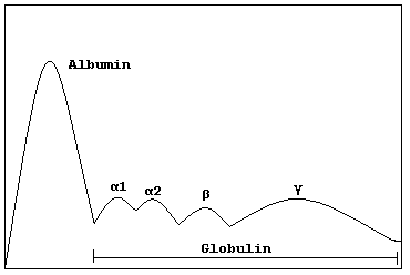
Schematic representation of a normal protein electrophoresis gel. A small spike would be present in the gamma (γ) band in MGUS

Monoclonal gammopathy of undetermined significance (MGUS) is a plasma cell dyscrasia in which plasma cells or other types of antibody-producing cells secrete a myeloma protein, i.e. an abnormal antibody, into the blood; this abnormal protein is usually found during standard laboratory blood or urine tests. MGUS resembles multiple myeloma and similar diseases, but the levels of antibodies are lower,[2] the number of plasma cells (white blood cells that secrete antibodies) in the bone marrow is lower, and it rarely has symptoms or major problems. However, since MGUS can progress to multiple myeloma, with a rate ranging from 0.5% to 1.5% per year depending on the risk category, yearly monitoring is recommended.

The progression from MGUS to multiple myeloma usually involves several steps. In rare cases, it may also be related with a slowly progressive symmetric distal sensorimotor neuropathy.[3]

Signs and symptoms

[edit]

People with monoclonal gammopathy generally do not experience signs or symptoms.[1] Some people may experience a rash or nerve problems, such as numbness or tingling.[1] MGUS is usually detected by chance when the patient has a blood test for another condition or as part of standard screening.[1]

Pathophysiology

[edit]

Pathologically, the lesion in MGUS is in fact very similar to that in multiple myeloma. There is a predominance of clonal plasma cells in the bone marrow with an abnormal immunophenotype (CD38+ CD56+ CD19−) mixed in with cells of a normal phenotype (CD38+ CD56− CD19+);[4][5] in MGUS, on average more than 3% of the clonal plasma cells have the normal phenotype, whereas in multiple myeloma, less than 3% of the cells have the normal phenotype.[6]

Diagnosis

[edit]

MGUS is a common, age-related medical condition characterized by an accumulation of bone marrow plasma cells derived from a single abnormal clone. Patients may be diagnosed with MGUS if they fulfill the following four criteria:[7]

  1. A monoclonal paraprotein band less than 30 g/L (< 3g/dL);
  2. Plasma cells less than 10% on bone marrow examination;
  3. No evidence of bone lesions, anemia, hypercalcemia, or chronic kidney disease related to the paraprotein, and
  4. No evidence of another B-cell proliferative disorder.

Differential diagnosis

[edit]

Several other illnesses can present with a monoclonal gammopathy, and the monoclonal protein may be the first discovery before a formal diagnosis is made:

Management

[edit]

MGUS occurs in over 3 percent of the White population over the age of 50, and is typically detected as an incidental finding when patients undergo a protein electrophoresis as part of an evaluation for a wide variety of clinical symptoms and disorders (e.g., peripheral neuropathy, vasculitis, hemolytic anemia, skin rashes, hypercalcemia, or elevated erythrocyte sedimentation rate). Although patients with MGUS have sometimes been reported to have peripheral neuropathy, a debilitating condition which causes bizarre sensory problems to painful sensory problems,[13] no treatment is indicated.[citation needed]

The protein electrophoresis test should be repeated annually, and if there is any concern for a rise in the level of monoclonal protein, then prompt referral to a hematologist is required. The hematologist, when first evaluating a case of MGUS, will usually perform a skeletal survey (X-rays of the proximal skeleton), check the blood for hypercalcemia and deterioration in renal function, check the urine for Bence Jones protein and perform a bone marrow biopsy. If none of these tests are abnormal, a patient with MGUS is followed up once every 6 months to a year with a blood test (serum protein electrophoresis).[citation needed]

Prognosis

[edit]

At the Mayo Clinic, MGUS transformed into multiple myeloma or similar lymphoproliferative disorders at the rate of about 0.5–2% a year depending on risk category. However, because they were elderly, most patients with MGUS died of something else and did not go on to develop multiple myeloma. When this was taken into account, only 11.2% developed lymphoproliferative disorders.[14]

Kyle et al. studied the prevalence of myeloma in the population as a whole (not clinic patients) in Olmsted County, Minnesota. They found that the prevalence of MGUS was 3.2% in people above 50, with a slight male predominance (4.0% vs. 2.7%). Prevalence increased with age: of people over 70 up to 5.3% had MGUS, while in the over-85 age group the prevalence was 7.5%. In the majority of cases (63.5%), the paraprotein level was <1 g/dL, while only a very small group had levels over 2 g/dL.[15]

A study of monoclonal protein levels conducted in Ghana showed a prevalence of MGUS of approximately 5.9% in African men over the age of 50.[16]

In 2009, prospective data demonstrated that all or almost all cases of multiple myeloma are preceded by MGUS.[17]

In addition to multiple myeloma, MGUS may also progress to Waldenström's macroglobulinemia or primary amyloidosis.[12]

See also

[edit]

References

[edit]

Further reading

[edit]
[edit]
Revisions and contributorsEdit on WikipediaRead on Wikipedia
from Grokipedia
Monoclonal gammopathy of undetermined significance (MGUS) is a benign, disorder characterized by the presence of a monoclonal protein) in the at a level below 3 g/dL, fewer than 10% clonal s in the , and no evidence of end-organ damage or symptoms related to a . This condition is typically discovered incidentally during routine tests, such as , and does not require immediate treatment but necessitates ongoing monitoring due to its potential to progress to more serious disorders like . MGUS is relatively common, affecting approximately 3% of individuals over 50 years of age and up to 5% of those over 70, with higher among individuals (about twice that of individuals) and a slight predominance in males; sensitive detection methods as of 2025 indicate rates exceeding 10% in those over 50. Risk factors include advancing age, male sex, race, family history of MGUS or related disorders, and possibly , though the exact remains unclear and likely involves a combination of genetic and environmental influences. There are three main subtypes—IgM MGUS, non-IgM (IgG or IgA) MGUS, and light-chain MGUS (with revised criteria as of 2025 narrowing )—each with varying risks of progression; for instance, IgM MGUS is more likely to evolve into or , while non-IgM types are associated with . Diagnosis involves confirming the M protein and ruling out myeloma-defining events, with management focusing on risk-adapted monitoring: low-risk patients require initial follow-up at 6 months and, if stable, every 2–3 years thereafter; more frequent for intermediate- and high-risk. The overall risk of progression to , , , or related conditions is about 1% per year, remaining constant over time, though individual risk can be more precisely estimated using models incorporating M protein size, type, and free light chain abnormalities. Early detection through vigilant monitoring allows for timely intervention, improving outcomes if develops.

Introduction and Epidemiology

Definition

Monoclonal gammopathy of undetermined significance (MGUS) is defined as a premalignant disorder characterized by the presence of a monoclonal protein (M-protein) in the serum or at a concentration less than 30 g/L, plasma cells comprising less than 10% of total nucleated cells, absence of end-organ damage attributable to the plasma cell proliferative disorder (as indicated by the criteria: hypercalcemia, renal insufficiency, anemia, and bone lesions), and lack of evidence for other such as , Waldenström macroglobulinemia, , or . The term MGUS was coined in 1978 by Robert A. Kyle and Philip R. Greipp to describe an gammopathy observed in 241 patients, where the monoclonal protein was detected without evidence of progression to malignancy at the time of diagnosis. This designation highlighted the benign nature of the condition in the majority of cases, distinguishing it from overt malignancies based on clinical and laboratory features. MGUS represents an clonal proliferation of plasma cells that can potentially progress to , , or , underscoring its premalignant status within the spectrum of . As of 2025, MGUS is increasingly viewed as part of a continuous spectrum of disorders, with conventional estimates at approximately 3.2% among adults over 50 years old in predominantly , though sensitive detection methods report higher rates up to 10% in some groups, reflecting advancements in detection methods and -based studies.

Monoclonal gammopathy of undetermined significance (MGUS) is a common premalignant condition, with estimates varying by age and demographics. In predominantly , the crude is approximately 3.2% among individuals older than 50 years, increasing to about 5.3% in those over 70 years and reaching 7.5% in those 85 years or older. Recent analyses from large cohorts confirm these figures remain stable at around 3% for those over 50, rising to about 5.3% over 70, as of 2025. The annual incidence of MGUS is estimated at 100-150 per 100,000 adults over 50 years, with rates increasing progressively with age—from about 120 per 100,000 at age 50 in men to 530 per 100,000 by age 90, and lower in women at 60-370 per 100,000. These incidence patterns have remained stable over decades, though improved routine screening has led to higher detection rates in recent years. Demographic risk factors strongly influence MGUS occurrence, with age as the most significant predictor—rare under 40 years—and a 1.5:1 male-to-female ratio. show a baseline prevalence of 3.2%, while exhibit nearly double the rate at 5.8-6.1% over 50, alongside higher progression risks. Geographic and ethnic variations highlight disparities, with a 5.84% prevalence reported in Ghanaian men over 50 in a 2007 study, compared to lower rates of about 2.1% in Japanese adults over 42. Prevalence ranges from 0.24% to 9% across global regions, with potential underdiagnosis in low-resource settings noted in 2025 updates. Asians generally experience lower rates than Western or African populations. Key risk factors include family , which confers a 2-3-fold increased in first-degree relatives of MGUS or patients. Emerging associations from recent meta-analyses link autoimmune diseases and chronic infections to higher MGUS prevalence, alongside , which raises odds by 73% after adjusting for confounders.

Pathophysiology and Classification

Pathophysiology

Monoclonal gammopathy of undetermined significance (MGUS) arises from abnormal differentiation of B cells into clonal plasma cells, leading to the production of monoclonal immunoglobulins without causing end-organ damage. This clonal proliferation is characterized by infiltration of less than 10% plasma cells, distinguishing MGUS from , where infiltration typically exceeds 10% and is associated with symptomatic disease. The process begins in the of lymphoid tissues, where post-germinal center B cells undergo and class-switch recombination, resulting in a stable but premalignant clone that secretes M-protein at levels below 3 g/dL. Genetic abnormalities in MGUS plasma cells include primary events such as hyperdiploidy, affecting chromosomes 5, 7, 9, 11, 15, and 19, and (IgH) translocations, notably t(11;14)(q13;q32) involving CCND1, which occur in approximately 25-50% of cases. Secondary genetic changes, including RAS mutations (primarily in NRAS and ) and TP53 mutations, are present in about 5-20% of MGUS cases, significantly lower than the 30-50% frequency observed in . Overall, MGUS exhibits a lower burden compared to , with fewer driver mutations and less genomic instability, contributing to its indolent nature. Immunophenotypically, MGUS plasma cells often display aberrant surface marker expression, such as CD56 positivity and negativity, which deviate from the normal plasma cell profile of +, ++, CD45 low, and CD56-. However, a key distinguishing feature is the persistence of greater than 5% phenotypically normal residual s in the of MGUS patients, in contrast to less than 5% in , reflecting a less dominant clonal population. The microenvironment in MGUS supports clonal survival through interactions with stromal cells, cytokines, and immune components, without promoting full . Dysregulated and immune evasion mechanisms, including reduced cytotoxic T-cell activity and altered myeloid-derived suppressor cells, foster a permissive niche that sustains low-level clonality. Recent studies highlight the emerging role of epigenetic modifications, such as altered patterns in genes like PHOX2A and CDH2, which show decreased methylation in MGUS compared to normal plasma cells and may contribute to clonal maintenance. Additionally, gut microbiome has been implicated in sustaining MGUS clonality, with microbiota-derived metabolites like influencing immune modulation and progression risk in precursor conditions.

Classification and Subtypes

Monoclonal gammopathy of undetermined significance (MGUS) is classified into distinct subtypes primarily based on the isotype of the monoclonal protein (M-protein), its size, and the free light chain (FLC) ratio, which guide clinical management and . The three main subtypes are non-IgM MGUS, IgM MGUS, and light-chain MGUS (LC-MGUS). Non-IgM MGUS, the most common subtype accounting for approximately 70% of cases, typically involves an intact immunoglobulin with IgG predominance (about 69%) over IgA (11%), and rarely IgD or IgE. IgM MGUS represents 15-20% of cases and is characterized by an IgM M-protein, often serving as a precursor to . LC-MGUS, comprising around 15% of MGUS, lacks a detectable intact immunoglobulin and features only abnormal free light chains, diagnosed via an elevated or FLC with an abnormal / ratio but without end-organ damage. Classification criteria emphasize the M-protein isotype for subtype assignment, alongside serum M-protein concentration below 30 g/L and a normal FLC ratio (typically 0.26-1.65) for non-LC subtypes, ensuring distinction from malignant conditions. A significant refinement occurred in 2025 with updated reference intervals for FLC assays in LC-MGUS diagnosis: the / ratio was revised to 0.44-2.16 for individuals under 70 years and 0.46-2.59 for those 70 years and older, based on large cohort studies with preserved function. This adjustment, derived from the iStopMM study, reduced false-positive LC-MGUS by 82% by improving assay specificity. Clinically, these subtypes differ in their predominant progression pathways, reflecting underlying pathogenic differences such as lymphoplasmacytic features in IgM MGUS. Non-IgM MGUS primarily advances to , while IgM MGUS is associated with progression to or . LC-MGUS carries risks for light-chain multiple myeloma or , underscoring the need for subtype-specific monitoring. Rare variants include biclonal MGUS, occurring in about 5% of cases with two distinct M-proteins (e.g., IgG and IgA), and oligoclonal patterns, which involve multiple bands often seen in immune-compromised states or post-therapy but are not true MGUS precursors. The 2025 JAMA Oncology study on LC-MGUS redefinition has further enhanced subtype specificity, promoting more precise risk stratification across populations.

Clinical Presentation

Signs and Symptoms

Monoclonal gammopathy of undetermined significance (MGUS) is typically , with the majority of cases discovered incidentally during routine blood tests for unrelated conditions. By definition, MGUS does not cause end-organ damage, distinguishing it from more advanced disorders. Rare direct manifestations may occur, particularly in specific subtypes. , presenting as numbness or tingling in the extremities, affects approximately 15–50% of patients with IgM MGUS and is often associated with anti-myelin-associated glycoprotein (anti-MAG) antibodies. Some individuals experience skin rashes or , though these are uncommon and typically linked to monoclonal gammopathies of rather than standard MGUS. MGUS lacks the characteristic CRAB features attributable to the disorder, including anemia, bone pain or lesions, renal failure, or hypercalcemia. Incidental findings such as fatigue or recurrent infections may arise in association with hypogammaglobulinemia, but these are not diagnostic criteria for MGUS and require evaluation for other causes.

Associated Conditions

Patients with monoclonal gammopathy of undetermined significance (MGUS) exhibit an increased risk of venous thromboembolism, with studies reporting a 1.3- to 3.4-fold elevation compared to individuals without MGUS. This prothrombotic state is attributed to factors such as elevated levels of factor VIII and reduced fibrinolytic activity in affected individuals. Additionally, MGUS is linked to a higher incidence of infections, including bacterial and viral types, due to underlying immune dysregulation that impairs effective pathogen clearance. Immune alterations, such as reduced vaccine responsiveness and polyclonal immunoglobulin suppression, are evident even in early MGUS stages. Associations between MGUS and autoimmune diseases have been observed in certain cohorts, with higher prevalence noted in conditions like and . However, population-based screening studies indicate no significant overall link, suggesting that clinical detection of MGUS may confound such associations in symptomatic patients. A key secondary effect of MGUS involves , which suppresses normal immunoglobulin production and predisposes individuals to recurrent infections, even without overt progression to . This immune suppression can manifest as frequent bacterial infections requiring medical intervention. Furthermore, MGUS is associated with rare osteoporotic fractures independent of development, with fracture risk elevated 1.4- to 2.5-fold due to subtle defects like increased bone volume and altered turnover. Non-malignant associations include heightened cardiovascular morbidity, encompassing conditions such as and . Recent analyses confirm a 20% increased risk of all-cause mortality in MGUS patients, persisting independently of progression to plasma cell disorders and linked to comorbidities like infections and cardiovascular events. In screening contexts, MGUS is incidentally detected in 3-5% of patients over age 50 evaluated for unrelated conditions, such as , highlighting its role in broader diagnostic workups. This overlap underscores the need to consider MGUS in neuropathy cases without direct symptomatic attribution.

Diagnosis

Diagnostic Criteria

The diagnosis of monoclonal gammopathy of undetermined significance (MGUS) is established according to the International Myeloma Working Group (IMWG) criteria, which require the presence of a monoclonal protein (M-protein) in serum at a concentration less than 30 g/L, or in urine less than 500 mg per 24 hours, along with bone marrow plasma cells comprising less than 10% of total cellularity, and the absence of myeloma-defining events or end-organ damage. These criteria, originally updated in 2014, were reaffirmed in subsequent IMWG guidelines through 2025 without substantive changes to the core thresholds for MGUS. Key exclusions include no evidence of CRAB features (hypercalcemia, renal insufficiency, , or bone lesions attributable to the disorder) and no myeloma-defining events, such as bone marrow s ≥60%, an involved-to-uninvolved serum free light (FLC) ≥100 (with the involved FLC level ≥100 mg/L), or more than one focal lesion on (MRI). Additionally, there must be no findings suggestive of other or , including , Waldenström macroglobulinemia, or , with normal , serum creatinine, and calcium levels to rule out associated complications. These criteria apply uniformly across MGUS subtypes, including IgM, non-IgM, and light-chain MGUS (LC-MGUS); for LC-MGUS specifically, diagnosis requires an abnormal serum FLC ratio with an elevated level of the involved serum FLC, urinary monoclonal protein below 500 mg per 24 hours, normal total immunoglobulin levels (or no immunoglobulin suppression), alongside the standard and exclusion requirements. Refinements emphasized in 2025 IMWG-related publications focus on preventing , particularly for low-risk incidental findings in screening contexts, by incorporating age-adjusted FLC reference intervals (e.g., κ/λ ratio of 0.44–2.16 for individuals under 70 years and 0.30–3.50 for those 70 years and older, with normal renal function) to reduce false positives in LC-MGUS by 82% and prioritizing multiparametric to confirm clonality in ambiguous cases.

Laboratory and Imaging Tests

The diagnosis of monoclonal gammopathy of undetermined significance (MGUS) relies on a series of laboratory tests to detect and characterize the monoclonal protein (M-protein) while excluding end-organ damage. Initial screening begins with (SPEP), which separates serum proteins to identify an M-protein spike, often denoted as an "M-spike" in laboratory reports. For example, a notation such as "M-spike 1 of 0.5" indicates a single monoclonal protein spike with a concentration of 0.5 g/dL (normal value is 0 g/dL), reflecting a small amount of abnormal M-protein typically seen in MGUS; levels below 1.5 g/dL, when combined with other favorable factors such as IgG subtype and normal serum free light chain ratio, are often indicative of low-risk MGUS. followed by to confirm its monoclonality and determine the heavy and light chain types. Urine protein electrophoresis (UPEP) on a 24-hour collection, combined with urine , is performed to detect Bence Jones proteins or light chain involvement not visible in serum. The serum free light chain (FLC) assay measures and light chains and their ratio, aiding in the identification of light chain MGUS (LC-MGUS) when an M-protein is absent on . Confirmatory laboratory tests include a (CBC) to assess for , serum creatinine to evaluate renal function, and serum calcium levels to rule out hypercalcemia, collectively helping exclude CRAB features (hypercalcemia, renal insufficiency, , bone lesions). biopsy and aspirate, with to quantify clonal plasma cells and cytogenetic analysis for chromosomal abnormalities, are recommended if the serum M-protein exceeds 1.5 g/dL (15 g/L) or the FLC ratio is abnormal, though they may be deferred in low-risk cases to avoid unnecessary procedures. Imaging studies are not routinely indicated for asymptomatic MGUS but are used to exclude bone involvement. A skeletal survey using plain X-rays of the , spine, , and long s detects lytic lesions, while low-dose whole-body CT is an alternative with higher sensitivity if symptoms such as are present. /computed tomography (PET/CT) is not standard for initial evaluation in patients due to its higher cost and without added benefit in low-risk MGUS. Quantitative assessment of the M-protein involves on the electrophoresis gel to measure its concentration in g/dL, ensuring it is below 3 g/dL for MGUS . Serum immunoglobulin levels (IgG, IgA, IgM) are also quantified to evaluate for suppression of uninvolved immunoglobulins, which provides additional context for the monoclonal expansion. Recent advancements as of 2025 include the use of , such as the MASS-FIX assay, for detecting low-level M-proteins with limits of detection as low as 0.001 g/L (0.0001 g/dL), improving sensitivity over traditional in cases of small or . For LC-MGUS, expanded FLC reference ranges adjusted for age—such as a / ratio of 0.44-2.16 for those under 70 with normal renal function—have been proposed to reduce misclassification and false positives by 82%; race-adjusted intervals have also been proposed, particularly for populations of African descent where traditional ranges lead to .

Differential Diagnosis

The differential diagnosis of monoclonal gammopathy of undetermined significance (MGUS) is crucial to exclude malignant or symptomatic conditions that may present with similar serum protein abnormalities, ensuring appropriate clinical management. Key distinctions rely on serum M-protein levels, findings, clinical symptoms, and specialized testing such as and . Malignant mimics include , characterized by an M-protein of ≥30 g/L and/or 10-59% clonal in the without end-organ damage (CRAB criteria: hypercalcemia, renal failure, , bone lesions). In contrast, solitary manifests as a single localized confirmed by and imaging, with absent or minimal systemic M-protein and no evidence of widespread involvement. Other gammopathies to consider are , which typically features an IgM M-protein, ≥10% infiltration by lymphoplasmacytic cells, and associated symptoms like hyperviscosity, , or . is differentiated by monoclonal light chain deposition causing organ dysfunction, verified through tissue demonstrating deposits and evidence of an underlying disorder. Benign or reactive conditions encompass polyclonal gammopathy, often arising from infections, autoimmune diseases, or , and identified by a broad, non-discrete elevation in gamma globulins on without a sharp monoclonal band. , appearing as multiple discrete protein bands, may occur in smoldering or as a post-therapy phenomenon in disorders, differing from the single dominant band of MGUS. Rare entities such as involve a alongside major criteria including , , endocrinopathy, and skin changes; elevated serum (VEGF) levels support differentiation from isolated MGUS. In 2025, updated free light chain (FLC) reference intervals for light-chain MGUS (LC-MGUS), derived from large cohort studies and adjusted for age, enhance differentiation from renal disease or by accounting for age and kidney function, reducing false-positive rates by 82% and minimizing .

Risk Assessment and Prognosis

Risk Stratification

Risk stratification in monoclonal gammopathy of undetermined significance (MGUS) involves assessing the likelihood of progression to or related disorders using validated models based on clinical and laboratory parameters. The most widely adopted approach is the risk stratification model, which identifies three key risk factors: serum M-protein concentration ≥1.5 g/dL, non-IgG M-protein isotype (IgA or IgM), and an abnormal serum free light chain (FLC) (κ/λ <0.26 or >1.65). Each factor is assigned one point, yielding a total score of 0 to 3 that categorizes patients into risk groups. For example, a serum M-protein concentration of 0.5 g/dL (often reported as an M-spike of 0.5 g/dL, such as "M-spike 1 of 0.5" indicating a single monoclonal spike) combined with IgG isotype and normal serum free light chain ratio results in zero risk factors and low-risk classification. Patients are classified as low-risk (0 factors), low-intermediate risk (1 factor), high-intermediate risk (2 factors), or high-risk (3 factors). The cumulative probability of progression at 20 years is 5% for low-risk, 21% for low-intermediate, 37% for high-intermediate, and 58% for high-risk patients, corresponding to approximate annual progression rates of 0.25%, 1%, 2.5%, and 5%, respectively. Overall, the annual risk of progression from MGUS to is about 1%, though this varies significantly by risk group. plasma cell percentage, while limited to <10% by MGUS diagnostic criteria, and the presence of immunoparesis (suppression of uninvolved immunoglobulins) can further refine in select cases. Recent advancements have incorporated genomic profiling to enhance prediction accuracy. The Dana-Farber genomic score, or "MM-like score," analyzes DNA mutations and genetic abnormalities in tumor samples to gauge disease severity and progression risk from MGUS, with higher scores predicting faster advancement to multiple myeloma; it was validated in a cohort of over 1,000 individuals, including those with MGUS. Mutations such as TP53 alterations, associated with genomic instability, are emerging as high-risk markers in precursor conditions like MGUS. Additionally, a 2025 predictive scoring model using clinical comorbidities (e.g., diabetes, chronic pulmonary disease) from a large Korean cohort has been developed to quantify progression risk, stratifying patients into low, intermediate, and high groups with hazard ratios up to 2.53 for the highest category. For intermediate- or high-risk patients, guidelines recommend repeating laboratory tests at 6 months to monitor for early signs of progression.

Prognosis

The prognosis for patients with monoclonal gammopathy of undetermined significance (MGUS) is generally favorable, as the condition remains stable in the majority of cases without progression to . The overall rate of progression to or a related disorder is approximately 1% per year, translating to a cumulative lifetime of 10% to 15%, with 80% to 90% of patients experiencing no progression over their lifetime. A long-term study reported cumulative progression rates of 12% at 10 years, 25% at 20 years, and 30% at 25 years, though more recent analyses accounting for competing of suggest a lower estimate of around 11% at 25 years. Progression risk varies significantly based on patient-specific factors identified through risk stratification models, with high-risk MGUS progressing 5 to 10 times faster than low-risk cases and a time to progression of 10 to 20 years. Subtype-specific rates, such as higher progression in IgM MGUS compared to non-IgM forms, further influence outcomes (as detailed in Classification and Subtypes). Early detection through vigilant monitoring allows for timely intervention, improving outcomes if develops. Survival among MGUS patients is near-normal, approximating 85% to 90% of age-matched population expectations, primarily because most individuals do not progress to symptomatic disease. However, there is a modest increase in non-hematologic mortality attributable to associated conditions, such as infections or cardiovascular issues. For those who do not progress, MGUS has minimal impact on , but ongoing monitoring can induce anxiety and psychological distress, as evidenced by studies showing reduced health-related and heightened cancer worry among affected patients.

Management

Monitoring Protocol

For patients diagnosed with monoclonal gammopathy of undetermined significance (MGUS), monitoring is essential to detect progression to symptomatic disorders such as or related conditions, as the condition remains asymptomatic by . The International Myeloma (IMWG) guidelines emphasize risk-stratified to balance detection of progression with minimizing unnecessary testing. Initial follow-up typically includes (SPEP) and serum free light chain (FLC) assay at 6 months post-diagnosis for all patients to confirm stability. Subsequent monitoring intervals are tailored based on risk stratification, which incorporates factors such as M-protein level, immunoglobulin type, and FLC ratio (as detailed in protocols). For stable low-risk MGUS (e.g., IgG type, M-protein <1.5 g/dL, normal FLC ratio), evaluation with SPEP, serum , FLC, (CBC), serum creatinine, and calcium every 2-3 years is recommended. In contrast, intermediate- or high-risk MGUS (e.g., non-IgG type, M-protein ≥1.5 g/dL, or abnormal FLC ratio) warrants assessment every 12 months using the same laboratory tests. For light-chain MGUS, urine protein (UPEP) and urine should be added at these intervals to monitor urinary M-protein. Escalation of monitoring occurs if there is significant change in M-protein levels, such as a doubling or a rise greater than 25% (or an absolute increase ≥0.5 g/dL), prompting repeat to assess clonal plasma cells. Imaging studies, such as skeletal survey, low-dose whole-body CT, or MRI, are not recommended routinely but should be pursued if new symptoms suggestive of CRAB features (hypercalcemia, renal insufficiency, , or bone lesions) emerge. is a key component, informing individuals about warning signs like persistent , unexplained , recurrent infections, or to facilitate prompt reporting. IMWG guidelines recommend that monitoring should be individualized by risk, with no routine advanced imaging unless CRAB symptoms are suspected, and emphasis on shared decision-making. is generally lifelong, though consensus allows de-escalation or discontinuation in elderly patients with low-risk MGUS and limited (e.g., <5 years), to reduce burden without compromising outcomes.

Intervention for Progression

Patients with monoclonal gammopathy of undetermined significance (MGUS) do not require treatment, as the condition is and early intervention has not demonstrated benefits in preventing progression or improving overall . Instead, active is the standard approach until evidence of progression emerges. Clinical trials, such as those evaluating lenalidomide-dexamethasone in precursor conditions, have shown prolonged but no overall advantage, reinforcing the lack of routine therapeutic intervention for MGUS itself. Intervention is initiated upon confirmed progression to smoldering multiple myeloma (SMM) or (MM), typically triggered by the development of features (hypercalcemia, renal insufficiency, , or bone lesions) or myeloma-defining events (MDEs), including bone marrow plasma cells ≥60%, an involved/uninvolved serum free light (FLC) ≥100 with involved FLC ≥100 mg/L, or more than one focal (≥5 mm) on MRI. Upon suspicion of progression, patients should be referred to a hematologist for confirmatory evaluation. For progression to high-risk SMM, treatment with approved regimens such as subcutaneous monotherapy (FDA-approved November 2025) is recommended, which significantly reduces the risk of progression to active MM compared to ; asymptomatic low-risk SMM continues . Progression to symptomatic MM prompts immediate . Treatment for progressed disease follows established protocols for SMM or MM. In high-risk SMM, risk-adapted approaches include subcutaneous monotherapy, which significantly reduces the risk of progression to active MM compared to observation. For active MM, frontline therapy typically involves proteasome inhibitors like combined with immunomodulatory agents (e.g., ) and dexamethasone (VRd regimen), often incorporating such as for enhanced efficacy. As of 2025, ongoing trials explore preventive strategies for high-risk precursors, including -based regimens to delay progression in select cases bordering SMM. Supportive care addresses complications upon progression. Bisphosphonates, such as , are used if bone lesions or hypercalcemia develop, to prevent skeletal-related events, though not routinely in non-progressed MGUS. In IgM MGUS cases progressing with associated , management may include intravenous immunoglobulin (IVIG) for short-term symptom relief or rituximab to target the monoclonal protein, particularly if anti-MAG antibodies are present.

References

Add your contribution
Related Hubs
User Avatar
No comments yet.