Recent from talks
Blood test
Knowledge base stats:
Talk channels stats:
Members stats:
Blood test
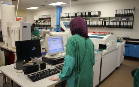
A blood test is a laboratory analysis performed on a blood sample that is usually extracted from a vein in the arm using a hypodermic needle, or via fingerprick. Multiple tests for specific blood components, such as a glucose test or a cholesterol test, are often grouped together into one test panel called a blood panel or blood work. Blood tests are often used in health care to determine physiological and biochemical states, such as disease, mineral content, pharmaceutical drug effectiveness, and organ function. Typical clinical blood panels include a basic metabolic panel or a complete blood count. Blood tests are also used in drug tests to detect drug abuse.
A venipuncture is useful as it is a minimally invasive way to obtain cells and extracellular fluid (plasma) from the body for analysis. Blood flows throughout the body, acting as a medium that provides oxygen and nutrients to tissues and carries waste products back to the excretory systems for disposal. Consequently, the state of the bloodstream affects or is affected by, many medical conditions. For these reasons, blood tests are the most commonly performed medical tests.
If only a few drops of blood are needed, a fingerstick is performed instead of a venipuncture.
Indwelling arterial, central venous and peripheral venous lines can also be used to draw blood.
Phlebotomists, laboratory practitioners and nurses are those in charge of extracting blood from a patient. This can be done in-clinic, in-office, or through mobile (home) phlebotomists. However, in special circumstances, and/or emergency situations, paramedics and physicians extract the blood. Also, respiratory therapists are trained to extract arterial blood to examine arterial blood gases.
A basic metabolic panel measures sodium, potassium, chloride, bicarbonate, blood urea nitrogen (BUN), magnesium, creatinine, glucose, and sometimes calcium. Tests that focus on cholesterol levels can determine LDL and HDL cholesterol levels, as well as triglyceride levels.
Some tests, such as those that measure glucose or a lipid profile, require fasting (or no food consumption) eight to twelve hours prior to the drawing of the blood sample.
For the majority of tests, blood is usually obtained from the patient's vein. Other specialized tests, such as the arterial blood gas test, require blood extracted from an artery. Blood gas analysis of arterial blood is primarily used to monitor carbon dioxide and oxygen levels related to pulmonary function, but is also used to measure blood pH and bicarbonate levels for certain metabolic conditions.
Hub AI
Blood test AI simulator
(@Blood test_simulator)
Blood test
A blood test is a laboratory analysis performed on a blood sample that is usually extracted from a vein in the arm using a hypodermic needle, or via fingerprick. Multiple tests for specific blood components, such as a glucose test or a cholesterol test, are often grouped together into one test panel called a blood panel or blood work. Blood tests are often used in health care to determine physiological and biochemical states, such as disease, mineral content, pharmaceutical drug effectiveness, and organ function. Typical clinical blood panels include a basic metabolic panel or a complete blood count. Blood tests are also used in drug tests to detect drug abuse.
A venipuncture is useful as it is a minimally invasive way to obtain cells and extracellular fluid (plasma) from the body for analysis. Blood flows throughout the body, acting as a medium that provides oxygen and nutrients to tissues and carries waste products back to the excretory systems for disposal. Consequently, the state of the bloodstream affects or is affected by, many medical conditions. For these reasons, blood tests are the most commonly performed medical tests.
If only a few drops of blood are needed, a fingerstick is performed instead of a venipuncture.
Indwelling arterial, central venous and peripheral venous lines can also be used to draw blood.
Phlebotomists, laboratory practitioners and nurses are those in charge of extracting blood from a patient. This can be done in-clinic, in-office, or through mobile (home) phlebotomists. However, in special circumstances, and/or emergency situations, paramedics and physicians extract the blood. Also, respiratory therapists are trained to extract arterial blood to examine arterial blood gases.
A basic metabolic panel measures sodium, potassium, chloride, bicarbonate, blood urea nitrogen (BUN), magnesium, creatinine, glucose, and sometimes calcium. Tests that focus on cholesterol levels can determine LDL and HDL cholesterol levels, as well as triglyceride levels.
Some tests, such as those that measure glucose or a lipid profile, require fasting (or no food consumption) eight to twelve hours prior to the drawing of the blood sample.
For the majority of tests, blood is usually obtained from the patient's vein. Other specialized tests, such as the arterial blood gas test, require blood extracted from an artery. Blood gas analysis of arterial blood is primarily used to monitor carbon dioxide and oxygen levels related to pulmonary function, but is also used to measure blood pH and bicarbonate levels for certain metabolic conditions.
Recent media