Hubbry Logo
NeuroscienceNeuroscienceMain
Open search
Neuroscience
Community hub
Neuroscience
logo
8 pages, 0 posts
0 subscribers
Be the first to start a discussion here.
Neuroscience
Neuroscience
from Wikipedia

Drawing by Santiago Ramón y Cajal (1899) of neurons in the pigeon cerebellum

Neuroscience is the scientific study of the nervous system (the brain, spinal cord, and peripheral nervous system), its functions, and its disorders.[1][2][3] It is a multidisciplinary science that combines physiology, anatomy, molecular biology, developmental biology, cytology, psychology, physics, computer science, chemistry, medicine, statistics, and mathematical modeling to understand the fundamental and emergent properties of neurons, glia, and neural circuits.[4][5][6][7][8] The understanding of the biological basis of learning, memory, behavior, perception, and consciousness has been described by Eric Kandel as the "epic challenge" of the biological sciences.[9]

The scope of neuroscience has broadened over time to include different approaches used to study the nervous system at different scales. The techniques used by neuroscientists have expanded enormously, from molecular and cellular studies of individual neurons to imaging of sensory, motor, and cognitive tasks in the brain.

History

[edit]
Illustration from Gray's Anatomy (1918) of a lateral view of the human brain, featuring the hippocampus among other neuroanatomical features

The earliest study of the nervous system dates to ancient Egypt. Trepanation, the surgical practice of either drilling or scraping a hole into the skull for the purpose of curing head injuries or mental disorders, or relieving cranial pressure, was first recorded during the Neolithic period. Manuscripts dating to 1700 BC indicate that the Egyptians had some knowledge about symptoms of brain damage.[10]

Early views on the function of the brain regarded it to be a "cranial stuffing" of sorts. In Egypt, from the late Middle Kingdom onwards, the brain was regularly removed in preparation for mummification. It was believed at the time that the heart was the seat of intelligence. According to Herodotus, the first step of mummification was to "take a crooked piece of iron, and with it draw out the brain through the nostrils, thus getting rid of a portion, while the skull is cleared of the rest by rinsing with drugs."[11]

The view that the heart was the source of consciousness was not challenged until the time of the Greek physician Hippocrates. He believed that the brain was not only involved with sensation—since most specialized organs (e.g., eyes, ears, tongue) are located in the head near the brain—but was also the seat of intelligence.[12] Plato also speculated that the brain was the seat of the rational part of the soul.[13] Aristotle, however, believed the heart was the center of intelligence and that the brain regulated the amount of heat from the heart.[14] This view was generally accepted until the Roman physician Galen, a follower of Hippocrates and physician to Roman gladiators, observed that his patients lost their mental faculties when they had sustained damage to their brains.[15]

Abulcasis, Averroes, Avicenna, Avenzoar, and Maimonides, active in the Medieval Muslim world, described a number of medical problems related to the brain. In Renaissance Europe, Vesalius (1514–1564), René Descartes (1596–1650), Thomas Willis (1621–1675) and Jan Swammerdam (1637–1680) also made several contributions to neuroscience.

The Golgi stain first allowed for the visualization of individual neurons.

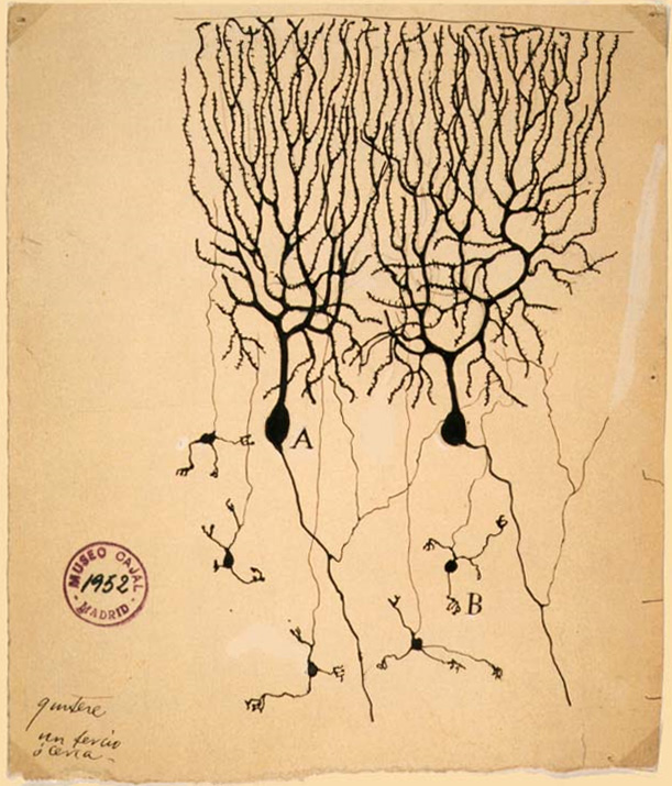
Luigi Galvani's pioneering work in the late 1700s set the stage for studying the electrical excitability of muscles and neurons. In 1843 Emil du Bois-Reymond demonstrated the electrical nature of the nerve signal,[16] whose speed Hermann von Helmholtz proceeded to measure,[17] and in 1875 Richard Caton found electrical phenomena in the cerebral hemispheres of rabbits and monkeys.[18] Adolf Beck published in 1890 similar observations of spontaneous electrical activity of the brain of rabbits and dogs.[19] Studies of the brain became more sophisticated after the invention of the microscope and the development of a staining procedure by Camillo Golgi during the late 1890s. The procedure used a silver chromate salt to reveal the intricate structures of individual neurons. His technique was used by Santiago Ramón y Cajal and led to the formation of the neuron doctrine, the hypothesis that the functional unit of the brain is the neuron.[20] Golgi and Ramón y Cajal shared the Nobel Prize in Physiology or Medicine in 1906 for their extensive observations, descriptions, and categorizations of neurons throughout the brain.

In parallel with this research, in 1815 Jean Pierre Flourens induced localized lesions of the brain in living animals to observe their effects on motricity, sensibility and behavior. Work with brain-damaged patients by Marc Dax in 1836 and Paul Broca in 1865 suggested that certain regions of the brain were responsible for certain functions. At the time, these findings were seen as a confirmation of Franz Joseph Gall's theory that language was localized and that certain psychological functions were localized in specific areas of the cerebral cortex.[21][22] The localization of function hypothesis was supported by observations of epileptic patients conducted by John Hughlings Jackson, who correctly inferred the organization of the motor cortex by watching the progression of seizures through the body. Carl Wernicke further developed the theory of the specialization of specific brain structures in language comprehension and production. Modern research through neuroimaging techniques, still uses the Brodmann cerebral cytoarchitectonic map (referring to the study of cell structure) anatomical definitions from this era in continuing to show that distinct areas of the cortex are activated in the execution of specific tasks.[23]

During the 20th century, neuroscience began to be recognized as a distinct academic discipline in its own right, rather than as studies of the nervous system within other disciplines. Eric Kandel and collaborators have cited David Rioch, Francis O. Schmitt, and Stephen Kuffler as having played critical roles in establishing the field.[24] Rioch originated the integration of basic anatomical and physiological research with clinical psychiatry at the Walter Reed Army Institute of Research, starting in the 1950s. During the same period, Schmitt established a neuroscience research program within the Biology Department at the Massachusetts Institute of Technology, bringing together biology, chemistry, physics, and mathematics. The first freestanding neuroscience department (then called Psychobiology) was founded in 1964 at the University of California, Irvine by James L. McGaugh.[25] This was followed by the Department of Neurobiology at Harvard Medical School, which was founded in 1966 by Stephen Kuffler.[26]

3-D sensory and motor homunculus models at the Natural History Museum, London

In the process of treating epilepsy, Wilder Penfield produced maps of the location of various functions (motor, sensory, memory, vision) in the brain.[27][28] He summarized his findings in a 1950 book called The Cerebral Cortex of Man.[29] Wilder Penfield and his co-investigators Edwin Boldrey and Theodore Rasmussen are considered to be the originators of the cortical homunculus.[30]

The understanding of neurons and of nervous system function became increasingly precise and molecular during the 20th century. For example, in 1952, Alan Lloyd Hodgkin and Andrew Huxley presented a mathematical model for the transmission of electrical signals in neurons of the giant axon of a squid, which they called "action potentials", and how they are initiated and propagated, known as the Hodgkin–Huxley model. In 1961–1962, Richard FitzHugh and J. Nagumo simplified Hodgkin–Huxley, in what is called the FitzHugh–Nagumo model. In 1962, Bernard Katz modeled neurotransmission across the space between neurons known as synapses. Beginning in 1966, Eric Kandel and collaborators examined biochemical changes in neurons associated with learning and memory storage in Aplysia. In 1981 Catherine Morris and Harold Lecar combined these models in the Morris–Lecar model. Such increasingly quantitative work gave rise to numerous biological neuron models and models of neural computation.

As a result of the increasing interest about the nervous system, several prominent neuroscience organizations have been formed to provide a forum to all neuroscientists during the 20th century. For example, the International Brain Research Organization was founded in 1961,[31] the International Society for Neurochemistry in 1963,[32] the European Brain and Behaviour Society in 1968,[33] and the Society for Neuroscience in 1969.[34] Recently, the application of neuroscience research results has also given rise to applied disciplines as neuroeconomics,[35] neuroeducation,[36] neuroethics,[37] and neurolaw.[38]

Over time, brain research has gone through philosophical, experimental, and theoretical phases, with work on neural implants and brain simulation predicted to be important in the future.[39]

Modern neuroscience

[edit]
Human nervous system

The scientific study of the nervous system increased significantly during the second half of the twentieth century, principally due to advances in molecular biology, electrophysiology, and computational neuroscience. This has allowed neuroscientists to study the nervous system in all its aspects: how it is structured, how it works, how it develops, how it malfunctions, and how it can be changed.

For example, it has become possible to understand, in much detail, the complex processes occurring within a single neuron. Neurons are cells specialized for communication. They are able to communicate with neurons and other cell types through specialized junctions called synapses, at which electrical or electrochemical signals can be transmitted from one cell to another. Many neurons extrude a long thin filament of axoplasm called an axon, which may extend to distant parts of the body and are capable of rapidly carrying electrical signals, influencing the activity of other neurons, muscles, or glands at their termination points. A nervous system emerges from the assemblage of neurons that are connected to each other in neural circuits, and networks.

The vertebrate nervous system can be split into two parts: the central nervous system (defined as the brain and spinal cord), and the peripheral nervous system. In many species—including all vertebrates—the nervous system is the most complex organ system in the body, with most of the complexity residing in the brain. The human brain alone contains around one hundred billion neurons and one hundred trillion synapses; it consists of thousands of distinguishable substructures, connected to each other in synaptic networks whose intricacies have only begun to be unraveled. At least one out of three of the approximately 20,000 genes belonging to the human genome is expressed mainly in the brain.[40]

Due to the high degree of plasticity of the human brain, the structure of its synapses and their resulting functions change throughout life.[41]

Making sense of the nervous system's dynamic complexity is a formidable research challenge. Ultimately, neuroscientists would like to understand every aspect of the nervous system, including how it works, how it develops, how it malfunctions, and how it can be altered or repaired. Analysis of the nervous system is therefore performed at multiple levels, ranging from the molecular and cellular levels to the systems and cognitive levels. The specific topics that form the main focus of research change over time, driven by an ever-expanding base of knowledge and the availability of increasingly sophisticated technical methods. Improvements in technology have been the primary drivers of progress. Developments in electron microscopy, computer science, electronics, functional neuroimaging, and genetics and genomics have all been major drivers of progress.

Advances in the classification of brain cells have been enabled by electrophysiological recording, single-cell genetic sequencing, and high-quality microscopy, which have combined into a single method pipeline called patch-sequencing in which all three methods are simultaneously applied using miniature tools.[42] The efficiency of this method and the large amounts of data that is generated has allowed researchers to make some general conclusions about cell types; for example that the human and mouse brain have different versions of fundamentally the same cell types.[43]

Molecular and cellular neuroscience

[edit]
Photograph of a stained neuron in a chicken embryo

Basic questions addressed in molecular neuroscience include the mechanisms by which neurons express and respond to molecular signals and how axons form complex connectivity patterns. At this level, tools from molecular biology and genetics are used to understand how neurons develop and how genetic changes affect biological functions.[44] The morphology, molecular identity, and physiological characteristics of neurons and how they relate to different types of behavior are also of considerable interest.[45]

Questions addressed in cellular neuroscience include the mechanisms of how neurons process signals physiologically and electrochemically. These questions include how signals are processed by neurites and somas and how neurotransmitters and electrical signals are used to process information in a neuron. Neurites are thin extensions from a neuronal cell body, consisting of dendrites (specialized to receive synaptic inputs from other neurons) and axons (specialized to conduct nerve impulses called action potentials). Somas are the cell bodies of the neurons and contain the nucleus.[46]

Another major area of cellular neuroscience is the investigation of the development of the nervous system.[47] Questions include the patterning and regionalization of the nervous system, axonal and dendritic development, trophic interactions, synapse formation and the implication of fractones in neural stem cells,[48][49] differentiation of neurons and glia (neurogenesis and gliogenesis), and neuronal migration.[50]

Computational neurogenetic modeling is concerned with the development of dynamic neuronal models for modeling brain functions with respect to genes and dynamic interactions between genes, on the cellular level (Computational Neurogenetic Modeling (CNGM) can also be used to model neural systems).[51]

Neural circuits and systems

[edit]
Proposed organization of motor-semantic neural circuits for action language comprehension. Adapted from Shebani et al. (2013).

Systems neuroscience research centers on the structural and functional architecture of the developing human brain, and the functions of large-scale brain networks, or functionally-connected systems within the brain. Alongside brain development, systems neuroscience also focuses on how the structure and function of the brain enables or restricts the processing of sensory information, using learned mental models of the world, to motivate behavior.

Questions in systems neuroscience include how neural circuits are formed and used anatomically and physiologically to produce functions such as reflexes, multisensory integration, motor coordination, circadian rhythms, emotional responses, learning, and memory.[52] In other words, this area of research studies how connections are made and morphed in the brain, and the effect it has on human sensation, movement, attention, inhibitory control, decision-making, reasoning, memory formation, reward, and emotion regulation.[53]

Specific areas of interest for the field include observations of how the structure of neural circuits effect skill acquisition, how specialized regions of the brain develop and change (neuroplasticity), and the development of brain atlases, or wiring diagrams of individual developing brains.[54]

The related fields of neuroethology and neuropsychology address the question of how neural substrates underlie specific animal and human behaviors.[55] Neuroendocrinology and psychoneuroimmunology examine interactions between the nervous system and the endocrine and immune systems, respectively.[56] Despite many advancements, the way that networks of neurons perform complex cognitive processes and behaviors is still poorly understood.[57]

Cognitive and behavioral neuroscience

[edit]

Cognitive neuroscience addresses the questions of how psychological functions are produced by neural circuitry. The emergence of powerful new measurement techniques such as neuroimaging (e.g., fMRI, PET, SPECT), EEG, MEG, electrophysiology, optogenetics and human genetic analysis combined with sophisticated experimental techniques from cognitive psychology allows neuroscientists and psychologists to address abstract questions such as how cognition and emotion are mapped to specific neural substrates. Although many studies hold a reductionist stance looking for the neurobiological basis of cognitive phenomena, recent research shows that there is an interplay between neuroscientific findings and conceptual research, soliciting and integrating both perspectives. For example, neuroscience research on empathy solicited an interdisciplinary debate involving philosophy, psychology and psychopathology.[58] Moreover, the neuroscientific identification of multiple memory systems related to different brain areas has challenged the idea of memory as a literal reproduction of the past, supporting a view of memory as a generative, constructive and dynamic process.[59]

Neuroscience is also allied with the social and behavioral sciences, as well as with nascent interdisciplinary fields. Examples of such alliances include neuroeconomics, decision theory, social neuroscience, and neuromarketing to address complex questions about interactions of the brain with its environment. A study into consumer responses for example uses EEG to investigate neural correlates associated with narrative transportation into stories about energy efficiency.[60]

Computational neuroscience

[edit]

Questions in computational neuroscience can span a wide range of levels of traditional analysis, such as development, structure, and cognitive functions of the brain. Research in this field utilizes mathematical models, theoretical analysis, and computer simulation to describe and verify biologically plausible neurons and nervous systems. For example, biological neuron models are mathematical descriptions of spiking neurons which can be used to describe both the behavior of single neurons as well as the dynamics of neural networks. Computational neuroscience is often referred to as theoretical neuroscience.

Neuroscience and medicine

[edit]

Clinical neuroscience

[edit]

Neurology, psychiatry, neurosurgery, psychosurgery, anesthesiology and pain medicine, neuropathology, neuroradiology, ophthalmology, otolaryngology, clinical neurophysiology, addiction medicine, and sleep medicine are some medical specialties that specifically address the diseases of the nervous system. These terms also refer to clinical disciplines involving diagnosis and treatment of these diseases.[61]

Neurology works with diseases of the central and peripheral nervous systems, such as amyotrophic lateral sclerosis (ALS) and stroke, and their medical treatment. Psychiatry focuses on affective, behavioral, cognitive, and perceptual disorders. Anesthesiology focuses on perception of pain, and pharmacologic alteration of consciousness. Neuropathology focuses upon the classification and underlying pathogenic mechanisms of central and peripheral nervous system and muscle diseases, with an emphasis on morphologic, microscopic, and chemically observable alterations. Neurosurgery and psychosurgery work primarily with surgical treatment of diseases of the central and peripheral nervous systems.[62]

Neuroscience underlies the development of various neurotherapy methods to treat diseases of the nervous system.[63][64][65]

Translational research

[edit]
An MRI of a human head showing benign familial macrocephaly (head circumference > 60 cm)

Recently, the boundaries between various specialties have blurred, as they are all influenced by basic research in neuroscience. For example, brain imaging enables objective biological insight into mental illnesses, which can lead to faster diagnosis, more accurate prognosis, and improved monitoring of patient progress over time.[66]

Integrative neuroscience describes the effort to combine models and information from multiple levels of research to develop a coherent model of the nervous system. For example, brain imaging coupled with physiological numerical models and theories of fundamental mechanisms may shed light on psychiatric disorders.[67]

Another important area of translational research is brain–computer interfaces (BCIs), or machines that are able to communicate and influence the brain. They are currently being researched for their potential to repair neural systems and restore certain cognitive functions.[68] However, some ethical considerations have to be dealt with before they are accepted.[69][70]

Major branches

[edit]

Modern neuroscience education and research activities can be very roughly categorized into the following major branches, based on the subject and scale of the system in examination as well as distinct experimental or curricular approaches. Individual neuroscientists, however, often work on questions that span several distinct subfields.

List of the major branches of neuroscience
Branch Description
Affective neuroscience Affective neuroscience is the study of the neural mechanisms involved in emotion, typically through experimentation on animal models.[71]
Behavioral neuroscience Behavioral neuroscience (also known as biological psychology, physiological psychology, biopsychology, or psychobiology) is the application of the principles of biology to the study of genetic, physiological, and developmental mechanisms of behavior in humans and non-human animals.[72]
Cellular neuroscience Cellular neuroscience is the study of neurons at a cellular level including morphology and physiological properties.[73]
Clinical neuroscience The scientific study of the biological mechanisms that underlie the disorders and diseases of the nervous system.[74]
Cognitive neuroscience Cognitive neuroscience is the study of the biological mechanisms underlying cognition.[74]
Computational neuroscience Computational neuroscience is the theoretical study of the nervous system.[75]
Cultural neuroscience Cultural neuroscience is the study of how cultural values, practices and beliefs shape and are shaped by the mind, brain and genes across multiple timescales.[76]
Developmental neuroscience Developmental neuroscience studies the processes that generate, shape, and reshape the nervous system and seeks to describe the cellular basis of neural development to address underlying mechanisms.[77]
Evolutionary neuroscience Evolutionary neuroscience studies the evolution of nervous systems.[78]
Molecular neuroscience Molecular neuroscience studies the nervous system with molecular biology, molecular genetics, protein chemistry, and related methodologies.[79]
Nanoneuroscience An interdisciplinary field that integrates nanotechnology and neuroscience.[80]
Neural engineering Neural engineering uses engineering techniques to interact with, understand, repair, replace, or enhance neural systems.[81]
Neuroanatomy Neuroanatomy is the study of the anatomy of nervous systems.[82]
Neurochemistry Neurochemistry is the study of how neurochemicals interact and influence the function of neurons.[83]
Neuroethology Neuroethology is the study of the neural basis of non-human animals behavior.
Neurogastronomy Neurogastronomy is the study of flavor and how it affects sensation, cognition, and memory.[84]
Neurogenetics Neurogenetics is the study of the genetical basis of the development and function of the nervous system.[85]
Neuroimaging Neuroimaging includes the use of various techniques to either directly or indirectly image the structure and function of the brain.[86]
Neuroimmunology Neuroimmunology is concerned with the interactions between the nervous and the immune system.[87]
Neuroinformatics Neuroinformatics is a discipline within bioinformatics that conducts the organization of neuroscience data and application of computational models and analytical tools.[88]
Neurolinguistics Neurolinguistics is the study of the neural mechanisms in the human brain that control the comprehension, production, and acquisition of language.[89][74]
Neuro-ophthalmology Neuro-ophthalmology is an academically oriented subspecialty that merges the fields of neurology and ophthalmology, often dealing with complex systemic diseases that have manifestations in the visual system.
Neurophysics Neurophysics is the branch of biophysics dealing with the development and use of physical methods to gain information about the nervous system.[90]
Neurophysiology Neurophysiology is the study of the structure and function of the nervous system, generally using physiological techniques that include measurement and stimulation with electrodes or optically with ion- or voltage-sensitive dyes or light-sensitive channels.[91]
Neuropsychology Neuropsychology is a discipline that resides under the umbrellas of both psychology and neuroscience, and is involved in activities in the arenas of both basic science and applied science. In psychology, it is most closely associated with biopsychology, clinical psychology, cognitive psychology, and developmental psychology. In neuroscience, it is most closely associated with the cognitive, behavioral, social, and affective neuroscience areas. In the applied and medical domain, it is related to neurology and psychiatry.[92]
Neuropsychopharmacology Neuropsychopharmacology, an interdisciplinary science related to psychopharmacology and fundamental neuroscience, is the study of the neural mechanisms that drugs act upon to influence behavior.[93]
Optogenetics Optogenetics is a biological technique to control the activity of neurons or other cell types with light.
Paleoneurobiology Paleoneurobiology is a field that combines techniques used in paleontology and archeology to study brain evolution, especially that of the human brain.[94]
Social neuroscience Social neuroscience is an interdisciplinary field devoted to understanding how biological systems implement social processes and behavior, and to using biological concepts and methods to inform and refine theories of social processes and behavior.[95]
Systems neuroscience Systems neuroscience is the study of the function of neural circuits and systems.[96]

Careers in neuroscience

[edit]

The career options for neuroscience graduates vary widely depending on the level of education. At the bachelor’s level, graduates often enter laboratory research, healthcare support, biotechnology, or science communication, though some pursue broader fields such as policy or nonprofit work. With a master’s degree, training may prepare individuals for applied health professions (e.g., occupational therapy, medicine -neurology, psychiatry, neuroimaging-, genetic counseling), research management, or public health. An advanced degree (PhD or equivalent) is usually required for independent research or university teaching. Source:[97]

Bachelor's Level

[edit]
Pharmaceutical Sales Residential Counselor
Laboratory Technician Regulatory Affairs Specialist
Psychometrist* Medical Technician*
Science Writer Clinical Research Assistant
Science Advocacy Special Education Assistant
Nonprofit Work Patient Care Assistant*
Health Educator Orthotic and Prosthetic Technician*
EEG Technologist* Lab Animal Care Technician
Medical and Healthcare Manager Sales Engineer
Forensic Science Technician Law Enforcement
Pharmacy Technician* Natural Sciences Manager
Public Policy Advertising/Marketing

Master's Level

[edit]
Nurse Practitioner Neuroimaging Technician
Physician's Assistant Teacher
Genetic Counselor Epidemiology
Occupational Therapist Biostatistician
Orthotist/Prosthetist Speech-Language Pathologist
Neural Engineer Public Health

Advanced Degree

[edit]
Medicine (MD, DO) Food Scientist
Research Scientist Pharmacist
Dentist Veterinarian
Physical Therapist Audiologist
Optometrist Lawyer
Clinical Psychologist Professor
Neuropsychologist Chiropractor

Neuroscience organizations

[edit]

The largest professional neuroscience organization is the Society for Neuroscience (SFN), which is based in the United States but includes many members from other countries. Since its founding in 1969 the SFN has grown steadily: as of 2010 it recorded 40,290 members from 83 countries.[98] Annual meetings, held each year in a different American city, draw attendance from researchers, postdoctoral fellows, graduate students, and undergraduates, as well as educational institutions, funding agencies, publishers, and hundreds of businesses that supply products used in research.

Other major organizations devoted to neuroscience include the International Brain Research Organization (IBRO), which holds its meetings in a country from a different part of the world each year, and the Federation of European Neuroscience Societies (FENS), which holds a meeting in a different European city every two years. FENS comprises a set of 32 national-level organizations, including the British Neuroscience Association, the German Neuroscience Society (Neurowissenschaftliche Gesellschaft), and the French Société des Neurosciences.[99] The first National Honor Society in Neuroscience, Nu Rho Psi, was founded in 2006. Numerous youth neuroscience societies which support undergraduates, graduates and early career researchers also exist, such as Simply Neuroscience[100] and Project Encephalon.[101]

In 2013, the BRAIN Initiative was announced in the US. The International Brain Initiative[102] was created in 2017,[103] currently integrated by more than seven national-level brain research initiatives (US, Europe, Allen Institute, Japan, China, Australia,[104] Canada,[105] Korea,[106] and Israel[107])[108] spanning four continents.

Public education and outreach

[edit]

In addition to conducting traditional research in laboratory settings, neuroscientists have also been involved in the promotion of awareness and knowledge about the nervous system among the general public and government officials. Such promotions have been done by both individual neuroscientists and large organizations. For example, individual neuroscientists have promoted neuroscience education among young students by organizing the International Brain Bee, which is an academic competition for high school or secondary school students worldwide.[109] In the United States, large organizations such as the Society for Neuroscience have promoted neuroscience education by developing a primer called Brain Facts,[110] collaborating with public school teachers to develop Neuroscience Core Concepts for K-12 teachers and students,[111] and cosponsoring a campaign with the Dana Foundation called Brain Awareness Week to increase public awareness about the progress and benefits of brain research.[112] In Canada, the Canadian Institutes of Health Research's (CIHR) Canadian National Brain Bee is held annually at McMaster University.[113]

Neuroscience educators formed a Faculty for Undergraduate Neuroscience (FUN) in 1992 to share best practices and provide travel awards for undergraduates presenting at Society for Neuroscience meetings.[114]

Neuroscientists have also collaborated with other education experts to study and refine educational techniques to optimize learning among students, an emerging field called educational neuroscience.[115] Federal agencies in the United States, such as the National Institute of Health (NIH)[116] and National Science Foundation (NSF),[117] have also funded research that pertains to best practices in teaching and learning of neuroscience concepts.

Engineering applications of neuroscience

[edit]

Neuromorphic computer chips

[edit]

Neuromorphic engineering is a branch of neuroscience that deals with creating functional physical models of neurons for the purposes of useful computation[118][119]. The emergent computational properties of neuromorphic computers are fundamentally different from conventional computers in the sense that they are complex systems, and that the computational components are interrelated with no central processor.[120]

One example of such a computer is the SpiNNaker supercomputer.[121]

Sensors can also be made smart with neuromorphic technology. An example of this is the Event Camera's BrainScaleS (brain-inspired Multiscale Computation in Neuromorphic Hybrid Systems), a hybrid analog neuromorphic supercomputer located at Heidelberg University in Germany. It was developed as part of the Human Brain Project's neuromorphic computing platform and is the complement to the SpiNNaker supercomputer, which is based on digital technology. The architecture used in BrainScaleS mimics biological neurons and their connections on a physical level; additionally, since the components are made of silicon, these model neurons operate on average 864 times (24 hours of real time is 100 seconds in the machine simulation) that of their biological counterparts.[122]

Recent advances in neuromorphic microchip technology have led a group of scientists to create an artificial neuron that can replace real neurons in diseases.[123][124]

[edit]
Year Prize field Image Laureate Lifetime Country Rationale Ref.
1904 Physiology Ivan Petrovich Pavlov 1849–1936 Russian Empire "in recognition of his work on the physiology of digestion, through which knowledge on vital aspects of the subject has been transformed and enlarged" [125]
1906 Physiology Camillo Golgi 1843–1926 Kingdom of Italy "in recognition of their work on the structure of the nervous system" [126]
Santiago Ramón y Cajal 1852–1934 Restoration (Spain)
1911 Physiology Allvar Gullstrand 1862– 1930 Sweden "for his work on the dioptrics of the eye" [127]
1914 Physiology Robert Bárány 1876–1936 Austria-Hungary "for his work on the physiology and pathology of the vestibular apparatus" [128]
1932 Physiology Charles Scott Sherrington 1857–1952 United Kingdom "for their discoveries regarding the functions of neurons" [129]
Edgar Douglas Adrian 1889–1977 United Kingdom
1936 Physiology Henry Hallett Dale 1875–1968 United Kingdom "for their discoveries relating to chemical transmission of nerve impulses" [130]
Otto Loewi 1873–1961 Austria
Germany
1938 Physiology Corneille Jean François Heymans 1892–1968 Belgium "for the discovery of the role played by the sinus and aortic mechanisms in the regulation of respiration" [131]
1944 Physiology Joseph Erlanger 1874–1965 United States "for their discoveries relating to the highly differentiated functions of single nerve fibres" [132]
Herbert Spencer Gasser 1888–1963 United States
1949 Physiology Walter Rudolf Hess 1881–1973 Switzerland "for his discovery of the functional organization of the interbrain as a coordinator of the activities of the internal organs" [133]
António Caetano Egas Moniz 1874–1955 Portugal "for his discovery of the therapeutic value of leucotomy in certain psychoses" [133]
1955 Chemistry Vincent du Vigneaud 1901–1978 United States "for his work on biochemically important sulphur compounds, especially for the first synthesis of a polypeptide hormone" (Oxytocin) [134]
1957 Physiology Daniel Bovet 1907–1992 Italy "for his discoveries relating to synthetic compounds that inhibit the action of certain body substances, and especially their action on the vascular system and the skeletal muscles" [135]
1961 Physiology Georg von Békésy 1899–1972 United States "for his discoveries of the physical mechanism of stimulation within the cochlea" [136]
1963 Physiology John Carew Eccles 1903–1997 Australia "for their discoveries concerning the ionic mechanisms involved in excitation and inhibition in the peripheral and central portions of the nerve cell membrane" [137]
Alan Lloyd Hodgkin 1914–1998 United Kingdom
Andrew Fielding Huxley 1917–2012 United Kingdom
1967 Physiology Ragnar Granit 1900–1991 Finland
Sweden
"for their discoveries concerning the primary physiological and chemical visual processes in the eye" [138]
Haldan Keffer Hartline 1903–1983 United States
George Wald 1906–1997 United States
1970 Physiology Julius Axelrod 1912–2004 United States "for their discoveries concerning the humoral transmittors in the nerve terminals and the mechanism for their storage, release and inactivation" [137]
Ulf von Euler 1905–1983 Sweden
Bernard Katz 1911–2003 United Kingdom
1973 Physiology Karl von Frisch 1886–1982 Austria "for their discoveries concerning organization and elicitation of individual and social behaviour patterns" [139]
Konrad Lorenz 1903–1989 Austria
Nikolaas Tinbergen 1907–1988 Netherlands
1977 Physiology Roger Guillemin 1924–2024 France "for their discoveries concerning the peptide hormone production of the brain" [140]
Andrew V. Schally 1926–2024 Poland
1981 Physiology Roger W. Sperry 1913–1994 United States "for his discoveries concerning the functional specialization of the cerebral hemispheres" [138]
David H. Hubel 1926–2013 Canada "for their discoveries concerning information processing in the visual system" [138]
Torsten N. Wiesel 1924– Sweden
1986 Physiology Stanley Cohen 1922–2020 United States "for their discoveries of growth factors" [141]
Rita Levi-Montalcini 1909–2012 Italy
1991 Physiology Erwin Neher 1944– Germany "For their discoveries concerning the function of single ion channels in cells" [142]
Bert Sakmann 1942– Germany
1997 Physiology Stanley B. Prusiner 1942– United States "for his discovery of Prions - a new biological principle of infection" [143]
1997 Chemistry Jens C. Skou 1918–2018 Denmark "for the first discovery of an ion-transporting enzyme, Na+, K+ -ATPase" [144]
2000 Physiology Arvid Carlsson 1923–2018 Sweden "for their discoveries concerning signal transduction in the nervous system" [145]
Paul Greengard 1925–2019 United States
Eric R. Kandel 1929– United States
2003 Chemistry Roderick MacKinnon Roderick MacKinnon 1956– United States "for discoveries concerning channels in cell membranes [...] for structural and mechanistic studies of ion channels" [146]
2004 Physiology Richard Axel 1946– United States "for their discoveries of odorant receptors and the organization of the olfactory system" [147]
Linda B. Buck 1947– United States
2012 Chemistry Robert Lefkowitz 1943– United States "for studies of G-protein-coupled receptors"" [148]
Brian Kobilka 1955– United States
2014 Physiology John O'Keefe 1939– United States
United Kingdom
"for their discoveries of place and grid cells that constitute a positioning system in the brain" [149]
May-Britt Moser 1963– Norway
Edvard I. Moser 1962– Norway
2017 Physiology Jeffrey C. Hall 1939– United States "for their discoveries of molecular mechanisms controlling the circadian rhythm" [150]
Michael Rosbash 1944– United States
Michael W. Young 1949– United States
2021 Physiology David Julius 1955– United States "for their discoveries of receptors for temperature and touch" [151]
Ardem Patapoutian 1967– Lebanon

United States

2024 Physics John Hopfield 1933– United States "for foundational discoveries and inventions that enable machine learning with artificial neural networks" [152]
Geoffrey Hinton 1947– United Kingdom

See also

[edit]

References

[edit]

Further reading

[edit]
[edit]
Revisions and contributorsEdit on WikipediaRead on Wikipedia
from Grokipedia
Neuroscience is the scientific study of the , encompassing the , , and networks of sensory, motor, and interneurons throughout the body. This interdisciplinary field integrates biology, psychology, chemistry, physics, and medicine to examine the structure, function, development, genetics, physiology, pharmacology, and pathology of neural systems. It seeks to explain how the nervous system enables perception, movement, learning, memory, emotion, and cognition, while investigating disorders such as Alzheimer's disease, Parkinson's disease, epilepsy, and psychiatric conditions. By bridging basic research and clinical applications, neuroscience informs treatments for neurological ailments affecting over a billion people globally and advances fields like brain mapping, neurorehabilitation, artificial intelligence, and neuroethics.

History of Neuroscience

Ancient and Early Observations

The roots of neuroscience trace back to ancient civilizations, including early descriptions in the Edwin Smith Surgical Papyrus from around 1700 BCE, which documented injuries and the role of the . In , of Kos (c. 460–370 BCE) challenged prevailing supernatural explanations for neurological disorders, particularly , which was often attributed to divine possession or sacred origins. In his treatise , part of the , he argued that arose from natural causes, such as imbalances in bodily humors originating in the , emphasizing heredity and phlegmatic constitutions as key factors rather than mystical interventions. This materialist perspective laid foundational ideas for understanding the as the organ responsible for mental functions, influencing subsequent medical thought. Building on Greek traditions, the Roman physician Galen (c. 129–c. 216 CE) advanced anatomical knowledge through extensive dissections, primarily of animals like oxen and apes, due to restrictions on cadavers. He proposed a ventricular theory of the , positing that the cerebral ventricles served as the seat of the rational soul, with sensory impressions processed in the anterior ventricle, imagination in the middle, and in the posterior one. Galen's observations of injuries in gladiators and animals reinforced his view of the as the center of the animal spirit, which he believed distributed sensations and movements via , though his reliance on non-human subjects introduced inaccuracies that persisted for centuries. During the medieval , scholars preserved and expanded Greek knowledge while integrating empirical methods. Ibn Sina (, 980–1037 CE) detailed the nervous system's structure in his , describing the , , and neural pathways as interconnected components originating from the during embryogenesis, with nerves extending from the to convey sensory and motor functions. He emphasized the 's role in processing impressions and generating animal spirits, while also noting the skin's nervous origin, linking it to the . Islamic physicians, including those in , conducted early vivisections on animals to study neural responses, as human dissection was generally prohibited, allowing observations of nerve function in living subjects that refined Galenic theories. The marked a pivotal shift toward direct anatomy, exemplified by (1514–1564) in his seminal work De Humani Corporis Fabrica (1543), which featured unprecedented illustrations of the based on personal dissections of cadavers. Vesalius corrected numerous Galenic errors, such as the portrayal of the 's ventricles and nerve origins, by depicting accurate cross-sections, sulci, and cranial nerve distributions, thereby challenging the animal-based assumptions of prior anatomists. These detailed engravings, attributed to artists like Jan van Calcar, facilitated a more precise understanding of topography and its relation to sensory pathways. In the , philosophical debates further intertwined anatomy with theories of mind, as (1596–1650) articulated mind-body dualism in works like (1641) and (1649). He proposed that the immaterial mind (res cogitans) interacted with the mechanical body (res extensa) via the , which he incorrectly located as a single structure in the brain's midline, serving as the principal seat for uniting sensory inputs into unified perceptions. This localization, drawn from anatomical studies, underscored the brain's centrality in while positing a non-physical soul, influencing dualistic views that persisted into modern neuroscience.

19th and 20th Century Foundations

The 19th and 20th centuries marked the transition of neuroscience from speculative to an empirical discipline, driven by advances in , electrical recording techniques, and clinical observations that established the cellular and functional basis of the . Key developments included the visualization of individual neurons, the measurement of their electrical activity, and the mapping of regions to specific functions, laying the groundwork for modern neurobiology. A pivotal advancement was the neuron doctrine, which posited that the nervous system comprises discrete cellular units rather than a continuous reticulum, as proposed by . In the late and , utilized Golgi's silver staining method to produce detailed histological observations of neural tissue, revealing neurons as independent cells with axons and dendrites that do not fuse but communicate across junctions. 's illustrations from rabbit and other tissues, published in works such as La Texture du Système Nerveux (1894–1904), provided compelling evidence against the reticular theory and earned him the 1906 Nobel Prize in Physiology or Medicine, shared with Golgi. These findings resolved longstanding debates originating from ancient misconceptions of a networked nerve substance, confirming the cellular nature of neural organization. Electrophysiology emerged as a cornerstone of neuroscience through experiments demonstrating the electrical basis of and muscle activity. In 1791, reported that electrical stimulation caused contractions in frog leg muscles, interpreting the phenomenon as "animal electricity" inherent to living tissue, detailed in his treatise De Viribus Electricitatis in Motu Musculari Commentarius. This work initiated quantitative studies of bioelectricity, influencing later researchers despite debates with over exogenous versus endogenous sources. Building on this, in the 1920s, developed vacuum tube amplifiers to record action potentials from single sensory and motor fibers in decerebrate cats, demonstrating their all-or-none nature and frequency coding for stimulus intensity. Adrian's recordings, which showed action potentials as brief voltage spikes of fixed , earned him the 1932 in or , shared with Charles Sherrington. Brain mapping advanced through clinical correlations of lesions and direct stimulation, localizing functions to specific cortical areas. In 1861, identified a left lesion in patient "Tan" (Louis Leborgne), who exhibited despite intact comprehension, establishing as critical for . This lesion-based approach supported cerebral localization, contrasting with holistic views. In the 1930s, refined intraoperative electrical stimulation during epilepsy surgeries at the Montreal Neurological Institute, mapping the by eliciting movements in awake patients, which informed the representation of body parts. Penfield's technique, using brief 60 Hz pulses, confirmed somatotopic organization and minimized risks, influencing surgical neuroscience. Charles Sherrington introduced the concept in 1906 to describe the functional junction between neurons, inferred from studies in spinal preparations where and inhibition occurred without direct continuity. In The Integrative Action of the Nervous System, Sherrington coined "" (from Greek , meaning "clasp") to explain delayed transmission and plasticity in neural circuits, integrating Cajal's doctrine with physiological data. This framework elucidated how coordinate behavior, earning Sherrington a share of the 1932 . A mathematical pinnacle was the 1952 Hodgkin-Huxley model, which quantitatively described action potential generation in squid giant axons using voltage-clamp recordings to isolate ionic currents. Alan Hodgkin and Andrew Huxley formulated differential equations for membrane potential VV, incorporating voltage-dependent sodium (Na+^+) and potassium (K+^+) conductances, with leak current, as follows: dVdt=1Cm(IgNam3h(VENa)gKn4(VEK)gL(VEL))\frac{dV}{dt} = \frac{1}{C_m} \left( I - g_{\text{Na}} m^3 h (V - E_{\text{Na}}) - g_{\text{K}} n^4 (V - E_{\text{K}}) - g_{\text{L}} (V - E_{\text{L}}) \right) where CmC_m is membrane capacitance, II is applied current, gg terms are maximal conductances, EE are reversal potentials, and m,h,nm, h, n are gating variables obeying first-order kinetics. This model predicted propagation via regenerative Na+^+ influx followed by K+^+ efflux, earning Hodgkin and Huxley the 1963 and enabling .

Post-2000 Breakthroughs

The completion of the in 2003 marked a pivotal advancement in neuroscience by providing the complete sequence of the , which facilitated the identification of over 20,000 protein-coding genes, many of which are expressed in neural tissues and implicated in function and disorders. This genomic blueprint accelerated the discovery of neural-specific genes, such as those involved in synaptic transmission and neurodevelopment, enabling researchers to link genetic variations to conditions like and through genome-wide association studies (GWAS). For instance, post-2003 analyses revealed hundreds of candidate genes for neurological traits, transforming from hypothesis-driven to data-rich inquiry. Building on this genomic foundation, the adaptation of gene-editing technology for neuroscience began around 2012, with initial demonstrations of precise editing in neuronal cells by 2014. This tool allowed targeted modifications of genes in post-mitotic neurons, overcoming previous limitations of viral vectors and enabling in vivo studies of neural circuits; early applications included knocking out genes like MeCP2 in mouse models of to dissect synaptic dysfunction. By the mid-2010s, CRISPR variants such as nickases and base editors were optimized for neuron-specific delivery via adeno-associated viruses (AAVs), achieving up to 80% editing efficiency in cortical regions without off-target effects, thus advancing therapeutic potential for genetic epilepsies and neurodegenerative diseases. Connectomics emerged as a major post-2000 frontier, with electron microscopy techniques enabling nanoscale mapping of neural wiring; efforts beginning with the 2018 electron microscopy imaging of the adult Drosophila melanogaster brain culminated in 2024 with the complete connectome revealing over 139,000 neurons and 50 million synapses, providing the first whole-brain connectome for a complex adult insect nervous system. Jeff Lichtman's laboratory advanced this field through serial section electron microscopy of mammalian brains, culminating in 2024 with the reconstruction of a 1-cubic-millimeter fragment of human cerebral cortex containing 57,000 cells, 230 millimeters of blood vessels, and 150 million synapses from 1.4 petabytes of imaging data. This human-scale connectomic dataset, processed via automated segmentation algorithms, illuminated cortical microcircuitry and variability across individuals, setting the stage for 2025 extensions toward larger volumes using machine learning-assisted proofreading. Large-scale collaborations further propelled multi-scale brain modeling in the . The U.S. , launched in 2013, prioritized technologies for circuit-scale mapping and dynamic brain activity recording, achieving milestones like the development of high-density arrays and optogenetic tools that enabled simultaneous monitoring of thousands of neurons across scales from synapses to networks. Complementing this, the European (HBP), also initiated in 2013 and concluding in 2023, integrated multiscale simulations on the EBRAINS platform, modeling neural activity from ion channels to whole-brain dynamics and validating predictions against empirical data in cognition tasks. These initiatives fostered interoperability of datasets, with HBP's virtual brain models simulating epileptic seizures at cellular-to-systems levels. In 2025, integrations of AlphaFold3 with neural proteomics have enabled AI-driven predictions of synaptic protein complexes, enhancing understanding of synapse formation and plasticity. Released in 2024, AlphaFold3 predicts structures and interactions of proteins, ligands, and nucleic acids with atomic accuracy, allowing reconstruction of synaptic protein structures and interactions. Applications in neuroscience include modeling assemblies, such as receptors in excitatory synapses, revealing conformational changes that underpin learning; this has informed studies predicting synaptic remodeling in Alzheimer's models. These advancements bridge and , accelerating for synaptic disorders.

Core Concepts in Neuroscience

Neuronal Structure and Function

Neurons serve as the fundamental signaling units of the , specialized cells capable of receiving, processing, and transmitting electrochemical signals to coordinate bodily functions. A typical neuron consists of a cell body, known as the soma, which houses the nucleus and organelles essential for protein synthesis and cellular maintenance; multiple branching dendrites that extend from the soma to receive incoming signals from other neurons; and a single elongated that conducts outgoing signals away from the soma toward target cells. The is often insulated by a sheath, a lipid-rich layer formed by glial cells that enhances the speed of signal propagation by reducing and increasing resistance along the membrane. Neurons exhibit diverse morphologies adapted to their specific roles in different brain regions. For instance, pyramidal neurons, which constitute the majority of excitatory projection neurons in the and hippocampus, feature a distinctive triangular soma with a prominent apical extending toward the cortical surface and basal branching laterally. In contrast, Purkinje cells in the cerebellar cortex possess highly elaborate, fan-like dendritic arbors that receive inputs from thousands of parallel fibers, enabling complex integration of signals. These structural variations underscore the functional specialization of neurons across neural circuits. At rest, neurons maintain a of approximately -70 mV, a negative voltage inside the cell relative to the , primarily due to unequal distributions of ions such as , sodium, and across the . This arises from the selective permeability of the membrane to ions and the action of ion pumps like the sodium- . The equilibrium potential for a given can be calculated using the : Eion=RTzFln([ion]out[ion]in)E_{\text{ion}} = \frac{RT}{zF} \ln \left( \frac{[\text{ion}]_{\text{out}}}{[\text{ion}]_{\text{in}}} \right) where RR is the gas constant, TT is the absolute temperature, zz is the ion's valence, FF is Faraday's constant, and [ion]out[\text{ion}]_{\text{out}} and [ion]in[\text{ion}]_{\text{in}} are the extracellular and intracellular ion concentrations, respectively. For potassium, this yields a value near -90 mV, which dominates the resting state. Supporting neurons are glial cells, which are present in the human brain at a ratio of approximately 1:1 to neurons, though this varies by region. , star-shaped , provide structural and metabolic support to neurons by regulating extracellular ion balance, recycling neurotransmitters, and forming the blood-brain barrier to maintain a stable microenvironment. , in turn, generate the sheath around neuronal axons in the , insulating multiple axons per cell to facilitate rapid signal conduction. These non-neuronal cells are indispensable for neuronal health and function. Despite comprising only about 2% of the body's mass, neurons and their supporting in the consume roughly 20% of the total ATP produced at rest, reflecting the high demands of maintaining gradients, synthesizing neurotransmitters, and sustaining constant signaling readiness. This disproportionate metabolic cost highlights the 's priority in for information processing. Neurons integrate inputs across their dendrites and soma before propagating outputs along the to communicate with other cells via synapses.

Synaptic Transmission

Synaptic transmission is the process by which communicate with one another, primarily through specialized junctions called synapses, where an in the presynaptic triggers the release of signaling molecules that influence the postsynaptic . There are two main types of synapses: chemical synapses, which are the most prevalent form of interneuronal communication in the vertebrate , and electrical synapses, which allow direct flow between cells via gap junctions composed of proteins. In chemical synapses, occurs via the release of neurotransmitters from synaptic vesicles in the presynaptic terminal, a process initiated by calcium influx through voltage-gated calcium channels when an depolarizes the terminal. This calcium-dependent ensures rapid and precise signaling, with synaptic vesicles docking at the active zone before fusing with the presynaptic membrane. The release cycle follows a quantal nature, where neurotransmitters are packaged into discrete vesicles that are released in fixed amounts, or quanta, upon calcium entry. This quantal release can be modeled using Poisson statistics, which describes the probabilistic nature of vesicle fusion, with the number of quanta released (quantal content) depending on the probability of release at each and the number of available sites. Each quantum generates a miniature postsynaptic potential, and the overall response reflects the summation of these events, allowing for reliable transmission under varying presynaptic activity levels. Upon release, neurotransmitters diffuse across the synaptic cleft to bind postsynaptic receptors, generating excitatory postsynaptic potentials (EPSPs) that depolarize the membrane or inhibitory postsynaptic potentials (IPSPs) that hyperpolarize it, thereby modulating the likelihood of an in the postsynaptic . These potentials undergo spatial summation, where inputs from multiple converge on the same postsynaptic site, or temporal summation, where successive inputs from the same synapse add up over time, enabling the integration of diverse signals to determine neuronal firing. A foundational principle underlying synaptic efficacy is the Hebbian rule, which posits that synapses strengthen when presynaptic and postsynaptic neurons are active simultaneously—"cells that fire together wire together"—providing a basis for associative learning. (Hebb, 1949) To terminate signaling and recycle neurotransmitters, mechanisms such as into the presynaptic or glial cells and enzymatic degradation are employed; for instance, serotonin is primarily cleared from the via the (SERT), a sodium-dependent carrier protein that facilitates its recapture for repackaging into vesicles. This process, first molecularly characterized through of the rat SERT, maintains synaptic and regulates serotonergic tone.

Neural Plasticity

Neural plasticity, or , encompasses the 's ability to reorganize its structure, functions, and connections in response to intrinsic or extrinsic stimuli, enabling to new experiences, learning, or recovery from injury. This dynamic process operates at multiple scales, from molecular changes at synapses to large-scale network remodeling, and persists throughout life, though its extent varies by developmental stage and brain region. Seminal studies have established that plasticity underlies both developmental refinement of neural circuits and compensatory mechanisms in adulthood, highlighting its role in maintaining neural stability and function. Key types of neural plasticity include synaptic, structural, and homeostatic forms. manifests as activity-dependent modifications in the efficacy of signal transmission between neurons, primarily through mechanisms like (LTP) and long-term depression (LTD), which rely on N-methyl-D-aspartate ( activation to strengthen or weaken connections, respectively. Structural plasticity involves physical alterations in neuronal architecture, such as the formation, stabilization, or elimination of dendritic spines, which serve as postsynaptic sites for excitatory synapses and can rapidly expand or contract in response to stimuli. Homeostatic scaling acts as a regulatory mechanism to preserve overall network balance, uniformly adjusting synaptic strengths across multiple inputs to counteract chronic changes in neuronal activity and prevent hyperexcitability or silencing. A core mechanism of is the induction of LTP, first demonstrated in the hippocampus by high-frequency stimulation of afferent pathways, which triggers robust, persistent enhancement of synaptic transmission. This process begins with coincident presynaptic glutamate release and postsynaptic , relieving the magnesium block on NMDA receptors and allowing calcium (Ca²⁺) influx into the postsynaptic ; the elevated Ca²⁺ then activates signaling cascades that promote the trafficking and insertion of α-amino-3-hydroxy-5-methyl-4-isoxazolepropionic acid () receptors into the synaptic , thereby increasing postsynaptic responsiveness. LTP and LTD, induced by high- and low-frequency stimulation patterns, respectively, provide a cellular basis for bidirectional control of synaptic weights. Neural plasticity is particularly pronounced during s, discrete developmental windows when neural circuits are highly susceptible to environmental inputs for proper organization. Classic experiments by Hubel and Wiesel in the 1960s revealed this in the of kittens: deprivation during the from eye opening to about three months of age caused a permanent shift in , with most cortical neurons becoming responsive exclusively to the non-deprived eye, demonstrating how shapes cortical maps through competitive plasticity mechanisms. These findings underscored the time-limited nature of developmental plasticity, where closure of the eye lid disrupts normal binocular input and leads to enduring amblyopia-like deficits. In the adult brain, plasticity supports recovery from neurological insults like , where perilesional areas exhibit remapping and of surviving pathways to restore lost functions, such as , through intensive rehabilitation that harnesses activity-dependent reorganization. Studies from the 2020s have further illuminated adult plasticity's potential via psychedelics; for instance, administration in preclinical models promotes rapid dendritic spine proliferation and in the , enhancing structural rewiring that correlates with behavioral flexibility and therapeutic outcomes in mood disorders. These effects stem from psychedelic-induced activation of plasticity-related and reversal of stress-induced synaptic deficits, opening avenues for targeted interventions in neurodegeneration.

Molecular and Cellular Mechanisms

Ion Channels and Action Potentials

Ion channels are integral membrane proteins that facilitate the selective passage of ions across the neuronal plasma membrane, enabling the rapid electrical signaling essential for neuronal communication. Voltage-gated ion channels, in particular, open or close in response to changes in , playing a central role in generating action potentials. These channels include voltage-gated sodium (Na⁺) channels, which permit Na⁺ influx to drive , and voltage-gated (K⁺) channels, which allow K⁺ efflux to promote . The discovery and characterization of these single-channel currents were made possible by the patch-clamp technique, developed by Erwin Neher and Bert Sakmann, who refined it in 1981 to achieve high-resolution recordings from cell-free membrane patches. Action potentials arise when the , typically maintained at a resting level of around -70 mV by ion gradients established via pumps like Na⁺/K⁺-ATPase, is depolarized beyond a threshold of approximately -55 mV. The rising phase occurs as voltage-gated Na⁺ channels open rapidly, allowing Na⁺ ions to enter the cell and further depolarize the to about +30 mV. This is followed by the falling phase, where Na⁺ channels inactivate and voltage-gated K⁺ channels open, leading to K⁺ efflux that repolarizes the toward the K⁺ equilibrium potential. The process concludes with a refractory period: an absolute phase during Na⁺ channel inactivation, preventing immediate re-excitation, and a relative phase where stronger stimuli can initiate another potential despite partial hyperpolarization. Action potentials propagate along without decrement, with speeds varying based on axon diameter and myelination. In myelinated , at nodes of Ranvier enables velocities from 1 to 100 m/s, far exceeding the 0.5–10 m/s in unmyelinated fibers, due to insulation that confines flow to these gaps. The biophysical mechanisms underlying these dynamics were quantitatively modeled in the seminal Hodgkin-Huxley equations, derived from voltage-clamp experiments on giant axons in 1952. The model describes the membrane current as the sum of leak, sodium, and components: I=CmdVdt+INa+IK+ILI = C_m \frac{dV}{dt} + I_{Na} + I_K + I_L where CmC_m is membrane capacitance, VV is membrane potential, and IL=gL(VEL)I_L = g_L (V - E_L) is the leak current with conductance gLg_L and reversal potential ELE_L. The sodium current is given by INa=gNam3h(VENa),I_{Na} = g_{Na} m^3 h (V - E_{Na}), with maximum conductance gNag_{Na} and gating variables mm (activation) and hh (inactivation) following first-order kinetics: dmdt=αm(1m)βmm,dhdt=αh(1h)βhh,\frac{dm}{dt} = \alpha_m (1 - m) - \beta_m m, \quad \frac{dh}{dt} = \alpha_h (1 - h) - \beta_h h, where rate constants α\alpha and β\beta are voltage-dependent. Similarly, the potassium current is IK=gKn4(VEK),I_K = g_K n^4 (V - E_K), with nn (activation) obeying dndt=αn(1n)βnn.\frac{dn}{dt} = \alpha_n (1 - n) - \beta_n n. These equations capture the nonlinear voltage- and time-dependent conductances that generate the 's characteristic shape and . Dysfunction in voltage-gated channels, known as channelopathies, can disrupt generation and , leading to neurological disorders. For instance, loss-of-function in the SCN1A gene, encoding the Naᵥ1.1 sodium channel α-subunit, cause , a severe infantile characterized by frequent seizures due to impaired neuronal excitability in inhibitory . Such highlight the precise role of channel kinetics in maintaining balanced neural signaling.

Neurotransmitter Systems

Neurotransmitter systems encompass the diverse chemical messengers that facilitate communication across synapses in the , enabling excitatory, inhibitory, and modulatory effects on neuronal activity. These systems involve the synthesis, release, and receptor binding of , which are small molecules or peptides stored in synaptic vesicles and discharged in response to presynaptic signals. Major neurotransmitter systems are broadly categorized into classical fast-acting transmitters and slower modulatory ones, each interacting with specific receptor subtypes to influence processes ranging from basic signal propagation to complex behaviors like reward and mood regulation. Classical neurotransmitter systems primarily handle rapid excitatory and inhibitory signaling in the central nervous system. Glutamate serves as the principal excitatory neurotransmitter, accounting for the majority of excitatory synaptic transmission in the brain; it binds to ionotropic receptors such as AMPA (α-amino-3-hydroxy-5-methyl-4-isoxazolepropionic acid) and NMDA (N-methyl-D-aspartate) subtypes, which mediate fast depolarization and calcium influx critical for synaptic plasticity. In contrast, GABA (gamma-aminobutyric acid) is the dominant inhibitory neurotransmitter, hyperpolarizing postsynaptic neurons to dampen excitability; it acts through GABAA receptors, which are ligand-gated chloride channels, and GABAB receptors, which indirectly inhibit via G-protein signaling. These systems ensure balanced neural activity, with glutamate promoting excitation and GABA counteracting it to prevent overstimulation. Modulatory neurotransmitter systems provide finer, longer-lasting regulation of neural circuits, often influencing diffuse brain networks involved in motivation and homeostasis. Dopamine, central to reward processing and motor control, primarily engages D1-like (Gs-coupled, excitatory) and D2-like (Gi-coupled, inhibitory) receptors in regions like the , where D1 activation facilitates goal-directed behaviors and D2 modulates inhibition of unwanted actions. Serotonin (5-hydroxytryptamine) modulates mood, anxiety, and through at least 14 receptor subtypes (5-HT1 to 5-HT7 families), many of which are G-protein coupled and distributed across cortical and limbic areas to fine-tune emotional responses. Acetylcholine plays a key role in peripheral neuromuscular transmission, binding to nicotinic receptors at the to trigger contraction via rapid cation influx. These modulatory systems often overlap with classical ones, integrating sensory inputs with behavioral outputs. Neurotransmitters are released from presynaptic terminals into the synaptic cleft, where they diffuse to bind postsynaptic receptors, initiating downstream effects. Receptors within these systems are divided into two main classes based on their signaling mechanisms: ionotropic receptors, which function as ligand-gated channels to produce fast, direct postsynaptic potentials through ion flux (e.g., glutamate's /NMDA or GABA's GABAA), and metabotropic receptors, which are seven-transmembrane G-protein-coupled receptors that activate intracellular cascades, such as the production of second messengers like (cyclic adenosine monophosphate), leading to slower, modulatory changes in neuronal excitability (e.g., GABAB, D1/D2, or most 5-HT subtypes). Ionotropic receptors enable millisecond-scale transmission essential for , while metabotropic ones sustain effects over seconds to minutes, influencing and plasticity. This dual receptor architecture allows systems to support both immediate reflexes and . Dysfunctions in systems underlie various neurological disorders, highlighting their clinical significance. In , progressive degeneration of neurons in the results in striatal depletion, disrupting motor circuits and leading to bradykinesia, rigidity, and as core symptoms. Serotonin imbalances are implicated in mood disorders, with studies as of 2025 indicating that influence central serotonin signaling via the microbiota-gut-brain axis; for instance, certain bacteria such as and can produce serotonin precursors, modulating brain activity through vagal and immune pathways and potentially contributing to anxiety and depression. These insights underscore the interconnectedness of systems with peripheral factors, paving the way for targeted interventions.

Gene Expression in Neurons

Gene expression in neurons is tightly regulated to support , , and adaptation to environmental stimuli, involving transcription factors, epigenetic modifications, and rapid induction of specific in response to neuronal activity. This process ensures that neurons maintain their specialized identity while responding dynamically to signals, such as those from synaptic inputs. Key mechanisms include the activation of transcription factors that bind to promoter regions and the alteration of states to facilitate or repress gene transcription. Transcription factors play a central role in controlling neuronal gene expression. The cAMP response element-binding protein (CREB) is a pivotal activator of activity-dependent genes in neurons, phosphorylated at Ser-133 in response to calcium influx triggered by synaptic stimulation, thereby promoting the transcription of genes essential for neuronal survival and plasticity. In contrast, the RE1-silencing transcription factor (REST), also known as NRSF, acts as a repressor of neuronal genes in non-neuronal cells and during early neural development, binding to RE1 motifs to silence genes involved in neuronal differentiation, ion channel function, and synaptic transmission until appropriate cellular contexts allow derepression. These factors collectively orchestrate the precise temporal and spatial expression patterns required for neuronal function. Epigenetic modifications further fine-tune in neurons, particularly in processes like formation. DNA typically represses gene activity by adding methyl groups to residues in CpG islands, but in neurons, activity-induced demethylation at promoters of plasticity-related genes enables their expression during . Histone acetylation, mediated by enzymes like (CBP), opens structure to enhance transcription; for instance, increased acetylation of histone H4 at lysine 12 in the hippocampus correlates with long-term potentiation and formation, as demonstrated in studies using inhibitors to boost memory performance. Activity-regulated genes are rapidly induced following neuronal stimulation, contributing to . The activity-regulated cytoskeleton-associated protein (Arc) and (BDNF) are prominent examples; Arc mRNA is transported to dendrites and localizes to active s to modulate trafficking and long-term depression, while BDNF supports dendritic growth and stabilization through TrkB receptor signaling. Immediate early genes (IEGs), such as c-fos and egr-1, are transcribed within minutes of stimulation via calcium-dependent pathways, serving as markers of neuronal activation and effectors of downstream plasticity changes, including those underlying learning and . Advances in the 2020s have illuminated the diversity of neuronal through single-cell RNA sequencing (scRNA-seq), revealing transcriptomic profiles of thousands of neuronal subtypes. The 2023 Allen Brain Cell Atlas for the mouse brain, integrating approximately 4 million single-cell transcriptomes, has mapped region-specific patterns, identifying novel subtypes based on markers like and ion channels, thereby enhancing understanding of neuronal heterogeneity and disease vulnerability. Later integrations, such as the 2024 atlas, extended this to over 11 million cells including human brains. Recent 2025 developments include integrations with scRNA-seq to map in 3D brain contexts, aiding disease modeling. These datasets underscore how varies across subtypes, informing targeted therapies for neurological disorders.

Systems-Level Organization

Sensory and Motor Pathways

Sensory pathways in the transmit information from peripheral receptors to the , enabling the processing of environmental stimuli across various modalities. These pathways are organized in a hierarchical manner, beginning with primary sensory neurons that detect stimuli and progressing through relay stations to cortical areas for higher-order analysis. Key examples include the visual and somatosensory pathways, which illustrate the precision of topographic mapping and modality-specific routing. The visual pathway originates in the retina, where photoreceptors transduce light into electrical signals processed by bipolar and ganglion cells. Retinal ganglion cell axons form the optic nerve, which partially decussates at the optic chiasm to segregate information from the left and right visual fields. The optic tract then carries this input to the lateral geniculate nucleus (LGN) of the thalamus, preserving retinotopic organization that maps the visual field spatially onto neural layers. From the LGN, axons project via the geniculostriate pathway through the optic radiations to the primary visual cortex (V1) in the occipital lobe, where initial feature detection for orientation, color, and motion occurs. This pathway maintains monocular input segregation in the LGN's layered structure, with magnocellular layers handling motion and parvocellular layers processing color and form. In contrast, the somatosensory pathway for , , and crude touch follows the . First-order neurons in dorsal root ganglia receive input from peripheral nociceptors and thermoreceptors, synapsing onto second-order neurons in the spinal cord's dorsal horn. These second-order neurons decussate immediately via the anterior white and ascend contralaterally in the anterolateral as the , forming the spinal lemniscus in the . The tract terminates in the (VPL) of the , from which third-order neurons project to the in the . This organization ensures rapid transmission of potentially harmful stimuli, with fibers conducting at velocities suited to their urgency. The serves as a critical gateway, relaying nearly all sensory information (except olfaction) to the through specific nuclei. For vision, the LGN receives retinal input and forwards it to V1, while for somatosensation, the VPL processes spinothalamic signals before projecting to the somatosensory cortex. This relay function involves gating and modulation, filtering irrelevant signals and enhancing salient ones based on cortical feedback, thereby shaping perceptual . Cross-modal integration occurs at subcortical sites like the , where sensory inputs from different modalities converge to produce enhanced behavioral responses. In integration, neurons respond more robustly to spatiotemporally aligned visual and auditory stimuli within overlapping receptive fields, often yielding a 100-150% response increase compared to unimodal inputs. This enhancement, mediated by competitive neural networks and nonlinearity, facilitates rapid orienting behaviors such as saccades, with spatial disparity leading to response depression. Such integration exemplifies how pathways interact beyond isolated modalities. Motor pathways exhibit a , coordinating movement from reflexive actions to voluntary, goal-directed behaviors. At the lowest level, spinal reflexes involve local circuits where sensory afferents directly activate alpha motor neurons via , enabling rapid responses like the knee-jerk reflex without supraspinal input. Higher levels incorporate nuclei for posture and locomotion, while the (M1) in the encodes movement parameters such as direction and force, firing 5-100 milliseconds prior to execution. The contribute to motor planning through cortico--thalamo-cortical loops, selecting and initiating appropriate motor programs. Inputs from motor and prefrontal cortices converge in the , which inhibits the internal segment to disinhibit thalamic projections back to the cortex, effectively gating movement onset. These loops integrate sensory and cognitive cues to refine action selection, with disruptions impairing as seen in . The refines via predictive internal models, compensating for sensory delays through forward and inverse computations. It receives cortical inputs via the pontine nuclei and climbs fibers, processing error signals to adjust timing in tasks like grip force modulation and saccades. Output through the dentatothalamic tract modulates excitability, ensuring smooth, accurate movements; lesions result in and due to impaired predictive control.

Neural Circuits and Networks

Neural circuits refer to interconnected ensembles of neurons that process and transmit information through coordinated activity, forming the building blocks of brain function. These circuits operate at multiple scales, from local microcircuits in specific brain regions to distributed large-scale networks spanning the brain. Microcircuits, such as those in the , rely on balanced excitatory and inhibitory interactions to shape neural responses. For instance, inhibition occurs when excitatory inputs activate inhibitory that suppress downstream pyramidal neurons, preventing overexcitation and refining signal timing. Feedback inhibition, in contrast, arises from recurrent connections where active pyramidal neurons recruit to dampen their own activity, stabilizing network dynamics. Canonical motifs like further enhance flexibility; in cortical layers, (VIP)-expressing inhibit somatostatin-positive , thereby releasing pyramidal neurons from suppression and gating information flow. This disinhibitory motif allows selective enhancement of specific pathways, as seen in where it modulates dendritic inputs to principal neurons. At larger scales, neural networks integrate multiple regions to support complex operations. The (DMN), comprising the medial , , and , activates during , self-referential thinking, and , showing anticorrelation with task-focused networks. This network's intrinsic connectivity facilitates internal mentation but deactivates during externally directed attention. Complementarily, the , anchored in the anterior insula and dorsal anterior cingulate cortex, detects behaviorally relevant stimuli and orchestrates rapid shifts in attention by toggling between the DMN and central executive network. For example, salient events like unexpected sounds trigger salience network activation, suppressing DMN activity to prioritize goal-directed processing. Neural oscillations synchronize circuit activity, enabling communication across scales. Theta oscillations (4-8 Hz) in the hippocampus coordinate spatial by phase-locking neuronal firing to environmental cues, as evidenced in intracranial recordings during virtual maze tasks. These rhythms, generated by interactions between hippocampal pyramidal cells and , support sequence encoding for path integration. Gamma oscillations (30-100 Hz), prevalent in cortical and hippocampal circuits, promote perceptual binding by synchronizing distributed s representing related features, such as linking visual contours into coherent objects. This high-frequency activity arises from excitatory-inhibitory loops, with 40 Hz gamma particularly implicated in cross-regional integration during . Recent efforts map these circuits' wiring, revealing architectural principles. The Caenorhabditis elegans boasts a fully reconstructed of 302 neurons and over 7,000 synapses, enabling detailed simulations of simple behaviors like . In contrast, mammalian brains yield partial maps; 2025 analyses of mouse visual cortex via electron microscopy describe dense local motifs, including thousands of inhibitory connections per column, but full-brain resolution remains elusive due to scale.

Brain Regions and Their Roles

The is anatomically divided into major regions, including the , , and , each contributing specialized functions essential for , , movement, and survival. The , encompassing the and subcortical structures, handles higher-order processing, while the and manage sensory relay, , and autonomic regulation. These regions interact through neural circuits to support integrated behavior, though their primary roles stem from distinct anatomical specializations. The includes the , a convoluted outer layer comprising about 80% of the brain's volume, which is responsible for advanced cognitive functions such as , , and integrating sensory information. Association areas within the cortex, particularly in the frontal and parietal lobes, synthesize diverse sensory inputs to form coherent perceptions and guide voluntary actions, enabling complex behaviors like problem-solving. The , a collection of forebrain structures including the and hippocampus, plays a central role in emotional processing and memory formation. The , located in the medial , rapidly evaluates emotional significance, particularly fear responses, by integrating sensory cues with affective valence to trigger adaptive reactions. In contrast, the hippocampus facilitates the consolidation of declarative memories, especially spatial navigation, by encoding contextual relationships between environmental features. Hemispheric lateralization further refines functions, with the left hemisphere typically dominant for processing in about 90% of right-handed individuals and 66% of left-handed ones. in the left coordinates speech production, while in the left supports comprehension, allowing articulate expression and interpretation of verbal information. The right hemisphere, conversely, excels in spatial tasks, such as visuospatial orientation and holistic , contributing to abilities like and musical processing. Subcortical structures in the , such as the and , underpin motor and homeostatic regulation. The form interconnected loops with the cortex and , modulating voluntary movement selection and execution through direct and indirect pathways. The direct pathway facilitates desired actions by disinhibiting thalamic projections to the , while the indirect pathway suppresses competing movements, ensuring precise motor program activation. The , a small region at the base of the , maintains bodily by monitoring and adjusting physiological parameters like hunger, thirst, and temperature. Nuclei such as the arcuate and ventromedial regulate feeding and energy balance, while the controls , and the paraventricular nucleus oversees release via the to sustain internal stability. The and collectively support basic motor and autonomic functions. The , a nucleus, initiates and modulates movement through dopaminergic projections to the basal ganglia's , enhancing the direct pathway to promote action vigor and reward-based selection. release from its neurons signals , facilitating the transition from intention to execution in voluntary behaviors. The , comprising the hindbrain's , , and portions, governs vital involuntary processes, including respiration and cardiovascular control. Respiratory rhythm is generated in the medullary , with pontine centers fine-tuning and rates, while vagal nuclei in the adjust to match metabolic demands. These mechanisms ensure continuous support for life-sustaining activities without conscious effort.

Cognitive and Behavioral Processes

Perception and Sensory Integration

Perception and sensory integration refer to the neural processes by which the combines and interprets inputs from multiple sensory modalities to form a unified and coherent representation of the external world. This integration occurs across hierarchical levels of the stream, where low-level features are detected and progressively combined into higher-order percepts. Fundamental to this is the detection of basic stimulus attributes, such as edges and orientations, in early sensory areas, followed by the synthesis of these features into recognizable objects and the cross-modal fusion of signals from different senses. Disruptions in these mechanisms can lead to perceptual illusions, highlighting the 's reliance on probabilistic inference to resolve ambiguities in sensory data. Feature detection begins in primary sensory cortices, where neurons respond selectively to specific stimulus properties. In the primary visual cortex (V1), neurons exhibit orientation selectivity, preferentially firing to edges at particular angles, as demonstrated by electrophysiological recordings in cats and . These simple and complex cells, identified through single-unit recordings, integrate inputs from the to detect oriented bars or contours, forming the building blocks of visual scene analysis. Further along the ventral visual stream, the inferotemporal cortex processes more abstract object features, with neurons responding to complex shapes like faces or tools regardless of exact position or size, enabling invariant essential for identifying entities in varied contexts. A central challenge in is the : how disparate features, such as color, shape, and motion from different neural populations, are unified into a single object representation. One influential theory posits that synchronous firing of neurons encodes these associations, with stimulus-specific oscillations in the gamma frequency range (30-80 Hz) synchronizing activity across cortical columns when features belong to the same object. This mechanism, observed in visual cortex during presentations of aligned gratings, facilitates feature integration by temporally linking relevant signals while desynchronizing unrelated ones, supporting the perceptual coherence of complex scenes. Multisensory convergence enhances perceptual robustness by integrating signals from vision, audition, and other modalities in association areas. The superior temporal sulcus (STS) plays a key role in this process, where neurons respond to combined audiovisual cues, such as the sight and sound of actions like hand movements, with enhanced firing rates compared to unisensory inputs. A classic demonstration is the McGurk effect, an illusion where conflicting visual lip movements alter the perceived auditory syllable—for instance, dubbing a video of /ga/ onto an audio /ba/ results in hearing /da/—revealing automatic cross-modal fusion in speech perception. Perceptual illusions further illustrate sensory integration's vulnerability to temporal and spatial congruency. The rubber hand illusion induces a of over a visible fake hand when it and the hidden real hand are stroked synchronously, shifting proprioceptive awareness toward the rubber hand and demonstrating how visuoproprioceptive conflicts are resolved in favor of visual dominance. This effect, measured via subjective reports and proprioceptive drift tasks, underscores the brain's multisensory Bayesian-like weighting of cues to maintain integrity.

Learning, Memory, and Plasticity

Learning and memory in neuroscience refer to the brain's capacity to acquire, store, and retrieve information through experience-dependent changes in neural circuits. These processes rely on underlying neural plasticity, which enables adaptive modifications in synaptic strength and connectivity across brain regions. Key memory systems distinguish between declarative memory, which involves conscious recollection of facts and events, and procedural memory, which governs unconscious skills and habits. Declarative memory, particularly episodic memory for personal experiences, depends heavily on the hippocampus for encoding and retrieval. In contrast, procedural memory formation and execution primarily engage the basal ganglia, including the striatum, which facilitates the automatization of motor sequences and cognitive routines. A central concept in memory storage is the engram, defined as the physical trace of a memory encoded by ensembles of neurons. Seminal optogenetic studies in mice have identified engram cells in the hippocampus that are activated during and can be reactivated to elicit memory recall. For instance, light stimulation of these engram cells sufficient to induce demonstrates their causal role in retrieving context-specific fear memories. This approach has revealed that engrams are sparse, distributed populations of neurons whose activity patterns represent specific experiences. Memory consolidation stabilizes initially labile traces into enduring forms through distinct temporal phases. Synaptic consolidation occurs rapidly, within hours, involving local strengthening of connections at the site of encoding to prevent decay. Systems consolidation, unfolding over days or longer, reorganizes memories across networks, gradually reducing hippocampal dependence as cortical representations strengthen. plays a in this process via replay of learning-related neural activity, where hippocampal sharp-wave ripples during reactivate engram ensembles to facilitate transfer to neocortical storage. Forgetting is not merely passive decay but an active neural process that promotes behavioral flexibility by selectively eliminating irrelevant information. Active forgetting mechanisms include targeted degradation of synaptic traces, such as through microglia-mediated that removes unused connections via complement-dependent pathways. (BDNF) modulates this pruning by regulating dendritic spine elimination and synaptic maturation, contributing to the refinement of circuits during development and learning. Disruptions in these processes can lead to pathological retention of maladaptive memories, as seen in neuropsychiatric disorders.

Emotion, Motivation, and Social Behavior

The plays a central role in processing , with key interactions between the and medial (mPFC) facilitating . The detects potential threats and initiates rapid defensive responses, while the mPFC modulates these reactions through inhibitory projections, enabling the of memories and contextual regulation of emotional responses. Seminal studies have demonstrated that lesions or disruptions in the amygdala-prefrontal circuit impair the acquisition and of conditioned , highlighting their partnership in adaptive emotional learning. Reward processing within the limbic circuit involves the (NAc), a key hub for encoding reward prediction errors (RPEs), which signal discrepancies between expected and actual rewards to guide motivational behavior. Dopaminergic inputs from the to the NAc compute these RPEs, driving and approach behaviors toward appetitive stimuli. Classic electrophysiological recordings in primates revealed that NAc dopamine responses shift from unpredicted rewards to reward-predicting cues, underscoring the circuit's role in updating value representations. Phasic bursts of in neurons, as identified in the , are critical for motivating goal-directed actions by invigorating effort and sustaining pursuit of rewards. These transient signals, elicited by unexpected rewards or salient cues, enhance neural plasticity in downstream targets like the NAc and , thereby linking emotional valence to behavioral drive. In parallel, oxytocin modulates social bonding by promoting affiliative behaviors and pair formation, particularly through interactions with the mesolimbic dopamine system in monogamous species like prairie voles. Intracerebral oxytocin administration facilitates partner preference formation post-mating, revealing its role in stabilizing long-term social attachments. Mirror neurons, discovered in the of monkeys during the , fire both during action execution and observation of similar actions in others, providing a neural basis for and social imitation. These neurons in area F5 encode goal-directed motor acts, allowing observers to map others' intentions onto their own motor repertoire, which supports interpersonal understanding and . Emotional experiences often integrate with formation, where affective valence from limbic circuits enhances consolidation of salient events, as seen in amygdala-modulated hippocampal plasticity.

Computational and Theoretical Approaches

Neural Modeling and Simulation

Neural modeling and simulation in neuroscience involve the development of mathematical and computational frameworks to replicate the dynamics of individual neurons and neural , enabling predictions about function without direct experimentation. These models abstract biological processes into equations that capture key mechanisms such as changes and synaptic interactions, facilitating the study of emergent behaviors in complex systems. By simulating neural activity at various scales, researchers can test hypotheses on how neural circuits—such as those underlying or motor control—give rise to cognitive phenomena. A foundational approach to single-neuron modeling is the integrate-and-fire (IF) framework, which simplifies the Hodgkin-Huxley equations by focusing on subthreshold voltage dynamics and instantaneous spiking. The leaky integrate-and-fire (LIF) variant incorporates passive properties, governed by the : dVdt=gL(VEL)C+I\frac{dV}{dt} = -\frac{g_L (V - E_L)}{C} + I where VV is the , gLg_L is the leak conductance, ELE_L the leak reversal potential, CC the capacitance, and II the input current; a spike occurs when VV reaches a threshold, after which it resets. This model, tracing back to early excitability studies, efficiently simulates spiking patterns in response to synaptic inputs while capturing temporal integration. For network-level simulations, mean-field models like the Wilson-Cowan equations describe the of excitatory (E) and inhibitory (I) neurons, balancing activity through recurrent connections. The core equations are: dEdt=E+f(IEwEEE+wIEI)\frac{dE}{dt} = -E + f(I_E - w_{EE} E + w_{IE} I) dIdt=I+f(II+wEIEwIII)\frac{dI}{dt} = -I + f(I_I + w_{EI} E - w_{II} I) where ff is a nonlinear firing-rate function (often sigmoidal), IEI_E and III_I are external inputs, and wijw_{ij} are connection strengths; these capture oscillations and stability in cortical networks. Originally formulated to model localized neural populations, they remain influential for analyzing rhythmicity and . Advanced simulation tools enable detailed multicompartment modeling of neuron morphology and biophysical properties. The software, a widely adopted platform, supports declarative specification of ionic channels, cable properties, and network topologies, allowing simulations from single cells to large-scale circuits on standard hardware. Complementary efforts, such as the Blue Brain Project's digital reconstructions in the 2020s, have produced morphologically and biophysically accurate models of neocortical columns, integrating thousands of neurons with validated synaptic rules to simulate microcircuit function.01145-3) Model validation is crucial and often achieved by comparing simulated responses to empirical , such as patch-clamp recordings that measure voltage dynamics and ionic currents in isolated neurons. For instance, parameter fitting ensures that LIF or multicompartment models replicate observed firing rates and under current injection, with discrepancies minimized through optimization techniques; this process confirms the models' to biological reality across cell types.

Information Processing in the Brain

The brain processes through schemes that transform sensory inputs and internal states into patterns of neuronal activity, enabling the encoding, transmission, and decoding of signals across neural circuits. These schemes operate at multiple scales, from individual neurons to populations, and are shaped by the biophysical constraints of spiking activity. Fundamental to this process is the idea that neurons do not merely relay signals but actively represent in ways that optimize and reliability. One prevalent coding scheme is rate coding, where the intensity of a stimulus is represented by the frequency of action potentials (spikes) fired by a neuron over a given time window, typically averaging activity to smooth out variability. For instance, in sensory systems like the auditory nerve, higher sound intensities correlate with increased firing rates, allowing the brain to gauge stimulus strength. This approach is computationally simple and robust to timing jitter but may lose fine temporal details. In contrast, temporal coding relies on the precise timing of individual spikes relative to an external event or internal rhythm, such as the phase of an oscillatory signal, to convey information. Evidence from the gustatory and visual systems shows that spike latencies or intervals can encode stimulus features like taste concentration or motion direction with higher resolution than rates alone, particularly for rapidly changing inputs. Complementing these single-neuron mechanisms, population coding integrates activity across groups of neurons, where the collective pattern represents complex variables; a classic example is the population vector model in motor cortex, in which the direction of arm movement is decoded from the weighted sum of preferred directions encoded by individual neurons' firing rates. This distributed representation enhances precision and robustness, as seen in primate studies where population vectors accurately predict three-dimensional reaching trajectories. The efficient coding hypothesis posits that neural systems evolve to minimize redundancy in sensory inputs, transmitting only essential to conserve metabolic resources and bandwidth. Proposed by Barlow in 1961, this suggests that early sensory stages, such as the , perform decorrelation to reduce statistical dependencies in natural signals, thereby maximizing information per spike. For example, retinal cells achieve redundancy reduction by whitening spatial correlations in visual scenes, aligning their responses with the statistics of natural images to efficiently encode edges and contrasts without wasteful repetition. This hypothesis has been validated through models showing that such adaptations improve signal reconstruction fidelity while limiting neural firing rates. Building on efficient coding, the Bayesian brain framework views information processing as probabilistic inference, where the brain maintains generative models to predict sensory data and updates beliefs to minimize prediction errors. Central to this is Karl Friston's from the , which formalizes the brain's drive to minimize variational free energy—a bound on surprise—as an approximation to Bayesian updating. Mathematically, free energy FF is expressed as: F=KL[QP]lnP(data)F = \mathrm{KL}[Q \parallel P] - \ln P(\mathrm{data}) where KL[QP]\mathrm{KL}[Q \parallel P] is the Kullback-Leibler divergence between the approximate posterior QQ and the true posterior PP, and lnP(data)-\ln P(\mathrm{data}) captures model evidence; minimizing FF enables predictive processing by adjusting internal models to better anticipate inputs, as evidenced in perceptual tasks where hierarchical cortical layers refine predictions layer by layer. This approach unifies , action, and learning under a single imperative: reducing about the environment. Neural information processing must also contend with inherent noise from synaptic variability, ion channel fluctuations, and external perturbations, yet this noise can paradoxically enhance reliability through phenomena like . In , moderate noise levels amplify weak subthreshold signals by increasing the probability of spiking, optimizing signal-to-noise ratios in nonlinear neural systems; for instance, in sensory neurons, added noise improves detection of faint auditory or visual stimuli, as demonstrated in psychophysical experiments where performance peaks at intermediate noise intensities. This mechanism contributes to the brain's adaptability, allowing noisy environments to facilitate rather than hinder information transmission across scales.

Brain-Inspired Algorithms

Brain-inspired algorithms draw from principles of neural computation to develop efficient, adaptive systems in and . These algorithms mimic aspects of biological neural , such as learning through synaptic adjustments and sparse, event-driven activity, to address challenges in where traditional von Neumann architectures fall short in energy efficiency and scalability. Artificial neural networks (ANNs) form the foundational class of brain-inspired algorithms, simulating interconnected neurons to process information in layers. The , introduced by in 1958, represents an early model of a single-layer neural unit capable of by weighting inputs and applying a threshold function, inspired by the brain's . This simple architecture laid the groundwork for but was limited to linearly separable problems. To overcome these limitations, the algorithm, developed by David Rumelhart, , and Ronald Williams in 1986, enabled training of multi-layer networks by propagating errors backward through layers using , allowing complex pattern recognition akin to hierarchical in the . revolutionized , powering applications from image recognition to . Spiking neural networks (SNNs) extend ANNs by incorporating temporal dynamics and energy-efficient spike-based communication, more closely emulating biological neurons that transmit discrete action potentials rather than continuous activations. The Izhikevich model, proposed by Eugene Izhikevich in 2003, provides a computationally simple yet biologically plausible framework for simulating diverse neuronal firing patterns, including regular spiking, , and , using just two differential equations. The model's dynamics are governed by: Cv˙=k(vvr)(vvt)u+IC \dot{v} = k (v - v_r)(v - v_t) - u + I u˙=a(b(vvr)u)\dot{u} = a (b (v - v_r) - u) where vv is the membrane potential, uu represents recovery current, II is synaptic input, and parameters aa, bb, CC, vrv_r, vtv_t tune the firing behavior; upon reaching a spike threshold (v30v \geq 30 mV), vv resets and uu updates. This quadratic integrate-and-fire variant balances accuracy and efficiency, enabling real-time simulation of large-scale networks on standard hardware. Neuromorphic hardware implements these algorithms in specialized chips that replicate neural architecture for low-power, parallel processing. IBM's TrueNorth chip, unveiled in , integrates 1 million digital and 256 million programmable synapses across 4096 cores, operating at 70 mW while handling asynchronous spike events in a scalable, brain-like manner. Building on this, Intel's Loihi 2 processor, released in 2021, advances on-chip learning with 128 neuromorphic cores supporting stateful models and microcode-programmable plasticity rules, achieving up to 10x improvements in energy efficiency for tasks like optimization and . Recent advances as of 2025 incorporate hybrid AI-neuroscience models that leverage analogs—biological processes eliminating weak connections during development—to enhance model efficiency in specialized domains. For instance, synaptic pruning-inspired regularization techniques prune redundant weights in deep networks, reducing and computational demands while improving , as demonstrated in biologically motivated dropout variants. In drug discovery, brain-inspired algorithms like the BiFDR (Brain-Inspired Federated Diffusion ) model integrate neural generative processes with to design novel molecules for , accelerating hit identification by mimicking sparse connectivity and adaptive refinement in neural circuits. These hybrids promise to streamline therapeutic development by combining neuromorphic sparsity with AI-driven prediction, potentially cutting discovery timelines by orders of magnitude.

Research Methods and Techniques

Neuroimaging and Mapping

Neuroimaging encompasses a suite of non-invasive techniques that allow visualization of structure, function, and connectivity, providing critical insights into neural organization and activity patterns in living subjects. These methods rely on physical properties of tissues, such as magnetic resonance or radioactive tracers, to generate images without requiring direct intervention. Structural neuroimaging focuses on anatomical details, while functional approaches capture dynamic processes, and connectivity analyses reveal networked interactions across regions. Limitations include spatial and temporal resolutions constrained by the underlying and instrumentation, typically operating at millimeter scales for macroscopic mapping. Structural (MRI) employs T1- and T2-weighting to delineate with high fidelity. T1-weighted images, acquired using short repetition and echo times, provide excellent gray-white matter contrast due to differences in longitudinal relaxation times, making them ideal for volumetric segmentation and cortical . T2-weighted images, with longer repetition and echo times, emphasize transverse relaxation, highlighting and pathological changes like or demyelination by accentuating water-rich tissues. These contrasts enable precise mapping of morphology, supporting studies of development, aging, and disorders. tensor imaging (DTI), a specialized MRI variant, quantifies water to trace white matter tracts; the (FA) metric, ranging from 0 (isotropic) to 1 (highly directional), serves as a of fiber integrity and organization, with values typically 0.4-0.8 in healthy tracts. Functional neuroimaging techniques infer neural activity through indirect physiological proxies. Functional MRI (fMRI) detects blood-oxygen-level-dependent (BOLD) signals arising from deoxyhemoglobin's paramagnetic effects on local magnetic fields; task-evoked or spontaneous fluctuations in oxygenation reflect heightened metabolic demands. The BOLD response follows a hemodynamic function peaking 4-6 seconds post-stimulus with a 2-5 second lag, limiting temporal resolution but offering whole-brain coverage at 1-3 mm voxels. Positron emission tomography (PET) measures regional cerebral metabolism via tracers like 18F-fluorodeoxyglucose (FDG), whose uptake reflects glucose utilization proportional to synaptic activity; FDG-PET thus maps energy consumption hotspots, with uptake rates calibrated against lumped constants derived from kinetic modeling. Connectivity mapping extends these modalities to elucidate large-scale networks. Resting-state fMRI exploits low-frequency BOLD correlations (<0.1 Hz) during unconstrained cognition to define intrinsic networks, such as the spanning medial prefrontal and posterior cingulate cortices; this approach reveals synchronized activity without tasks, though millimeter resolution (e.g., 2-4 mm voxels) precludes subcellular detail and confounds from motion or physiology persist. Such analyses have mapped canonical circuits, including sensorimotor and salience networks, informing models of brain-wide integration. Recent advancements as of 2025 include methods to track brain-wide synaptic protein changes, integrating PET and advanced MRI for higher-resolution mapping of synaptic dynamics. Recent advancements in 2025 leverage ultra-high-field 7T MRI for enhanced signal-to-noise ratios, enabling layer-specific structural and at sub-millimeter resolutions. At 7T, T1/T2 contrasts resolve laminar cortical architecture, distinguishing superficial from deep layers in regions like primary , and support high-resolution with improved FA sensitivity. These capabilities, demonstrated in human studies of , promise refined mapping of microcircuitry while mitigating distortions through advanced shimming and sequences.

Electrophysiological Recordings

Electrophysiological recordings provide high temporal precision for measuring electrical activity in neural tissues, enabling the study of ion channel function, synaptic transmission, and network dynamics at the cellular level. These invasive techniques involve direct electrode insertion into brain tissue or cell cultures to capture voltage changes associated with neuronal signaling, contrasting with non-invasive methods by offering sub-millisecond resolution of events like action potentials. Developed in the late 20th century, such recordings have revolutionized neuroscience by allowing real-time observation of single neurons and populations in vivo and in vitro. The patch-clamp technique, pioneered by Neher and Sakmann, uses a glass micropipette to form a high-resistance seal with the , isolating ionic currents for precise measurement. In whole-cell mode, the pipette interior dialyzes the cell's , permitting recording of total membrane currents under current-clamp for changes or voltage-clamp for controlled voltage steps to study channel kinetics. This method has been instrumental in elucidating voltage-gated sodium and potassium currents underlying action potentials, with resolutions down to picoampere levels. Voltage-clamp configurations, including cell-attached patches, further enable single-channel recordings without disrupting cellular . Extracellular multi-electrode arrays (MEAs) consist of planar or 3D grids of microelectrodes that detect field potentials from multiple neurons simultaneously without penetrating cell membranes, facilitating long-term monitoring of network activity in cultured slices or organoids. These arrays, with electrode densities up to thousands per square millimeter, capture extracellular and local oscillations, providing insights into synchronized firing patterns across populations. Advances in CMOS-based MEAs have scaled recordings to over channels, enhancing throughput for drug screening and circuit mapping. In vivo applications often employ tetrode recordings, bundles of four fine wires that improve single-unit isolation by comparing waveforms across channels to localize neuron positions in 3D space. Tetrodes yield higher isolation rates—up to several units per shank—compared to single electrodes, enabling chronic implantation in freely moving animals to track place cells or motor neurons over extended periods. Complementing this, local field potentials (LFPs) derived from low-pass filtered tetrode signals reflect population-level synaptic currents and subthreshold membrane fluctuations, indexing oscillatory rhythms like theta or gamma waves that coordinate neural ensembles. These recordings capture action potentials as brief extracellular voltage transients generated by rapid ion fluxes during depolarization and repolarization. Optrode hybrids integrate optical fibers or LEDs with recording electrodes, merging for targeted neuronal control with for readout. By delivering light to activate channelrhodopsin-expressing cells while simultaneously recording evoked spikes, optrodes enable of circuit motifs, such as excitatory-inhibitory balance in the cortex. Devices like silicon-based optrodes minimize tissue damage and heat artifacts, supporting multisite stimulation in behaving rodents. Recordings are prone to artifacts from movement, electromagnetic , or overlapping spikes, necessitating robust analysis pipelines. Spike sorting disentangles multi-unit signals by extracting features like waveform principal components via PCA, which reduces dimensionality while preserving variance for clustering into putative units based on shape similarity. Traditional PCA-based methods, effective for data, achieve isolation accuracies above 90% in low-noise conditions but struggle with drifts. In the , AI-driven approaches, including deep neural networks like convolutional autoencoders, have advanced real-time decoding by automating detection and sorting on high-density arrays, reducing time to milliseconds and improving yield in dense recordings. As of 2025, NeuroAI integrations further enhance analysis of large-scale datasets from these arrays.

Genetic and Molecular Tools

Genetic and molecular tools have revolutionized neuroscience by enabling precise manipulation of genes and molecules in living neurons, allowing researchers to dissect causal relationships in neural circuits and functions. These techniques facilitate targeted , conditional genetic modifications, and activity control through light or chemical actuators, providing spatiotemporal specificity that traditional methods lack. By integrating with , scientists can express or silence specific proteins in defined neuronal populations, advancing understanding of development, plasticity, and disorders. Viral vectors, particularly adeno-associated viruses (AAVs), serve as efficient vehicles for in the due to their low , long-term expression, and ability to transduce both dividing and non-dividing cells like neurons. AAVs can carry transgenes up to 4.7 kb, with serotypes such as AAV9 crossing the blood-brain barrier for systemic delivery, enabling widespread or region-specific expression in and models. The system complements AAV delivery by allowing conditional knockouts, where loxP-flanked DNA segments are excised only in cells expressing , driven by neuron-specific promoters like CaMKIIα for precise spatiotemporal control of ablation in the . This approach has been instrumental in studying functions without embryonic , as demonstrated in models of and neurodegeneration. Optogenetics employs light-sensitive proteins to control neuronal activity with millisecond precision, pioneered by the introduction of Channelrhodopsin-2 (ChR2) in 2005, a algal-derived cation channel that depolarizes neurons upon blue light illumination, enabling reliable spiking in targeted cells. For inhibition, , a light-driven pump from , hyperpolarizes neurons under yellow light, suppressing action potentials and synaptic transmission without altering endogenous signaling pathways. These tools, expressed via viral vectors, have mapped neural circuits by activating or silencing specific populations, such as neurons in reward pathways. Chemogenetics uses designer receptors exclusively activated by designer drugs (DREADDs), engineered G-protein-coupled receptors unresponsive to natural ligands but activated by inert compounds like clozapine-N-oxide, allowing remote modulation of neuronal excitability or signaling. First developed in 2007 from muscarinic receptors, DREADDs enable excitation (via Gq-coupled hM3Dq) or inhibition (via Gi-coupled hM4Di) of targeted neurons, with applications in probing behavioral circuits like anxiety and . Their chemical activation provides longer timescales than , suitable for chronic studies. Recent advances in CRISPR-based base editing, as of 2025, offer precise neural mutations without double-strand breaks (DSBs), reducing off-target effects and genomic instability compared to traditional CRISPR-. Base editors fuse deaminases to catalytically dead , converting C-to-T or A-to-G in DNA, enabling single-nucleotide corrections in neuronal genes linked to disorders like . In neuroscience, these tools have facilitated editing of synaptic proteins in models, enhancing precision for studying mutation-specific phenotypes without indels, with applications demonstrated in rare neurodevelopmental disorders.

Clinical Neuroscience and Disorders

Neurodegenerative Diseases

Neurodegenerative diseases encompass a group of progressive neurological disorders characterized by the gradual loss of neurons and synaptic connections in the , leading to cognitive, motor, and functional impairments. These conditions, including (AD) and (PD), arise from complex interactions between genetic, environmental, and aging-related factors, resulting in protein misfolding, aggregation, and cellular toxicity. Unlike acute injuries, neurodegeneration unfolds over years or decades, often beginning with subtle pathological changes before manifesting clinically. Alzheimer's disease, the most common neurodegenerative disorder, is defined by the accumulation of extracellular amyloid-beta (Aβ) plaques and intracellular neurofibrillary tangles composed of hyperphosphorylated , which disrupt neuronal function and promote cell death. These hallmarks primarily affect the hippocampus and cortex, impairing and . Additionally, AD involves a significant cholinergic deficit due to degeneration of neurons in the , reducing signaling essential for learning and attention. Parkinson's disease features the selective death of neurons in the , leading to depletion in the and motor circuit dysfunction. Pathologically, it is marked by intraneuronal inclusions known as Lewy bodies, primarily consisting of aggregated protein, which spreads prion-like through neural networks and exacerbates neuronal vulnerability. This pathology extends beyond the to other brainstem and cortical regions in advanced stages. Epidemiologically, aging is the primary risk factor for neurodegenerative diseases, with AD prevalence reaching approximately 11% among individuals over 65 in the United States, affecting an estimated 7.2 million people aged 65 and older in 2025. For PD, prevalence is about 1% in those over 60, with nearly 1.1 million cases in the U.S. and around 90,000 new diagnoses annually. Genetic factors significantly modulate risk; the APOE ε4 allele is the strongest known genetic contributor to late-onset AD, present in 15-25% of the population and increasing disease odds by 3- to 15-fold depending on copy number. By 2025, blood-based biomarkers have advanced diagnostics for neurodegenerative diseases, particularly through assays detecting phosphorylated species like p-tau217, which correlate strongly with tangle burden in and enable early, non-invasive detection with high specificity. These plasma markers distinguish from other dementias and show promise in PD for identifying comorbid , facilitating presymptomatic screening in at-risk populations.

Neuropsychiatric Conditions

Neuropsychiatric conditions encompass a range of disorders characterized by disruptions in mood, thought processes, and , arising from complex interactions within neural circuits rather than primary neuronal loss. These conditions often involve imbalances in systems and altered stress responses, leading to symptoms such as hallucinations, persistent , or social withdrawal. Research highlights how genetic, environmental, and developmental factors contribute to these dysfunctions, with implications for understanding plasticity and resilience. Schizophrenia is a prototypical neuropsychiatric disorder marked by positive symptoms (e.g., delusions, hallucinations), negative symptoms (e.g., apathy, social isolation), and cognitive deficits. The dopamine hypothesis posits that hyperactivity in the contributes to positive symptoms, while hypoactivity in the underlies negative and cognitive symptoms, supported by evidence from studies showing elevated synthesis in the of affected individuals. Additionally, hypofunction of NMDA glutamate receptors disrupts cortical excitatory-inhibitory balance, leading to widespread circuit dysconnectivity; this model is evidenced by the ability of NMDA antagonists like to induce schizophrenia-like symptoms in healthy volunteers. Major depressive disorder involves pervasive low mood, anhedonia, and psychomotor changes, linked to deficiencies. The monoamine depletion hypothesis suggests reduced serotonin, norepinephrine, and levels impair mood regulation, as demonstrated by rapid mood worsening following acute depletion of these transmitters in remitted patients. exacerbates this through dysregulation of the hypothalamic-pituitary-adrenal (HPA) axis, resulting in sustained hypersecretion that atrophies hippocampal neurons and prefrontal circuits, with postmortem studies revealing elevated resistance in depressed brains. Autism spectrum disorder (ASD) features challenges in social communication and repetitive behaviors, rooted in early neurodevelopmental anomalies. Deficits in during fail to refine cortical connectivity, leading to excessive local circuits and impaired long-range integration, as shown in of adolescents with ASD displaying enlarged cortical surface areas.00613-5) Genetic variants in the oxytocin pathway, such as single polymorphisms in the OXTR gene, diminish social bonding and empathy, with intranasal oxytocin administration improving eye gaze and trust behaviors in ASD individuals during functional MRI tasks. Emerging insights as of 2025 underscore the gut-brain axis's role in anxiety disorders, where dysbiosis modulates signaling to amplify hyperactivity and prefrontal hypoactivation. Clinical trials demonstrate that fecal transplantation from healthy donors reduces anxiety symptoms by restoring vagal anti-inflammatory pathways, with abolishing these effects in models. These conditions often intersect with emotional circuits, influencing and through dysregulated limbic-prefrontal networks.

Diagnosis and Treatment Strategies

Diagnosis in clinical neuroscience relies on a combination of clinical evaluation, imaging, and specialized laboratory tests to identify neurological and psychiatric disorders such as , , , and . (EEG) is a cornerstone for diagnosing , capturing abnormal electrical activity in the brain that manifests as interictal epileptiform discharges or ictal patterns during seizures. For MS, to analyze (CSF) for provides evidence of intrathecal immunoglobulin synthesis, supporting the diagnosis when combined with MRI findings of demyelination. plays a pivotal role in confirming hereditary conditions like , where expansion of CAG repeats in the HTT gene beyond 36 repeats confirms the diagnosis and predicts disease onset. Symptomatic treatments target core manifestations of these disorders to improve and reduce symptom severity. In , antiepileptic drugs like are widely used for their broad-spectrum efficacy against both generalized and focal seizures, modulating neuronal excitability through enhancement of transmission and blockade. For , particularly treatment-resistant cases, stands out as the most effective , reducing positive and negative symptoms via multi-receptor antagonism, including strong D4 and 5-HT2A serotonin blockade, with superior outcomes in reducing relapse rates compared to other agents. Ongoing monitoring enhances management by enabling proactive interventions. In the 2020s, wearable devices integrated with algorithms have advanced prediction in by analyzing physiological signals like and from wrist-based sensors, achieving sensitivity rates up to 80% in prospective studies for forecasting hours in advance. Key challenges in diagnosis and treatment include overcoming the blood-brain barrier (BBB), which restricts over 98% of small-molecule drugs from reaching therapeutic concentrations in the due to tight endothelial junctions and efflux transporters like . Additionally, approaches via address inter-individual variability in drug response; for instance, polymorphisms in genes like influence metabolism of antipsychotics in , guiding dosing to minimize adverse effects and optimize efficacy in neurodegenerative disorders.

Translational and Applied Neuroscience

Drug Development and Pharmacology

Drug development in neuroscience focuses on identifying and validating molecular targets within the (CNS) to address disorders such as (AD) and (ALS), while overcoming barriers like the blood-brain barrier (BBB). The process typically spans 12 to 15 years from discovery to regulatory approval, emphasizing rigorous validation to mitigate high failure rates in CNS therapeutics. Key stages include target identification, preclinical testing, and phased clinical trials, with innovations in delivery systems and pharmacodynamics enhancing efficacy. Target identification begins with elucidating disease mechanisms, such as hyperphosphorylated in , which forms neurofibrillary tangles disrupting neuronal function. Kinase inhibitors targeting , like those inhibiting kinase-3 (GSK-3), have emerged as promising candidates by reducing aggregation in preclinical models. For instance, small-molecule inhibitors of kinases have demonstrated reduced in cellular assays of . These efforts prioritize high-impact targets like aggregation initiators, informed by genetic and proteomic studies. Preclinical testing relies heavily on models to evaluate , , and before trials. Transgenic models expressing mutant or amyloid-beta mimic AD pathology, allowing assessment of drug effects on plaque formation and cognitive deficits. Similarly, SOD1 mutant models replicate ALS degeneration, testing compounds for . These models, while not perfect predictors of outcomes, validate target engagement and dosing, with recent advancements incorporating multi-organoid systems for better translational relevance. Despite limitations in replicating complex CNS environments, translation from preclinical models to efficacy in neuropsychiatric drugs remains low, with overall success rates around 9%. Clinical development proceeds through Phase I trials, which assess safety and in 20-100 healthy volunteers or patients, focusing on CNS penetration and tolerability for neurodrugs. Phase II trials evaluate efficacy in 100-300 patients with specific disorders, using biomarkers like to measure target engagement, such as reduced tau levels in AD. Phase III confirmatory trials, involving hundreds to thousands, confirm therapeutic benefits against , powering for subtle cognitive endpoints common in neuroscience. Challenges include high responses and patient heterogeneity, contributing to only 53% success from Phase III to approval in . Innovations like adaptive trial designs have accelerated CNS drug evaluation. Advancing is critical due to the BBB's restriction of 98% of small molecules from the CNS. Nanoparticles, such as lipid-based or polymeric carriers (1-200 nm), enhance BBB crossing via receptor-mediated , delivering therapeutics like anti-amyloid agents directly to neurons with minimal systemic exposure. For , intranasal routes bypass the BBB via olfactory pathways, enabling rapid brain uptake; insulin nasal sprays, for example, reach memory-associated regions like the hippocampus, improving in early trials. These strategies reduce off-target effects and improve for biologics. Pharmacodynamics in neuroscience examines how drugs interact with CNS targets, particularly receptors. Receptor affinity, quantified by the KdK_d, measures binding strength; low KdK_d values (e.g., nanomolar range) indicate high affinity, as seen in dopamine D2 receptor antagonists for Parkinson's, ensuring potent modulation at therapeutic doses. Allosteric modulation provides subtype selectivity by binding sites distinct from orthosteric ligands, altering receptor conformation to enhance or inhibit efficacy without direct competition. For NMDA receptors in , positive allosteric modulators like D-cycloserine boost affinity, prolonging channel opening for . This approach preserves physiological signaling, reducing side effects compared to orthosteric drugs. As of 2025, the neuroscience features advanced therapies, notably antisense oligonucleotides (ASOs) targeting mutations in , which account for 2% of cases and cause toxic . (Qalsody), an intrathecal ASO, reduces mRNA by over 70% in , slowing disease progression in Phase III trials and gaining approvals in the (2023) and (July 2025) for - patients. Ongoing trials, including presymptomatic carriers (NCT04856982, estimated completion 2027), aim to enable early intervention, marking a shift toward precision pharmacotherapeutics. Other candidates, like AAV-based silences, complement ASOs in the .

Neurorehabilitation and Therapies

Neurorehabilitation encompasses a range of non-pharmacological interventions aimed at restoring function and promoting recovery following neurological injury or disease, leveraging the brain's inherent capacity for neural plasticity. These therapies target motor, cognitive, and behavioral impairments by facilitating adaptive changes in neural circuits through repetitive practice, environmental modifications, and targeted stimulation. Physical therapies form a cornerstone of neurorehabilitation, particularly for motor deficits resulting from conditions like . (CIMT) involves restricting the unaffected limb to force intensive use of the impaired one, typically over 2-6 weeks of daily sessions, which has been shown to improve upper extremity function in chronic survivors. A seminal demonstrated that CIMT leads to sustained gains in motor performance and , with effects persisting up to a year post-treatment. Complementing CIMT, (tDCS) applies low-intensity electrical currents to the scalp to modulate cortical excitability, enhancing motor learning during rehabilitation exercises. Meta-analyses indicate that anodal tDCS over the improves motor skill acquisition by 10-20% in healthy individuals and patients, with optimal effects when combined with physical practice. Cognitive rehabilitation addresses deficits in attention, memory, and executive function, often following (TBI). Computerized cognitive training programs, such as those using adaptive tasks to target selective , have proven effective in mitigating post-TBI attentional impairments. Randomized trials show improvements in sustained on standardized tests like the Test of Everyday Attention following 8-12 weeks of training, with benefits transferring to real-world tasks such as driving safety. These interventions exploit neural plasticity by promoting synaptic strengthening and network reorganization in frontoparietal attention circuits. Emerging cellular therapies, including implants, offer promise for structural repair in neurodegenerative diseases. (iPSC)-derived neurons, generated from patient-specific cells and differentiated into neurons, have entered clinical trials for in the 2020s. A phase I/II trial initiated in 2021 demonstrated safe transplantation of iPSC-derived neurons into the , with 2025 results confirming graft survival, production, and no tumor formation, alongside modest improvements in motor symptoms as measured by the Unified Rating Scale up to 18 months post-implantation. These approaches aim to replace lost neurons and restore circuit integrity, though long-term efficacy and scalability remain under investigation in ongoing multicenter studies. Neurofeedback techniques enable patients to self-regulate brain activity, providing a behavioral tool for managing neuropsychiatric symptoms. EEG-based trains individuals to modulate specific frequency bands, such as theta/beta ratios, through real-time visual or auditory feedback during sessions. In attention-deficit/hyperactivity disorder (ADHD), meta-analyses of randomized trials show that 20-40 sessions of reduce core symptoms by 0.5-0.8 standard deviations on ADHD rating scales, comparable to medications in short-term effects, with gains in and sustained for 6-12 months. This method fosters volitional control over dysregulated neural oscillations, particularly in prefrontal regions, without invasive procedures.

Brain-Computer Interfaces

Brain-computer interfaces (BCIs) enable direct communication between the and external devices, allowing individuals to control computers, prosthetics, or other systems through neural signals alone. These interfaces translate brain activity—often measured via electrophysiological recordings—into actionable commands, bypassing traditional motor pathways. BCIs have evolved from experimental tools to therapeutic applications, particularly for restoring function in patients with , , or neurological disorders. Early developments focused on decoding motor intentions, while recent advances emphasize high-resolution, implantable systems for real-time interaction. Invasive BCIs involve surgical implantation of electrodes into the brain to record or stimulate neural activity with high precision. A seminal example is the use of Utah electrode arrays in the motor cortex of rhesus monkeys during the early 2000s, led by Miguel Nicolelis and colleagues. In these experiments, ensembles of cortical neurons were recorded to predict and control robotic arm movements, enabling monkeys to grasp objects or navigate virtual environments solely through thought. For instance, in a 2000 study, real-time decoding of neural signals allowed a monkey to operate a robotic arm to retrieve food rewards, demonstrating the feasibility of multi-neuron population recordings for prosthetic control. Subsequent work in 2003 extended this to bimanual reaching and grasping, where monkeys learned to use brain signals to manipulate two robotic arms simultaneously, achieving accuracies comparable to natural movements. These Utah array-based systems, with up to 100 electrodes penetrating the cortex, provided the foundational proof-of-concept for translating animal models to human applications in motor restoration. Non-invasive BCIs rely on external sensors to detect brain signals without , prioritizing safety and accessibility despite lower signal resolution. (EEG)-based spellers, such as the P300 paradigm, exemplify this approach by eliciting event-related potentials from visual stimuli. Introduced by Farwell and Donchin in 1988, the P300 speller presents a matrix of characters on a screen, flashing rows and columns to provoke a P300 response—a positive voltage peak around 300 milliseconds post-stimulus—when the target letter is highlighted. Users focus on desired characters, and algorithms classify the EEG signals to spell words at rates of 5-10 characters per minute with accuracies exceeding 90% in able-bodied individuals. This system has empowered communication for those with severe motor impairments, like patients, who can select letters independently. Complementing EEG, (fMRI) neurofeedback BCIs allow voluntary modulation of brain activity through real-time visualization of hemodynamic responses. Pioneered in the early 2000s, these interfaces train users to regulate regional activation, such as in the for pain control, by providing feedback on blood-oxygen-level-dependent signals; studies have shown participants achieving up to 40% modulation of target activity after short sessions, aiding in rehabilitation for conditions like . Closed-loop BCIs integrate sensing and stimulation in a feedback cycle, adapting interventions based on detected neural patterns to treat disorders like . The Responsive Neurostimulation (RNS) System, developed by NeuroPace, exemplifies this by detecting abnormal activity via implanted depth or cortical strip electrodes and delivering targeted electrical pulses to interrupt . Approved by the FDA in November 2013 as an adjunctive for adults with drug-resistant focal originating from one or two foci, the device reduced frequency by a of 37.9% after one year and over 50% by the seventh year in long-term trials involving 191 patients. Unlike continuous stimulation, RNS operates responsively, delivering over 100,000 personalized detections annually per patient, with safety profiles showing low complication rates (e.g., 3.2% serious device-related adverse events). This closed-loop paradigm has transformed management by minimizing side effects and optimizing through stored data for review. By 2025, progress in high-channel invasive BCIs has accelerated, particularly with Neuralink's fully implantable, wireless systems designed for paralysis restoration. Neuralink's N1 implant, featuring over 1,000 electrodes on 64 flexible threads inserted by a , records and stimulates up to 1,024 channels simultaneously from the . As of September 2025, 12 individuals with conditions including quadriplegia from injuries have received implants, enabling cursor control on computers at speeds rivaling able-bodied users (e.g., up to 8 bits per second in thought-to-text tasks for some patients). These advancements build on prior decoding techniques, with ongoing clinical trials aiming for broader applications in speech and mobility restoration.

Interdisciplinary Extensions

Neuroengineering and Devices

Neuroengineering integrates principles of electrical, mechanical, and materials engineering with neuroscience to develop devices that interface with neural tissue, aiming to restore or enhance neurological function in cases of impairment. These devices typically employ electrodes, sensors, or actuators to stimulate or record from neurons, mimicking natural neural signaling pathways while minimizing tissue damage. Key advancements have focused on , , and operation to enable long-term implantation. Prosthetic devices represent a cornerstone of neuroengineering, particularly for sensory restoration. Cochlear implants, for instance, bypass damaged hair cells in the by using multi-electrode arrays to deliver patterned electrical pulses directly to the , enabling in profoundly deaf individuals. Introduced clinically in the , these implants process sound via external microphones and speech processors, converting acoustic signals into biphasic current pulses that activate surviving neurons. More than 1 million devices have been implanted worldwide as of 2022, with outcomes enabling open-set in over 75% of adult recipients. Retinal prostheses similarly target sensory deficits, addressing outer retinal degeneration in conditions like or age-related . These epiretinal or subretinal implants use microelectrode arrays to stimulate surviving inner retinal cells, such as bipolar or cells, in response to patterns captured by an external camera. The Argus II system, approved by the FDA in 2013, features 60 electrodes delivering phosphene-based vision, allowing users to detect large objects and navigate obstacles with 20/1260 acuity equivalent. Engineering challenges include achieving high electrode density for improved resolution, with recent designs incorporating flexible polymers to conform to the curved . Neuromodulation devices modulate neural activity through chronic electrical , offering reversible alternatives to ablative surgeries. (DBS) for , with origins in the 1970s, targets the subthalamic nucleus or to suppress and bradykinesia by delivering high-frequency pulses (typically 130 Hz) via implanted leads connected to a chest pacemaker. Early experiments in the late 1970s, such as those by Brice and McLellan for control, demonstrated that could mimic effects without permanent damage, paving the way for modern systems approved in 1997. DBS reduces motor symptoms by 40-60% in advanced Parkinson's patients, with over 200,000 procedures performed globally as of 2024. Biohybrid systems combine synthetic with biological tissues to create minimally invasive neural interfaces. Neural , developed in the 2010s, consists of millimeter-scale, sensors powered by that record and stimulate peripheral or central neurons without tethers. These piezoelectric devices, first prototyped in 2013, use backscattered for data transmission, enabling dense arrays for distributed monitoring in organs like the or . In studies, neural dust motes have achieved single-neuron resolution recordings with power consumption under 1 μW, facilitating chronic implantation without foreign body reactions. As of 2025, innovations in have advanced interfaces, integrating flexible actuators with to restore locomotion after . These bioinspired devices use hydrogel-based or soft robots to deliver targeted epidural while conforming to the 's contours, reducing inflammation and improving signal fidelity. A March 2025 proof-of-concept study demonstrated that combining soft robotic exoskeletons with spinal in humans with incomplete enhanced voluntary control of leg muscles and improved symmetry compared to traditional rigid interfaces. Such systems prioritize tissue-safe materials like composites, enabling closed-loop feedback for adaptive therapy.

Neuroethics and Societal Implications

Neuroenhancement, the use of pharmacological or technological interventions to augment cognitive abilities in healthy individuals, has ignited intense ethical debates within , particularly around the concept of "cognitive doping." , a wakefulness-promoting agent initially approved for treating , exemplifies this issue, as its among students and professionals to enhance focus, memory, and executive function raises questions of fairness in competitive environments like academics and workplaces. Critics argue that such enhancements create an uneven playing field, pressuring non-users to adopt similar measures or face disadvantages, while proponents highlight potential societal benefits like increased . Equity concerns further complicate the discourse, as access to these enhancers remains stratified by , potentially widening gaps in educational and professional opportunities and reinforcing existing inequalities. Privacy challenges in neuroscience have escalated with the proliferation of wearable devices that capture neural signals, transforming brain activity into quantifiable vulnerable to and exploitation. These technologies, including consumer-grade EEG headsets, can infer , intentions, and cognitive states, blurring the line between mental and external accessibility. In the , regulatory frameworks have evolved to address this, with extensions to the EU's (GDPR) classifying neural as a special category of sensitive information, mandating explicit consent and robust security measures for its collection and processing. Similar protections have emerged globally, emphasizing the need for "neurorights" to prevent unauthorized decoding of signals that could reveal deeply personal information. Neuroscience's exploration of decision-making has profound implications for moral responsibility, most notably through Benjamin Libet's seminal 1980s experiments, which revealed that a brain's readiness potential precedes conscious awareness of voluntary actions by several hundred milliseconds. This temporal dissociation suggests unconscious neural processes may initiate behavior, challenging traditional views of free will and raising questions about individual accountability in ethical and legal domains. In legal contexts, these findings have influenced debates on criminal responsibility, with some jurisdictions considering neuroscientific evidence to argue for reduced culpability in cases of impulsivity or neurological impairment, though interpretations remain contested and have not universally altered doctrines of mens rea. As of , emerging concerns in center on AI biases in neurodiagnostics and the dual-use risks of advanced techniques like . AI-driven tools for analyzing scans and predicting disorders often perpetuate biases from underrepresented datasets, leading to disparate diagnostic accuracy across racial and ethnic groups and exacerbating health inequities in . Initiatives promoting inclusive AI development seek to counteract these issues by prioritizing diverse training data and algorithmic transparency. Meanwhile, —enabling light-based control of genetically modified neurons—presents dual-use dilemmas, as its therapeutic precision could be repurposed for non-consensual applications in warfare, such as modulating soldier or enemy , necessitating international ethical guidelines to mitigate misuse.

Neuroinformatics and Data Science

Neuroinformatics encompasses the development of computational tools, databases, and analytical methods to manage, integrate, and analyze vast neuroscience datasets, bridging empirical observations with quantitative insights. This discipline addresses the of data from techniques like , , and , enabling researchers to uncover patterns in structure and function that would be intractable through manual analysis alone. in neuroscience extends these efforts by applying statistical modeling and algorithms to extract meaningful information, fostering and interdisciplinary collaboration. Key databases exemplify neuroinformatics infrastructure. The Allen Brain Atlas, maintained by the for Brain Science, offers high-resolution maps of in the human and mouse brain, integrating and data to reveal spatial and temporal patterns of neural transcripts. Launched in 2010, the provides diffusion magnetic resonance imaging (dMRI) datasets from over 1,200 healthy adults, facilitating the reconstruction of fiber tracts and connectivity profiles through high-angular resolution diffusion imaging protocols. These resources support downstream analyses, such as correlating genetic markers with anatomical variations. Big data tools have transformed neural data processing. Machine learning approaches, particularly deep neural networks, automate spike sorting by classifying action potentials from multi-electrode array recordings, achieving higher accuracy and scalability than traditional clustering methods on dense datasets. Graph theory underpins network analysis, where adjacency matrices encode binary or weighted connections between brain regions derived from imaging or electrophysiological data, enabling quantification of network properties like small-world topology and modularity. Standardization efforts enhance data usability. The Brain Imaging Data Structure (BIDS) organizes raw and derived files in a consistent, self-describing format, promoting across software pipelines and facilitating large-scale meta-analyses. Complementing this, the FAIR principles—emphasizing findability, accessibility, , and reusability—guide neuroscience data curation to maximize scientific impact while minimizing silos. As of 2025, challenges in handling distributed brain data have spotlighted , a privacy-preserving framework that trains models across multiple sites without centralizing sensitive raw data, such as multi-institutional or electrophysiological records. This approach mitigates privacy risks under regulations like GDPR, allowing collaborative insights into population-level neural variations while keeping data localized.

Education, Careers, and Recognition

Academic Training Pathways

Undergraduate programs in neuroscience typically offer bachelor's degrees such as a Bachelor of Science (BS) in Neuroscience or Neurobiology, providing foundational training in brain structure, function, and related biological principles. Core coursework emphasizes disciplines like biology, chemistry, psychology, and mathematics, with specific courses covering neuroanatomy, cellular neurophysiology, biopsychology, and molecular biology of the nervous system. For instance, programs often require studies in anatomy and physiology to build understanding of neural systems, alongside quantitative skills for data analysis in neuroscience experiments. Hands-on laboratory research is a key component of undergraduate training, allowing students to apply classroom knowledge through experiments in neural circuits, behavioral analysis, or imaging techniques. Many programs integrate research opportunities via summer internships or capstone projects, fostering skills in experimental design and data interpretation. A prominent pathway is the National Science Foundation's Research Experiences for Undergraduates (REU) program, which funds intensive 8-10 week research projects in neuroscience labs, often focusing on topics like or , and is available to students from diverse institutions. Graduate training in neuroscience primarily occurs through PhD programs, which span 5-7 years and emphasize original research leading to a dissertation. These programs typically include 1-2 years of and laboratory rotations to identify a topic, such as mechanisms in learning or models, followed by 3-5 years of independent research under faculty mentorship. Students complete a defending novel contributions to the field, often involving techniques like or , with an average completion time of about 5.5 years. Interdisciplinary tracks within PhD programs increasingly incorporate or neuroengineering, blending with or to address complex questions in brain modeling and AI-inspired neural . During graduate studies, trainees acquire proficiency in methods, including ethical experimental practices and statistical , essential for advancing neuroscience inquiries. Programs often provide stipends and tuition support to enable full-time focus on . Postdoctoral training serves as a bridge from graduate research to independent academic careers, typically lasting 2-5 years in neuroscience labs where fellows conduct advanced studies to build expertise and publication records. This period involves leading projects on specialized topics, such as neurodegenerative diseases or , while mentoring junior researchers and applying for grants. Funding often comes through fellowships like the NIH Ruth L. Kirschstein National Research Service Award (F32), which supports up to three years of mentored research for promising postdocs, providing stipends around $61,000–$76,000 annually depending on experience as of fiscal year 2025. As of 2025, neuroscience education trends include expanded access to online Massive Open Online Courses (MOOCs) for introductory topics like neural signaling and brain imaging, offered by platforms such as and in partnership with universities, enabling global learners to build foundational knowledge flexibly. Additionally, (DEI) initiatives are prominent in training programs, with efforts like workshops and targeted recruitment to support underrepresented groups, as highlighted by National Academies reports and advocacy groups addressing systemic barriers in neuroscience. These trends aim to broaden participation and adapt to evolving research demands.

Professional Roles and Industries

In the pharmaceutical sector, neuroscientists often serve as designers and coordinators, where they apply expertise in function to structure studies evaluating novel therapeutics for neurological disorders. For instance, professionals in this role at companies like Bristol-Myers Squibb lead global program teams to design and execute integrated strategies for neuroscience drug candidates, from preclinical testing to regulatory submission. In biotech startups focused on neurotech, roles emphasize innovation in areas such as and precision medicine; employees at firms like Alzheon develop oral drugs like ALZ-801 for , involving oversight and funding acquisition through series investments. Government and policy positions provide neuroscientists with opportunities to influence national research agendas and regulatory frameworks. At the National Institute of Neurological Disorders and Stroke (NINDS), program officers administer scientific programs, oversee grant portfolios, and guide applicants on funding mechanisms while contributing to research priority setting. These roles involve pre- and post-award management, including policy clarification on and human subjects research. In the Food and Drug Administration's (FDA) Office of Neuroscience, drug reviewers in divisions such as I and evaluate applications for neuroscience products, assessing , , and toxicology for biologics and small molecules targeting conditions like and depression. Beyond core research sectors, neuroscientists engage in science communication and consulting. In science communication, roles include editing and writing for high-impact journals like , where professionals translate complex findings into accessible formats for broad audiences, often drawing on skills from academic training in experimental design and data interpretation. Neuroethics consulting involves advising on ethical implications of neuroscience advancements, such as risk management in brain-computer interfaces or policy for deployment, with consultants providing expertise on judgment and societal impacts. As of 2025, the neuroscience job market shows strong demand for specialists integrating (AI) with neural , particularly in remote roles focused on bioinformatics and for . AI adoption in biopharma is increasing, fueling needs for computational neuroscientists who analyze large datasets from fMRI or EEG to accelerate diagnostics and personalized therapies. This trend, amid a U.S. life sciences of approximately 2.1 million, highlights a persistent skills gap in AI-neuro expertise, with hubs like driving growth in neurotech startups.

Awards, Prizes, and Milestones

Neuroscience has been recognized through prestigious awards that honor groundbreaking discoveries in understanding the brain and . The in Physiology or has frequently acknowledged neuroscientific achievements, beginning with the award to and for their work on the neuron doctrine and the structure of the . A pivotal early recognition came in 1963, when , , and John Eccles received the prize for their discoveries concerning the ionic mechanisms involved in excitation and inhibition in the peripheral and central portions of the nerve cell membrane, laying the foundation for modern understanding of neuronal signaling. Subsequent Nobel Prizes have continued to highlight key advances, such as the 1986 award to and Stanley Cohen for their discoveries of growth factors, including (NGF), which demonstrated critical roles in neural development and maintenance; Levi-Montalcini's work exemplifies the contributions of women pioneers in the field. Other notable laureates include in 2000 for in the , and Edvard and in 2014 for their discoveries of cells that constitute a in the , further advancing spatial navigation research. Beyond the Nobel, the Brain Prize, awarded annually since 2011 by the Lundbeck Foundation, recognizes outstanding contributions to neuroscience with a €1.3 million award; for instance, in 2025, it was given to Michelle Monje and Frank Winkler for establishing the field of cancer neuroscience through their discovery of functional synapses between neurons and cells. The in Neuroscience, awarded biennially since 2008 by the Kavli Foundation with a $1 million purse, honors advances in function; the 2024 recipients, , Winrich Freiwald, and Doris Tsao, were recognized for discovering a specialized system for face recognition using functional brain imaging. Key milestones have also marked the field's progress. The Decade of the Brain, proclaimed by the U.S. Congress from 1990 to 2000, significantly increased federal funding and public awareness of brain research, leading to advances in and treatment of neurological disorders. Launched in 2013, the has driven innovations in neurotechnologies, with the 2023 BRAIN 2.0 report outlining priorities for human neuroscience and integration of data across scales; as of 2025, Phase I goals are complete, with transitions to Phase II focusing on broader therapeutic applications.

References

Add your contribution
Related Hubs
User Avatar
No comments yet.