
Blood type
View on Wikipedia
A blood type (also known as a blood group) is a classification of blood based on the presence and absence of antibodies and inherited antigenic substances on the surface of red blood cells (RBCs). These antigens may be proteins, carbohydrates, glycoproteins, or glycolipids, depending on the blood group system. Some of these antigens are also present on the surface of other types of cells of various tissues. Several of these red blood cell surface antigens can stem from one allele (or an alternative version of a gene) and collectively form a blood group system.[1]
Blood types are inherited and represent contributions from both parents of an individual. As of June 2025, a total of 48 human blood group systems are recognized by the International Society of Blood Transfusion (ISBT).[2] The two most important blood group systems are ABO and Rh; they determine someone's blood type (A, B, AB, and O, with + or − denoting RhD status) for suitability in blood transfusion.
Blood group systems
[edit]A complete blood type would describe each of the 48 blood groups, and an individual's blood type is one of many possible combinations of blood-group antigens.[3] Almost always, an individual has the same blood group for life, but very rarely an individual's blood type changes through addition or suppression of an antigen in infection, malignancy, or autoimmune disease.[3][4][5][6] Another more common cause of blood type change is a bone marrow transplant. Bone-marrow transplants are performed for many leukemias and lymphomas, among other diseases. If a person receives bone marrow from someone of a different ABO type (e.g., a type O patient receives a type A bone marrow), the patient's blood type should eventually become the donor's type, as the patient's hematopoietic stem cells (HSCs) are destroyed, either by ablation of the bone marrow or by the donor's T-cells. Once all the patient's original red blood cells have died, they will have been fully replaced by new cells derived from the donor HSCs. Provided the donor had a different ABO type, the new cells' surface antigens will be different from those on the surface of the patient's original red blood cells.[7]
Some blood types are associated with the inheritance of other diseases; for example, the Kell antigen is sometimes associated with McLeod syndrome.[8] Certain blood types may affect susceptibility to infections, such as the resistance to specific malaria species seen in individuals lacking the Duffy antigen.[9] The Duffy antigen, presumably as a result of natural selection, is less common in population groups from areas having a high incidence of malaria.[10]
ABO blood group system
[edit]

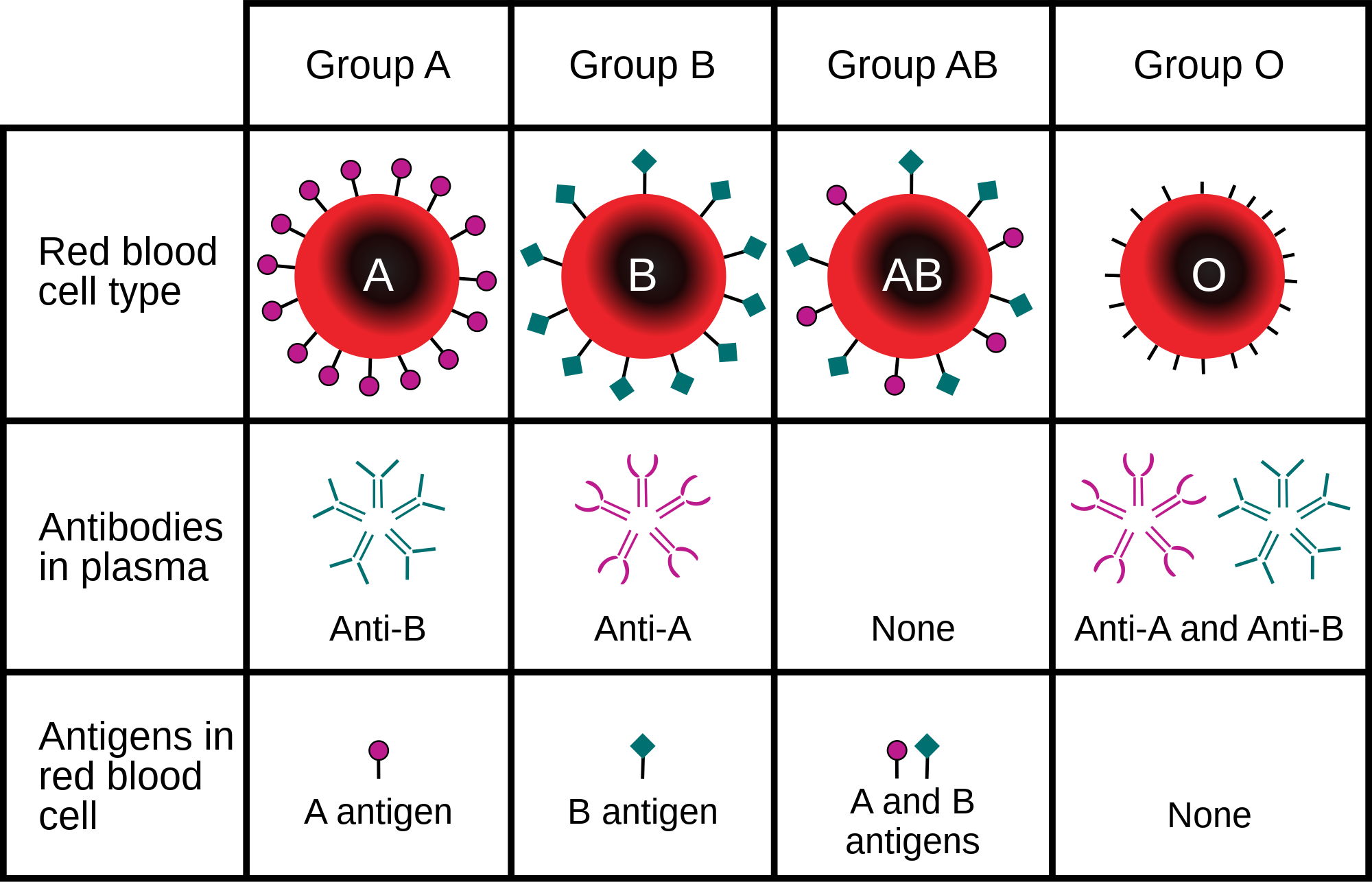
The ABO blood group system involves two antigens and two antibodies found in human blood. The two antigens are antigen A and antigen B. The two antibodies are A and B. The antigens are present on the red blood cells, and the antibodies in the serum. Regarding the antigen property of the blood, all human beings can be classified into four groups: those with antigen A (group A), those with antigen B (group B), those with both antigen A and B (group AB), and those with neither antigen (group O). The antibodies present together with the antigens are found as follows:[citation needed]
- Antigen A with antibody B
- Antigen B with antibody A
- Antigen AB with neither antibody A nor B
- Antigen null (group O) with both antibodies A and B
There is an agglutination reaction between similar antigen and antibody (for example, antigen A agglutinates the antibody A, and antigen B agglutinates the antibody B). Thus, transfusion can be considered safe as long as the serum of the recipient does not contain antibodies for the blood cell antigens of the donor.[citation needed]
The ABO system is the most important blood-group system in human-blood transfusion. The associated anti-A and anti-B antibodies are usually immunoglobulin M, abbreviated IgM, antibodies. It has been hypothesized that ABO IgM antibodies are produced in the first years of life by sensitization to environmental substances such as food, bacteria, and viruses.[11] The original terminology used by Karl Landsteiner in 1901 for the classification was A/B/C; in later publications "C" became "O".[12] Type O is often called 0 (zero, or null) in other languages.[12][13]
| Phenotype | Alleles |
|---|---|
| A | ABO*A1.01 |
| B | ABO*B1.01 |
| AB | ABO*A1.01, ABO*B1.01 |
| O | Two nonfunctional ABO genes |
Rh blood group system
[edit]The Rh system (Rh meaning Rhesus) is the second most significant blood-group system in human blood transfusion, with currently 50 antigens. The most significant Rh antigen is the D antigen, because it is the most likely to provoke an immune system response of the five main Rh antigens. It is common for D-negative individuals not to have any anti-D IgG or IgM antibodies, because anti-D antibodies are not usually produced by sensitization against environmental substances. However, D-negative individuals can produce IgG anti-D antibodies following a sensitizing event: possibly a fetomaternal transfusion of blood from a fetus in pregnancy or occasionally a blood transfusion with D-positive RBCs.[14][15] Rh negative blood types are much less common in Asian populations (0.3%) than they are in European populations (15%).[16]
The presence or absence of the Rh(D) antigen is signified by the + or − sign, so that, for example, the A− group is ABO type A and does not have the Rh (D) antigen.[17]
ABO and Rh distribution by country
[edit]As with many other genetic traits, the distribution of ABO and Rh blood groups varies significantly between populations.[citation needed][18] While theories are still debated in the scientific community as to why blood types vary geographically and why they emerged in the first place, evidence suggests that the evolution of blood types may be driven by genetic selection for those types whose antigens confer resistance to particular diseases in certain regions – such as the prevalence of blood type O in malaria-endemic countries where individuals of blood type O exhibit the highest rates of survival.[19]
Other blood group systems
[edit]As of June 2025, 48 blood-group systems have been identified and are recognized by the International Society for Blood Transfusion.[2] Thus, in addition to the ABO antigens and Rh antigens, many other antigens are expressed on the RBC surface membrane. For example, an individual can be AB, D positive, and at the same time M and N positive (MNS system), K positive (Kell system), Lea or Leb negative (Lewis system). Many of the blood group systems were named after the patients in whom the corresponding antibodies were initially encountered. Blood group systems other than ABO and Rh pose a potential, yet relatively low, risk of complications upon mixing of blood from different people.[20]

Following is a comparison of clinically relevant characteristics of antibodies against the main human blood group systems:[21]
| ABO | Rh | Kell | Duffy | Kidd | |
|---|---|---|---|---|---|
| Naturally occurring | Yes | No | No | No | No |
| Most common in immediate hemolytic transfusion reactions | A | Yes | Fya | Jka | |
| Most common in delayed hemolytic transfusion reactions | E, D, C | Jka | |||
| Most common in hemolytic disease of the newborn | Yes | D, C | Yes | ||
| Commonly produce intravascular hemolysis | Yes | Yes |
Clinical significance
[edit]Blood transfusion
[edit]Transfusion medicine is a specialized branch of hematology that is concerned with the study of blood groups, along with the work of a blood bank that provides a transfusion service with blood and other blood products. Across the world, blood products must be prescribed by a medical doctor (licensed physician or surgeon) in a similar way as medicines.[citation needed]

Much of the routine work of a blood bank involves testing blood from both donors and recipients to ensure that every individual recipient is given blood that is compatible and as safe as possible. If a unit of incompatible blood is transfused between a donor and recipient, a severe acute hemolytic reaction with hemolysis (RBC destruction), kidney failure and shock is likely to occur, and death is a possibility.[24] Antibodies can be highly active and can attack RBCs and bind components of the complement system to cause massive hemolysis of the transfused blood.[25]
Patients should ideally receive their own blood or type-specific blood products to minimize the chance of a transfusion reaction. It is also possible to use the patient's own blood for transfusion. This is called autologous blood transfusion, which is always compatible with the patient. The procedure of washing a patient's own red blood cells goes as follows: The patient's lost blood is collected and washed with a saline solution. The washing procedure yields concentrated washed red blood cells. The last step is reinfusing the packed red blood cells into the patient. There are multiple ways to wash red blood cells. The two main ways are centrifugation and filtration methods. This procedure can be performed with microfiltration devices. Risks can be further reduced by cross-matching blood, but this may be skipped when blood is required for an emergency. The oldest form of cross-matching involves mixing a sample of the recipient's serum with a sample of the donor's red blood cells and checking if the mixture agglutinates or forms clumps. If agglutination is not obvious by direct vision, a blood bank technologist may check for agglutination with a microscope. If agglutination occurs, that donor's blood cannot be transfused to that particular recipient. In a bank transfusion service, all blood specimens must be correctly identified, so labelling has been standardized using a barcode system known as ISBT 128.
The blood group may be included on identification tags or historically on tattoos worn by military personnel, in case they should need an emergency blood transfusion. Frontline German Waffen-SS had blood group tattoos during World War II.
Rare blood types can cause supply problems for blood banks and hospitals. For example, Duffy-negative blood occurs much more frequently in people of African origin,[26] and the rarity of this blood type in the rest of the population can result in a shortage of Duffy-negative blood for these patients. Similarly, for RhD negative people there is a risk associated with travelling to parts of the world where supplies of RhD-negative blood are rare, particularly East Asia, where blood services may endeavour to encourage Westerners to donate blood.[27]
Hemolytic disease of the newborn (HDN)
[edit]A pregnant woman may carry a fetus with a blood type that is different from her own. Typically, this is an issue if a RhD negative mother has a child with a RhD positive father, and the fetus ends up being Rh positive.[28] In those cases, the mother can make IgG blood group antibodies. This can happen if some of the fetus' blood cells pass into the mother's blood circulation (e.g., a small fetomaternal hemorrhage at the time of childbirth or obstetric intervention), or sometimes after a therapeutic blood transfusion. This can cause hemolytic disease of the newborn (HDN) in the current pregnancy and/or subsequent pregnancies. Sometimes this is lethal for the fetus; in these cases it is called hydrops fetalis.[29] If a pregnant woman is known to have anti-D antibodies, the RhD blood type of a fetus can be tested by analysis of fetal DNA in maternal plasma to assess the risk to the fetus of Rh disease.[30] Cell-free DNA testing can determine the fetal RHD genotype in a sample of material plasma after 10 weeks of gestation. One of the major advances of twentieth-century medicine was to prevent this disease by stopping the formation of anti-D antibodies by D-negative mothers with an injectable medication called Rho(D) immune globulin.[31][32] Antibodies associated with some blood groups can cause severe HDN, others can only cause mild HDN and others are not known to cause HDN.[29]
Blood products
[edit]To provide maximum benefit from each blood donation and to extend shelf-life, blood banks fractionate some whole blood into several products. The most common of these products are RBCs, plasma, platelets, cryoprecipitate, and fresh frozen plasma (FFP). FFP is quick-frozen to retain the labile clotting factors V and VIII, which are usually administered to patients who have a potentially fatal clotting problem caused by a condition such as advanced liver disease, overdose of anticoagulant, or disseminated intravascular coagulation (DIC).[citation needed]
Units of packed red cells are made by removing as much of the plasma as possible from whole blood units.
Clotting factors synthesized by modern recombinant methods are now in routine clinical use for hemophilia, as the risks of infection transmission that occur with pooled blood products are avoided.
Red blood cell compatibility
[edit]- Blood group AB individuals have both A and B antigens on the surface of their RBCs, and their blood plasma does not contain any antibodies against either A or B antigen. Therefore, an individual with type AB blood can receive blood from any group (with AB being preferable), but cannot donate blood to any group other than AB. They are known as universal recipients.
- Blood group A individuals have the A antigen on the surface of their RBCs, and blood serum containing IgM antibodies against the B antigen. Therefore, a group A individual can receive blood only from individuals of groups A or O (with A being preferable), and can donate blood to individuals with type A or AB.
- Blood group B individuals have the B antigen on the surface of their RBCs, and blood serum containing IgM antibodies against the A antigen. Therefore, a group B individual can receive blood only from individuals of groups B or O (with B being preferable), and can donate blood to individuals with type B or AB.
- Blood group O individuals have no A or B antigens on the surface of their RBCs, and their blood serum contains IgM anti-A and anti-B antibodies. Therefore, a group O individual can receive blood only from a group O individual, but can donate blood to individuals of any ABO blood group (i.e., A, B, O or AB). If a patient needs an urgent blood transfusion, and if the time taken to process the recipient's blood would cause a detrimental delay, O-negative blood can be used. Because it is compatible with anyone, there are some concerns that O-negative blood is often overused and consequently is always in short supply.[33] According to the Association for the Advancement of Blood and Biotherapies (AABB) and the British Chief Medical Officer's National Blood Transfusion Committee, the use of group O RhD negative red cells should be restricted to persons with O negative blood, women who might be pregnant, and emergency cases in which blood-group testing is genuinely impracticable.[33]

In addition to donating to the same blood group, type O blood donors can give to A, B, and AB; blood donors of types A and B can give to AB.
Donor[1] Recipient |
O− | O+ | A− | A+ | B− | B+ | AB− | AB+ |
|---|---|---|---|---|---|---|---|---|
| O− | 
|

|
|
|
|
|
|
|
| O+ |
|
|
|
|
|
|
|
|
| A− |
|
|
|
|
|
|
|
|
| A+ |
|
|
|
|
|
|
|
|
| B− |
|
|
|
|
|
|
|
|
| B+ |
|
|
|
|
|
|
|
|
| AB− |
|
|
|
|
|
|
|
|
| AB+ |
|
|
|
|
|
|
|
|
Table note
1. Assumes absence of atypical antibodies that would cause an incompatibility between donor and recipient blood, as is usual for blood selected by cross matching.
An Rh D-negative patient who does not have any anti-D antibodies (never been previously sensitized to D-positive RBCs) can receive a transfusion of D-positive blood, but this may cause sensitization to the D antigen, and a female patient could become at risk for hemolytic disease of the newborn. If a D-negative patient has developed anti-D antibodies, a subsequent exposure to D-positive blood could lead to a potentially dangerous transfusion reaction. Rh D-positive blood should never be given to D-negative women of childbearing age or to patients with D antibodies, so blood banks must conserve Rh-negative blood for these patients. In extreme circumstances, such as for a major bleed when stocks of D-negative blood units are very low at the blood bank, D-positive blood might be given to D-negative females above childbearing age or to Rh-negative males, provided that they did not have anti-D antibodies, to conserve D-negative inventory in the blood bank. The converse is not true; Rh D-positive patients do not react to D-negative blood.
This same matching is done for other antigens of the Rh system as C, c, E, and e, and for other blood group systems with a known risk for alloimmunization, such as the Kell system, particularly for chronically transfused patients.
Plasma compatibility
[edit]
In addition to donating to the same blood group, plasma from type AB can be given to A, B, and O; plasma from types A, B, and AB can be given to O.
Blood plasma compatibility is the inverse of red blood cell compatibility.[36] Type AB plasma carries neither anti-A nor anti-B antibodies and can be transfused to individuals of any blood group; but type AB patients can only receive type AB plasma. Type O carries both antibodies, so individuals of blood group O can receive plasma from any blood group, but type O plasma can be used only by type O recipients.
Donor Recipient
|
O | A | B | AB |
|---|---|---|---|---|
| O |
|
|
|
|
| A |
|
|
|
|
| B |
|
|
|
|
| AB |
|
|
|
|
Table note
1. Assuming the absence of strong atypical antibodies in donor plasma
Universal donors and universal recipients
[edit]
In transfusions of red blood cells, individuals with type O Rh D-negative blood are often called universal donors. Those with type AB Rh D-positive blood are called universal recipients. However, these terms are only generally true with respect to possible reactions of the recipient's anti-A and anti-B antibodies to transfused red blood cells, and also possible sensitization to Rh D antigens. One exception is individuals with hh antigen system (also known as the Bombay phenotype) who can only receive blood safely from other hh donors, because they form antibodies against the H antigen present on all red blood cells.[38][39]
Blood donors with exceptionally strong anti-A, anti-B, or any atypical blood group antibody may be excluded from blood donation of high plasma volume blood products. In general, while the plasma fraction of a blood transfusion may carry donor antibodies not found in the recipient, a significant reaction is unlikely because of dilution.
Additionally, red blood cell surface antigens other than A, B and Rh D might cause adverse reactions and sensitization, if they can bind to the corresponding antibodies to generate an immune response. Transfusions are further complicated because platelets and white blood cells (WBCs) have their own systems of surface antigens, and sensitization to platelet or WBC antigens can occur as a result of transfusion.
For transfusions of plasma, this situation is reversed. Type O plasma, containing both anti-A and anti-B antibodies, can only be given to O recipients. The antibodies will attack the antigens of any other blood type. Conversely, AB plasma can be given to patients of any ABO blood group, because it does not contain any anti-A or anti-B antibodies.
Blood typing
[edit]Typically, blood type tests are performed by adding a blood sample to a solution containing antibodies corresponding to each antigen. The presence of an antigen on the surface of the blood cells is indicated by agglutination.
Blood group genotyping
[edit]In addition to the current practice of serologic testing of blood types, the progress in molecular diagnostics allows the increasing use of blood group genotyping,[40] commonly known as red cell genotyping.[41][42][43] In contrast to serologic tests reporting a direct blood type phenotype, genotyping allows the prediction of a phenotype based on the knowledge of the molecular basis of the currently known antigens. This allows a more detailed determination of the blood type and therefore a better match for transfusion, which can be crucial in particular for patients with needs for many transfusions to prevent alloimmunization.[44]
History
[edit]Blood types were first discovered by an Austrian physician, Karl Landsteiner, working at the Pathological-Anatomical Institute of the University of Vienna (now Medical University of Vienna). In 1900, he found that blood sera from different persons would clump together (agglutinate) when mixed in test tubes, and not only that, some human blood also agglutinated with animal blood.[45] He wrote a two-sentence footnote:
The serum of healthy human beings not only agglutinates animal red cells, but also often those of human origin, from other individuals. It remains to be seen whether this appearance is related to inborn differences between individuals or is the result of some damage of a bacterial kind.[46]
This was the first evidence that blood variation exists in humans. The next year, in 1901, he made a definitive observation that the blood serum of an individual would agglutinate with only those of certain individuals. Based on this, he classified human blood into three groups, namely group A, group B, and group C. He defined that group A blood agglutinates with group B, but never with its own type. Similarly, group B blood agglutinates with group A. Group C blood is different in that it agglutinates with both A and B.[47] This was the discovery of blood groups for which Landsteiner was awarded the Nobel Prize in Physiology or Medicine in 1930. (C was later renamed to O after the German Ohne, meaning without, or zero, or null.[48]) Another group (later named AB) was discovered a year later by Landsteiner's students Adriano Sturli and Alfred von Decastello without designating the name (simply referring it to as "no particular type").[49][50][51] Thus, after Landsteiner, three blood types were initially recognised, namely A, B, and C.[51]
Czech serologist Jan Janský was the first to recognise and designate four blood types in 1907 that he published in a local journal,[52] using the Roman numerical I, II, III, and IV (corresponding to modern O, A, B, and AB respectively).[53] Unknown to Janský, American physician William L. Moss introduced an almost identical classification in 1910,[54] but with Moss's I and IV corresponding to Janský's IV and I.[55] Thus the existence of two systems immediately created confusion and potential danger in medical practice. Moss's system was adopted in Britain, France, and the US, while Janský's was preferred in most other European countries and some parts of the US. It was reported that "The practically universal use of the Moss classification at that time was completely and purposely cast aside. Therefore, in place of bringing order out of chaos, chaos was increased in the larger cities."[56] To resolve the confusion, the American Association of Immunologists, the Society of American Bacteriologists, and the Association of Pathologists and Bacteriologists made a joint recommendation in 1921 that the Jansky classification be adopted based on priority.[57] But it was not followed particularly where Moss's system had been used.[58]
In 1927, Landsteiner, who had moved to the Rockefeller Institute for Medical Research in New York, and as a member of a committee of the National Research Council concerned with blood grouping suggested to substitute Janský's and Moss's systems with the letters O, A, B, and AB. There was another confusion on the use of O, which was introduced by Polish physician Ludwik Hirszfeld and German physician Emil von Dungern in 1910.[59] It was never clear whether it was meant for the figure 0, German null for zero or the upper case letter O for ohne, meaning without; Landsteiner chose the letter.[60]
In 1928 the Permanent Commission on Biological Standardization adopted Landsteiner's proposal and stated:
The Commission learns with satisfaction that, on the initiative of the Health Organization of the League of Nations, the nomenclature proposed by von Dungern and Hirszfeld for the classification of blood groups has been generally accepted, and recommends that this nomenclature shall be adopted for international use as follows: 0 A B AB. To facilitate the change from the nomenclature hitherto employed, the following is suggested:
- Jansky ....O(I) A(II) B(III) AB(IV)
- Moss ... O(IV) A(II) B(III) AB(I)[61]
This classification became widely accepted and after the early 1950s it was universally followed.[62][63]
Hirszfeld and Dungern discovered the inheritance of blood types as Mendelian genetics in 1910 and the existence of sub-types of A in 1911.[59][64] In 1927, Landsteiner, with Philip Levine, discovered the MN blood group system,[65] and the P system.[66] Development of the Coombs test in 1945,[67] the advent of transfusion medicine, and the understanding of ABO hemolytic disease of the newborn led to discovery of more blood groups. As of June 2025[update], the International Society of Blood Transfusion (ISBT) recognizes 48 blood groups.[2]
Society and culture
[edit]A popular pseudoscientific belief in Eastern Asian countries (especially in Japan and South Korea[68]) is that a person's ABO blood type is predictive of their personality, character, and compatibility with others.[69] Researchers have established no scientific basis exists for blood type personality categorization, and studies have found no "significant relationship between personality and blood type, rendering the theory 'obsolete' and concluding that no basis exists to assume that personality is anything more than randomly associated with blood type."[68]
See also
[edit]References
[edit]- ^ Maton, Anthea; Jean Hopkins; Charles William McLaughlin; Susan Johnson; Maryanna Quon Warner; David LaHart; Jill D. Wright (1998). Human Biology and Health. Englewood Cliffs NJ: Prentice Hall. ISBN 0-13-981176-1.
- ^ a b c "'Gwada negative': French scientists find new blood type in woman". www.lemonde.fr. Retrieved 2025-06-21.
- ^ a b Dean 2005, The ABO blood group "... A number of illnesses may alter a person's ABO phenotype ..."
- ^ Stayboldt C, Rearden A, Lane TA (1987). "B antigen acquired by normal A1 red cells exposed to a patient's serum". Transfusion. 27 (1): 41–4. doi:10.1046/j.1537-2995.1987.27187121471.x. PMID 3810822.
- ^ Matsushita S, Imamura T, Mizuta T, Hanada M (November 1983). "Acquired B antigen and polyagglutination in a patient with gastric cancer". The Japanese Journal of Surgery. 13 (6): 540–2. doi:10.1007/BF02469500. PMID 6672386.
- ^ Kremer Hovinga, Idske CL; Koopmans, Marije; de Heer, Emile; Bruijn, Jan A; Bajema, Ingeborg M (January 2007). "Change in blood group in systemic lupus erythematosus". The Lancet. 369 (9557): 186–187. doi:10.1016/S0140-6736(07)60099-3. PMID 17240276.
- ^ Worel N (January 2016). "ABO-Mismatched Allogeneic Hematopoietic Stem Cell Transplantation". Transfus Med Hemother. 43 (1): 3–12. doi:10.1159/000441507. PMC 4797460. PMID 27022317.
- ^ Chown B.; Lewis M.; Kaita K. (October 1957). "A new Kell blood-group phenotype". Nature. 180 (4588): 711. Bibcode:1957Natur.180..711C. doi:10.1038/180711a0. PMID 13477267.
- ^ Miller LH, Mason SJ, Clyde DF, McGinniss MH (August 1976). "The resistance factor to Plasmodium vivax in blacks. The Duffy-blood-group genotype, FyFy". The New England Journal of Medicine. 295 (6): 302–4. doi:10.1056/NEJM197608052950602. PMID 778616.
- ^ Kwiatkowski DP (August 2005). "How Malaria Has Affected the Human Genome and What Human Genetics Can Teach Us about Malaria". American Journal of Human Genetics. 77 (2): 171–92. doi:10.1086/432519. PMC 1224522. PMID 16001361.
The different geographic distributions of α thalassemia, G6PD deficiency, ovalocytosis, and the Duffy-negative blood group are further examples of the general principle that different populations have evolved different genetic variants to protect against malaria
- ^ "Position statement: Red blood cell transfusion in newborn infants". Canadian Pediatric Society. April 14, 2014. Archived from the original on 19 May 2018.
- ^ a b Schmidt, P.; Okroi, M. (2001). "Also sprach Landsteiner – Blood Group 'O' or Blood Group 'NULL'". Transfusion Medicine and Hemotherapy. 28 (4): 206–208. doi:10.1159/000050239.
- ^ "Your blood – a textbook about blood and blood donation" (PDF). p. 63. Archived from the original (PDF) on June 26, 2008. Retrieved 2008-07-15.
- ^ Talaro, Kathleen P. (2005). Foundations in microbiology (5th ed.). New York: McGraw-Hill. pp. 510–1. ISBN 0-07-111203-0.
- ^ Moise, Kenneth J. (July 2008). "Management of Rhesus Alloimmunization in Pregnancy". Obstetrics & Gynecology. 112 (1): 164–176. doi:10.1097/AOG.0b013e31817d453c. PMID 18591322.
- ^ "Rh血型的由來". Hospital.kingnet.com.tw. Archived from the original on 2009-12-11. Retrieved 2010-08-01.
- ^ Levy, Jerrold H.; McKee, Andrew (2007). "Bleeding, Hemostasis, and Transfusion Medicine". Cardiothoracic Critical Care. pp. 437–460. doi:10.1016/B978-075067572-7.50033-3. ISBN 978-0-7506-7572-7.
- ^ Mohamud, Mohamed Hayir Tahill; Aweis, Abdullah Dahir H; Adam, Abdiwahab Sheikh Elmi; Mohamed, Farhia Abdullahi; Fidow, Safia Qasim; Mohamed, Lul Mohamud (30 January 2022). "Distribution and Frequency of ABO and Rhesus (D) Blood Groups in Somalia: A Retrospective Study on Students of Jazeera University, Mogadishu-Somalia". BioMed Research International. 2022 (7981325): 1–5. doi:10.1155/2022/7981325. PMC 8818412. PMID 35136827.
- ^ Cserti, Christine M.; Dzik, Walter H. (1 October 2007). "The ABO blood group system and Plasmodium falciparum malaria". American Society of Hematology. 110 (7): 2250–2258. doi:10.1182/blood-2007-03-077602. PMID 17502454.
- ^ Goodell, Pamela P.; Uhl, Lynne; Mohammed, Monique; Powers, Amy A. (August 2010). "Risk of Hemolytic Transfusion Reactions Following Emergency-Release RBC Transfusion". American Journal of Clinical Pathology. 134 (2): 202–206. doi:10.1309/AJCP9OFJN7FLTXDB. PMID 20660321.
- ^ Mais, Daniel (2014). Quick compendium of clinical pathology. United States: American Society for Clinical Pathology Press. ISBN 978-0-89189-615-9. OCLC 895712380.[page needed]
- ^ Possible Risks of Blood Product Transfusions Archived 2009-11-05 at the Wayback Machine from American Cancer Society. Last Medical Review: 03/08/2008. Last Revised: 01/13/2009
- ^ 7 adverse reactions to transfusion Archived 2015-11-07 at the Wayback Machine Pathology Department at University of Michigan. Version July 2004, Revised 11/5/08
- ^ Rout, Preeti; Harewood, Janine; Ramsey, Adam; Master, Samip R. (2024). "Hemolytic Transfusion Reaction". StatPearls. StatPearls Publishing. PMID 28846280.
- ^ Fujii Y (2011). "Prevention of ABO-incompatible transfusion". Masui. 60 (1): 47–54. PMID 21348250.
- ^ Nickel RG; Willadsen SA; Freidhoff LR; et al. (August 1999). "Determination of Duffy genotypes in three populations of African descent using PCR and sequence-specific oligonucleotides". Human Immunology. 60 (8): 738–42. doi:10.1016/S0198-8859(99)00039-7. PMID 10439320.
- ^ Bruce, MG (May 2002). "BCF – Members – Chairman's Annual Report". The Blood Care Foundation. Archived from the original on April 10, 2008. Retrieved 2008-07-15.
As Rhesus Negative blood is rare amongst local nationals, this Agreement will be of particular value to Rhesus Negative expatriates and travellers
- ^ Freeborn, Donna. "Hemolytic Disease of the Newborn (HDN)". University of Rochester Medical Center. Archived from the original on 19 September 2016. Retrieved 30 November 2020.
- ^ a b E.A. Letsky; I. Leck; J.M. Bowman (2000). "Chapter 12: Rhesus and other haemolytic diseases". Antenatal & neonatal screening (2nd ed.). Oxford University Press. ISBN 978-0-19-262826-8.
- ^ Daniels G, Finning K, Martin P, Summers J (September 2006). "Fetal blood group genotyping: present and future". Annals of the New York Academy of Sciences. 1075 (1): 88–95. Bibcode:2006NYASA1075...88D. doi:10.1196/annals.1368.011. PMID 17108196.
- ^ "Use of Anti-D Immunoglobulin for Rh Prophylaxis". Royal College of Obstetricians and Gynaecologists. May 2002. Archived from the original on December 30, 2008.
- ^ "Pregnancy – routine anti-D prophylaxis for D-negative women". NICE. May 2002. Archived from the original on 2022-02-05. Retrieved 2022-02-11.
- ^ a b American Association of Blood Banks (24 April 2014), "Five Things Physicians and Patients Should Question", Choosing Wisely: an initiative of the ABIM Foundation, American Association of Blood Banks, archived from the original on 24 September 2014, retrieved 25 July 2014, which cites
- The Chief Medical Officer's National Blood Transfusion Committee (c. 2008). "The appropriate use of group O RhD negative red cells" (PDF). National Health Service. Archived from the original (PDF) on 9 August 2014. Retrieved 25 July 2014.
- ^ "RBC compatibility table". American National Red Cross. December 2006. Archived from the original on 2008-09-13. Retrieved 2008-07-15.
- ^ Blood types and compatibility Archived 2010-04-19 at the Wayback Machine bloodbook.com
- ^ "Blood Component ABO Compatibility Chart Red Blood Cells and Plasma". Blood Bank Labsite. University of Michigan. Archived from the original on 16 June 2019. Retrieved 16 December 2014.
- ^ "Plasma Compatibility". Matching Blood Groups. Australian Red Cross. Archived from the original on 7 May 2020. Retrieved 19 June 2020.
- ^ Fauci, Anthony S.; Eugene Braunwald; Kurt J. Isselbacher; Jean D. Wilson; Joseph B. Martin; Dennis L. Kasper; Stephen L. Hauser; Dan L. Longo (1998). Harrison's Principals of Internal Medicine. McGraw-Hill. p. 719. ISBN 0-07-020291-5.
- ^ "Universal acceptor and donor groups". Webmd.com. 2008-06-12. Archived from the original on 2010-07-22. Retrieved 2010-08-01.
- ^ Flegel; Wagner; Müller; Gassner (1998). "Rh phenotype prediction by DNA typing and its application to practice". Transfusion Medicine. 8 (4): 281–302. doi:10.1046/j.1365-3148.1998.00173.x. ISSN 0958-7578. Retrieved 2025-07-20.
- ^ Avent ND (1998). "Molecular genetic methods: principles and feasibility in transfusion medicine". Vox Sang. 74: 275–84. doi:10.1111/j.1423-0410.1998.tb05432.x. PMID 9704457.
- ^ Anstee DJ (2009). "Red cell genotyping and the future of pretransfusion testing". Blood. 114 (2): 248–56. doi:10.1182/blood-2008-11-146860. PMID 19411635.
- ^ Flegel, Willy A; Gottschall, Jerome L; Denomme, Gregory A (2015). "Integration of red cell genotyping into the blood supply chain: a population-based study". The Lancet Haematology. 2 (7): e282 – e288. doi:10.1016/S2352-3026(15)00090-3. PMC 4508019. PMID 26207259. Retrieved 2025-07-20.
- ^ Avent ND (2009). "Large-scale blood group genotyping: clinical implications". Br J Haematol. 144 (1): 3–13. doi:10.1111/j.1365-2141.2008.07285.x. PMID 19016734.
- ^ Landsteiner K (1900). "Zur Kenntnis der antifermentativen, lytischen und agglutinierenden Wirkungen des Blutserums und der Lymphe". Zentralblatt für Bakteriologie, Parasitenkunde und Infektionskrankheiten. 27: 357–362.
- ^ Kantha, S.S. (1995). "The blood revolution initiated by the famous footnote of Karl Landsteiner's 1900 paper". The Ceylon Medical Journal. 40 (3): 123–125. PMID 8536328.
- ^ Kantha, S. S. (September 1995). "The blood revolution initiated by the famous footnote of Karl Landsteiner's 1900 paper". The Ceylon Medical Journal. 40 (3): 123–125. ISSN 0009-0875. PMID 8536328.
- ^ Landsteiner, Karl (1961-01-02). "On Agglutination of Normal Human Blood". Transfusion. 1 (1): 5–8. doi:10.1111/j.1537-2995.1961.tb00005.x. ISSN 0041-1132. PMID 13758692.
- ^ Farhud, Dariush D.; Zarif Yeganeh, Marjan (2013). "A brief history of human blood groups". Iranian Journal of Public Health. 42 (1): 1–6. ISSN 2251-6085. PMC 3595629. PMID 23514954.
- ^ Von Decastello, A.; Sturli, A. (1902). "Concerning isoagglutinins in serum of healthy and sick humans". Munchener Medizinische Wochenschrift. 26: 1090–1095.
- ^ a b Farr AD (April 1979). "Blood group serology—the first four decades (1900–1939)". Medical History. 23 (2): 215–26. doi:10.1017/s0025727300051383. PMC 1082436. PMID 381816.
- ^ Janský J. (1907). "Haematologické studie u psychotiků". Sborník Klinický (in Czech). 8: 85–139.
- ^ Garratty, G.; Dzik, W.; Issitt, P.D.; Lublin, D.M.; Reid, M.E.; Zelinski, T. (April 2000). "Terminology for blood group antigens and genes—historical origins and guidelines in the new millennium". Transfusion. 40 (4): 477–489. doi:10.1046/j.1537-2995.2000.40040477.x. PMID 10773062.
- ^ Moss W.L. (1910). "Studies on isoagglutinins and isohemolysins". Bulletin of the Johns Hopkins Hospital. 21: 63–70.
- ^ Farr, A. D. (April 1979). "Blood group serology—the first four decades (1900–1939)". Medical History. 23 (2): 215–226. doi:10.1017/S0025727300051383. PMC 1082436. PMID 381816.
- ^ Kennedy, James A. (23 February 1929). "BLOOD GROUP CLASSIFICATIONS USED IN HOSPITALS IN THE UNITED STATES AND CANADA: FINAL REPORT". Journal of the American Medical Association. 92 (8): 610. doi:10.1001/jama.1929.02700340010005.
- ^ Garratty, G.; Dzik, W.; Issitt, P.D.; Lublin, D.M.; Reid, M.E.; Zelinski, T. (April 2000). "Terminology for blood group antigens and genes—historical origins and guidelines in the new millennium". Transfusion. 40 (4): 477–489. doi:10.1046/j.1537-2995.2000.40040477.x. PMID 10773062.
- ^ Doan, Charles A. (1927). "The Transfusion Problem". Physiological Reviews. 7 (1): 1–84. doi:10.1152/physrev.1927.7.1.1.
- ^ a b Okroi, Mathias; McCarthy, Leo J. (July 2010). "The Original Blood Group Pioneers: The Hirszfelds". Transfusion Medicine Reviews. 24 (3): 244–246. doi:10.1016/j.tmrv.2010.03.006. PMID 20656191.
- ^ Schmidt, P.; Okroi, M. (2001). "Also sprach Landsteiner – Blood Group 'O' or Blood Group 'NULL'". Transfusion Medicine and Hemotherapy. 28 (4): 206–208. doi:10.1159/000050239.
- ^ Goodman, Neville M. (1940). "Nomenclature of Blood Groups". British Medical Journal. 1 (4123): 73. doi:10.1136/bmj.1.4123.73-a. PMC 2176232.
- ^ Goodman, N. M. (1940-01-13). "Nomenclature of Blood Groups". BMJ. 1 (4123): 73. doi:10.1136/bmj.1.4123.73-a. ISSN 0959-8138.
- ^ Garratty, G.; Dzik, W.; Issitt, P.D.; Lublin, D.M.; Reid, M.E.; Zelinski, T. (April 2000). "Terminology for blood group antigens and genes—historical origins and guidelines in the new millennium". Transfusion. 40 (4): 477–489. doi:10.1046/j.1537-2995.2000.40040477.x. PMID 10773062.
- ^ Dungern, E.; Hirschfeld, L. (December 1911). "Über Vererbung gruppenspezifischer Strukturen des Blutes". Zeitschrift für Induktive Abstammungs- und Vererbungslehre. 5 (1): 196–197. doi:10.1007/BF01798027.
- ^ Landsteiner, K.; Levine, P. (1927). "A New Agglutinable Factor Differentiating Individual Human Bloods". Experimental Biology and Medicine. 24 (6): 600–602. doi:10.3181/00379727-24-3483.
- ^ Landsteiner, K.; Levine, P. (1927). "Further Observations on Individual Differences of Human Blood". Experimental Biology and Medicine. 24 (9): 941–942. doi:10.3181/00379727-24-3649.
- ^ Coombs RR, Mourant AE, Race RR (1945). "A new test for the detection of weak and incomplete Rh agglutinins". Br J Exp Pathol. 26 (4): 255–66. PMC 2065689. PMID 21006651.
- ^ a b "Despite scientific debunking, in Japan you are what your blood type is". MediResource Inc. Associated Press. 2009-02-01. Archived from the original on September 28, 2011. Retrieved 2011-08-13.
- ^ Nuwer, Rachel. "You are what you bleed: In Japan and other east Asian countries some believe blood type dictates personality". Scientific American. Archived from the original on 10 January 2012. Retrieved 16 Feb 2011.
Further reading
[edit]- Dean, Laura (2005). Blood Groups and Red Cell Antigens, a guide to the differences in our blood types that complicate blood transfusions and pregnancy. Bethesda MD: National Center for Biotechnology Information. ISBN 1-932811-05-2. NBK2261.
- Mollison PL, Engelfriet CP, Contreras M (1997). Blood Transfusion in Clinical Medicine (10th ed.). Oxford UK: Blackwell Science. ISBN 0-86542-881-6.
External links
[edit]

- BGMUT Blood Group Antigen Gene Mutation Database at NCBI, NIH has details of genes and proteins, and variations thereof, that are responsible for blood types
- Online Mendelian Inheritance in Man (OMIM): ABO Glycosyltransferase; ABO - 110300
- Online Mendelian Inheritance in Man (OMIM): Rhesus Blood Group, D Antigen; RHD - 111680
- "Blood group test". Gentest.ch GmbH. Archived from the original on 2017-03-24. Retrieved 2017-03-23.
- "Blood Facts – Rare Traits". LifeShare Blood Centers. Archived from the original on September 26, 2006. Retrieved September 15, 2006.
- "Modern Human Variation: Distribution of Blood Types". Dr. Dennis O'Neil, Behavioral Sciences Department, Palomar College, San Marcos, California. 2001-06-06. Archived from the original on 2001-06-06. Retrieved November 23, 2006.
- "Racial and Ethnic Distribution of ABO Blood Types – BloodBook.com, Blood Information for Life". bloodbook.com. Archived from the original on 2010-03-04. Retrieved September 15, 2006.
- "Molecular Genetic Basis of ABO". Archived from the original on December 7, 2008. Retrieved July 31, 2008.
| International | |
|---|---|
| National | |
| Other | |

Blood type
View on GrokipediaBlood Group Systems
ABO Blood Group System
The ABO blood group system is the most clinically significant blood grouping mechanism, characterized by the presence or absence of A and B antigens on the surface of red blood cells (RBCs). These antigens are complex carbohydrate structures attached to proteins or lipids on the RBC membrane, derived from a precursor known as the H antigen. The H antigen serves as the foundational structure, consisting of a chain of sugars ending in fucose attached via an α1,2 linkage to a galactose residue. In individuals with blood type O, the H antigen remains unmodified and is prominently expressed on RBCs.[2] The A and B antigens are formed by the addition of specific terminal sugar residues to the H antigen precursor through the action of glycosyltransferase enzymes encoded by alleles at the ABO locus on chromosome 9. The A allele encodes an α1,3-N-acetylgalactosaminyltransferase enzyme that transfers N-acetylgalactosamine from UDP-N-acetylgalactosamine to the terminal galactose of the H antigen, creating the A antigen. Similarly, the B allele encodes an α1,3-galactosyltransferase that adds galactose from UDP-galactose to the same position, forming the B antigen. The O allele results in a nonfunctional enzyme due to a frameshift mutation, preventing A or B antigen synthesis and leaving the H antigen intact. These enzymatic differences determine the four primary ABO phenotypes: type A (presence of A antigen), type B (presence of B antigen), type AB (presence of both A and B antigens), and type O (absence of A and B antigens).[2][6] Naturally occurring antibodies in plasma provide immune surveillance against mismatched ABO antigens, developing early in life due to exposure to similar carbohydrate structures in the environment. Individuals with blood type B produce anti-A antibodies (primarily IgM and some IgG), those with type A produce anti-B antibodies, and type O individuals produce both anti-A and anti-B antibodies, which can cause hemagglutination and hemolysis upon incompatible transfusion. Type AB individuals lack these anti-A and anti-B antibodies. Anti-H antibodies are generally absent in most people, as the H antigen is ubiquitous, but rare exceptions occur. The Bombay phenotype, resulting from a homozygous recessive hh genotype at the separate FUT1 locus on chromosome 19, prevents H antigen formation by inactivating the fucosyltransferase enzyme, thus blocking A and B antigen expression despite IA or IB alleles; affected individuals appear as type O but produce anti-H antibodies in addition to anti-A and anti-B.[2][7] Globally, the ABO phenotypes exhibit varying frequencies across populations, with type O being the most prevalent at approximately 46%, followed by type A (around 40%), type B (about 11%), and type AB as the rarest at roughly 4%; these distributions reflect evolutionary pressures and genetic drift in different ethnic groups.[2][8]Rh Blood Group System
The Rh blood group system is one of the most clinically significant blood group systems, characterized by a complex array of antigens expressed on the surface of red blood cells. The primary antigens are carried by two homologous transmembrane proteins, RhD and RhCE, which are encoded by the closely linked RHD and RHCE genes on chromosome 1. These proteins span the red blood cell membrane multiple times and form a complex with Rh-associated glycoprotein (RhAG), contributing to the structural integrity of the membrane and the transport of ammonium and carbon dioxide. The RhCE protein expresses the antigens C, c, E, and e through allelic variations, while the RhD protein specifically carries the D antigen. Over 50 distinct Rh antigens have been identified, but the D antigen (Rh1) is the most immunogenic, capable of eliciting strong immune responses in individuals lacking it.[9][10][11] The presence or absence of the RhD antigen determines an individual's Rh status, classifying them as Rh-positive or Rh-negative, respectively. Approximately 85% of Caucasians are Rh-positive due to the expression of functional RhD protein, whereas the remaining 15% lack this antigen because of a deletion or silencing of the RHD gene. Rh-negative individuals can produce anti-D antibodies upon exposure to RhD-positive blood, such as through transfusion or pregnancy, leading to potential hemolytic reactions. In combination with the ABO system, this results in eight main blood types, such as O+ and A-. The RhD antigen's high immunogenicity makes it the primary focus of Rh typing in clinical practice.[3][10] Variants of the RhD antigen, including weak D and partial D, arise from mutations in the RHD gene that alter antigen expression or structure. Weak D variants involve reduced quantitative expression of the RhD protein, often due to missense mutations, resulting in weaker reactions with anti-D reagents in serological testing but generally not eliciting anti-D formation in recipients. Partial D variants, in contrast, feature qualitative changes, such as amino acid substitutions or hybrid alleles with RHCE, leading to altered epitopes that can react variably with different anti-D antibodies and potentially immunize Rh-negative individuals if the missing epitopes are present on donor cells. These variants require molecular genotyping for accurate identification to guide safe transfusion practices.[12][13] The Rh null phenotype represents an extremely rare condition where all Rh antigens are absent due to mutations in the RHAG gene (regulator type) or homozygous inheritance of silent RHD and RHCE alleles (amorph type), affecting fewer than 50 individuals worldwide. This absence disrupts the red blood cell membrane structure, leading to chronic hemolytic anemia characterized by spherocytosis, increased osmotic fragility, and shortened red cell survival. Affected individuals often require supportive transfusions, though their lack of Rh antigens makes them universal donors for Rh typing but increases risks of alloimmunization to other antigens. Rh incompatibility, particularly involving the D antigen, can also contribute to hemolytic disease of the newborn if an Rh-negative mother is sensitized by an Rh-positive fetus.[14][15][16]Other Blood Group Systems
Beyond the ABO and Rh systems, the International Society of Blood Transfusion (ISBT) recognizes 48 blood group systems comprising 367 red cell antigens as of August 2025.[17] These systems are defined by genetically determined antigens on red blood cell surfaces, with clinical relevance primarily in transfusion medicine due to their potential to elicit alloantibodies causing hemolytic reactions.[18] While ABO and Rh dominate routine compatibility testing, extended phenotyping panels incorporate antigens from these other systems to mitigate risks in sensitized patients.[19] The Kell blood group system (ISBT 006) includes 38 antigens carried on a type II transmembrane glycoprotein, with K (KEL1) and k (KEL2) being the most prevalent and immunogenic after those in ABO and Rh.[19] The K antigen is highly immunogenic, capable of provoking strong IgG alloantibodies that cause severe hemolytic transfusion reactions and hemolytic disease of the fetus and newborn (HDFN).[20] The rare Kell-null (K0) phenotype, lacking all Kell antigens due to mutations in the KEL gene, is associated with reduced Kell protein expression but no overt hematologic abnormalities in most cases.[19] The Duffy blood group system (ISBT 008) features FY^A and FY^B antigens on a glycoprotein that also functions as a chemokine receptor (DARC).[21] The Fy(a-b-) phenotype, resulting from a FY*B^ES allele silencing expression in erythrocytes, predominates in populations of African ancestry (up to 70%) and confers resistance to Plasmodium vivax malaria invasion by eliminating the parasite's receptor.[21] Duffy antibodies, typically IgG, can cause delayed hemolytic transfusion reactions and mild HDFN, though their clinical impact is often less severe than Kell.[22] In the Kidd blood group system (ISBT 009), JK^A and JK^B antigens are expressed on a urea transporter protein (UT-B), with anti-JK^A and anti-JK^B antibodies notorious for causing delayed hemolytic transfusion reactions due to their ability to evade detection by standard methods and re-emerge post-transfusion.[23] These IgG antibodies, often complement-activating, lead to extravascular hemolysis days to weeks after incompatible transfusion, emphasizing the need for Kidd-compatible units in antibody-positive patients.[24] The MNS blood group system (ISBT 002), one of the most complex with over 50 antigens, includes high-prevalence M (MNS1), N (MNS2), S (MNS4), and s (MNS5) determinants on glycophorins A and B.[25] Anti-M and anti-N are usually IgM and clinically benign, but anti-S and anti-s (IgG) pose risks of acute or delayed hemolytic reactions and HDFN, particularly in multiparous women.[25] The Lutheran blood group system (ISBT 005) encompasses 28 antigens, primarily Lu^A (LU1) and Lu^B (LU2) on the basal lamina glycoprotein (LU), with Lu^B expressed in nearly all individuals.[26] Lutheran antibodies are uncommon and typically cause mild delayed hemolytic transfusion reactions, though rare cases of severe HDFN have been reported; the dominant inhibitor (In(Lu)) phenotype suppresses expression without clinical sequelae.[26] Among rarer systems, the Diego blood group (ISBT 010) includes DI^A and DI^B antigens on the anion exchanger band 3 (AE1), with DI^A prevalent in East Asians and Native Americans (up to 50% in some groups).[27] Anti-DI^A can induce HDFN or hemolytic transfusion reactions, as seen in severe neonatal cases requiring exchange transfusion.[28] The P1PK system (ISBT 003), involving glycosphingolipid antigens P1, P^k, and NOR1, features the rare p phenotype (lacking all three) associated with increased norovirus susceptibility but minimal transfusion risks beyond anti-PP1P^k antibodies causing occasional intravascular hemolysis.[29]Genetics and Inheritance
Genetic Basis of ABO and Rh Systems
The ABO blood group system is determined by a single genetic locus on the long arm of chromosome 9 at position 9q34.1-q34.2.[2] This locus encodes a glycosyltransferase enzyme responsible for adding specific sugar residues to the H antigen precursor on red blood cell surfaces. The three principal alleles—A, B, and O—arise from variations in this gene, with the A and B alleles differing by seven nucleotide substitutions, four of which result in amino acid changes that alter enzyme specificity.[2] The A allele encodes an enzyme that transfers N-acetylgalactosamine, while the B allele transfers galactose; both A and B alleles exhibit codominance, leading to the AB phenotype when inherited together.[2] In contrast, the O allele is characterized by a frameshift mutation—a single guanine deletion at nucleotide position 261 in exon 6 (tagged by the single nucleotide polymorphism rs8176719)—which introduces a premature stop codon and produces a nonfunctional enzyme, rendering the O allele recessive to A and B.[2][30] Expression of ABO antigens requires the H antigen precursor, synthesized by the fucosyltransferase encoded by the FUT1 gene on chromosome 19 at 19q13.3.[31] This gene catalyzes the addition of fucose to a precursor glycolipid or glycoprotein, forming the H structure essential for subsequent A or B antigen modification.[31] In cases of homozygous FUT1 mutations (h/h genotype), as seen in the rare Bombay phenotype, no H antigen is produced, resulting in an epistatic interaction that prevents ABO antigen expression regardless of the ABO genotype, phenotypically mimicking blood type O.[31] The Rh blood group system is governed by the RH locus on the short arm of chromosome 1 at 1p36.11, comprising two highly homologous genes, RHD and RHCE, arranged in tandem and separated by approximately 35 kb.[3] The RHD gene encodes the RhD protein, responsible for the D antigen, while RHCE encodes the RhCE protein, which carries the C/c and E/e antigens through alternative splicing and polymorphisms.[3] The RhD-negative phenotype in most populations, particularly Europeans, results from a complete deletion of the RHD gene, occurring via unequal homologous recombination between flanking "Rhesus boxes"—noncoding sequences that bookend RHD.[3] Variants such as the rare D-- phenotype arise from hybrid RHD-RHCE genes, where recombination replaces portions of RHCE exons with RHD sequences, altering antigen expression and potentially leading to partial D or weakened epitopes.[3] Allele frequencies for both ABO and Rh systems vary significantly by ancestry, reflecting evolutionary pressures and genetic drift. In the ABO system, the frequency of the B antigen is notably higher in Asian populations (27%) compared to Europeans (9%) or Africans (20%), contributing to elevated B and AB phenotypes in East and South Asian groups.[2] For the Rh system, the RHD deletion allele (associated with Rh-negative) reaches frequencies of 0.35–0.40 in Europeans but is rare (less than 0.05) in Asians and Africans, where alternative mutations like pseudogenes or hybrids predominate.[3]Inheritance Patterns and Phenotypes
The inheritance of blood types follows Mendelian principles, with the ABO system governed by a single gene on chromosome 9 featuring three alleles: I^A (producing A antigen), I^B (producing B antigen), and i (producing no antigen, resulting in type O).[2] The I^A and I^B alleles exhibit codominance, meaning both antigens are expressed if present together, while the i allele is recessive to both.[2] This leads to four main phenotypes: type A (genotypes I^A I^A or I^A i), type B (I^B I^B or I^B i), type AB (I^A I^B), and type O (i i).[2] To illustrate, consider parents with genotypes I^A i (type A) and I^B i (type B). A Punnett square predicts the offspring phenotypes as follows:| I^B | i | |
|---|---|---|
| I^A | I^A I^B (AB) | I^A i (A) |
| i | I^B i (B) | i i (O) |
Global Distribution and Prevalence
Worldwide Prevalence of ABO and Rh Types
The worldwide prevalence of ABO and Rh blood types exhibits significant variation, influenced by genetic drift, migration, and historical population movements, with no single uniform global distribution but approximate averages derived from large-scale blood donor surveys and population studies. Based on compilations from international blood services, the most common type is O positive, accounting for approximately 42% of the global population, followed by A positive at 31%, B positive at 15%, AB positive at 5%, O negative at 3%, A negative at 2.5%, B negative at 1% (relatively rare in many populations), and AB negative at 0.5%.[36] These figures reflect data aggregated from diverse regions up to 2023, including surveys by organizations like the American Red Cross and World Population Review, though exact percentages can shift slightly by study methodology.[37] Overall, type O dominates globally at around 45% across ABO groups alone, underscoring its ancestral prevalence in early human populations.[38] Regional patterns highlight stark contrasts: in indigenous populations of the Americas, type O reaches up to 90-100%, a near-monomorphic trait likely stemming from founder effects during ancient migrations across Beringia.[39] In contrast, type A predominates in Europe, comprising 35-40% of the population, particularly in northern regions where it may have conferred selective advantages against certain pathogens.[37] For the Rh factor, negative status is rare globally at about 7%, but it peaks among the Basques at 35-50%, possibly due to genetic isolation in prehistoric Iberian refugia, while it is nearly absent in Asian populations at less than 1%.[40][41] Evolutionary pressures have shaped these distributions, with type O associated with lower thrombosis risk compared to A and B types, which carry higher von Willebrand factor levels and thus elevated clotting tendencies.[42] Conversely, A and B antigens may offer protection against specific infections, while blood type O may confer higher susceptibility to others such as norovirus, contributing to balanced polymorphism over millennia.[43] Global migration may lead to shifts in local blood type distributions in multicultural urban centers.| Blood Type | Approximate Global Prevalence (%) |
|---|---|
| O+ | 42 |
| A+ | 31 |
| B+ | 15 |
| AB+ | 5 |
| O- | 3 |
| A- | 2.5 |
| B- | 1 |
| AB- | 0.5 |
Regional and Ethnic Variations
Blood type distributions exhibit significant regional and ethnic variations, shaped by genetic drift, founder effects, and historical migrations. In India, the ABO frequencies show a predominance of group B, reflecting South Asian genetic patterns, with overall nationwide data indicating O at 37.12%, B at 32.26%, A at 22.88%, and AB at 7.74%; Rh-positive individuals comprise 94.61% of the population.[44] These proportions vary regionally, with B more common in the north and O prevailing elsewhere, influenced by ancient population movements. In Japan, group A is the most frequent at approximately 40%, followed by O at 30%, B at 20%, and AB at 10%, while Rh-negative is rare at less than 0.5%.[45] Nigeria demonstrates a strong prevalence of group O at 52.93%, with A at 22.77%, B at 20.64%, AB at 3.66%, and Rh-positive at 94.90%, consistent with West African profiles where O offers potential selective advantages against certain infections.[46] Ethnic groups further highlight these differences. Among Ashkenazi Jews, group A frequencies are elevated compared to some non-European populations, aligning closer to Northern European patterns at around 40%, though exact distributions vary by subgroup due to historical admixture.[47] Native American populations show near-exclusive dominance of group O, with frequencies exceeding 90% in many indigenous groups, and virtually all being Rh-positive, a pattern attributed to founder effects from ancient Beringian migrations.[48] Historical migrations have profoundly influenced these variations. The B allele's spread across Central Asia and into South Asia is linked to ancient trade routes like the Silk Road, where it appears at higher frequencies in Turkic and related populations, facilitating gene flow from East to West.[49] Similarly, the Rh-negative allele, prevalent in Europeans at 15-40%, likely arose from genetic bottlenecks during prehistoric population expansions and isolations, such as in the Basque region, where drift amplified its frequency to 47.2%.[40] Recent surveys from the 2020s underscore how urbanization affects donor pools, with urban areas showing higher donation rates but potential biases in blood type representation due to demographic shifts toward younger, more mobile populations, potentially reducing diversity in rural-sourced types like higher O in indigenous groups.[50]| Region/Ethnic Group | O (%) | A (%) | B (%) | AB (%) | Rh- (%) |
|---|---|---|---|---|---|
| India (national) | 37.1 | 22.9 | 32.3 | 7.7 | 5.4 |
| Japan | 30 | 40 | 20 | 10 | <0.5 |
| Nigeria | 52.9 | 22.8 | 20.6 | 3.7 | 5.1 |
| Ashkenazi Jews | ~34 | ~40 | ~16 | ~10 | ~15 |
| Native Americans | >90 | <5 | <5 | <1 | <1 |
Clinical Significance
Blood Transfusion and Compatibility
Blood transfusion compatibility is essential to prevent immune-mediated reactions that can lead to hemolysis, organ damage, or death. The primary systems governing compatibility are ABO and Rh, where red blood cells (RBCs) from the donor must not express antigens against which the recipient has pre-existing antibodies. In the ABO system, individuals produce antibodies against the A and/or B antigens they lack: type A has anti-B antibodies, type B has anti-A, type AB has neither, and type O has both. Thus, donor RBCs must lack the antigens targeted by the recipient's antibodies to avoid agglutination and complement activation.[51] The compatibility rules for ABO types are as follows:| Recipient | Compatible Donors (RBCs) |
|---|---|
| A | A, O |
| B | B, O |
| AB | AB, A, B, O (universal recipient) |
| O | O only |