Hubbry Logo
LymphomaLymphomaMain
Open search
Lymphoma
Community hub
Lymphoma
logo
8 pages, 0 posts
0 subscribers
Be the first to start a discussion here.
Lymphoma
Lymphoma
from Wikipedia

Lymphoma
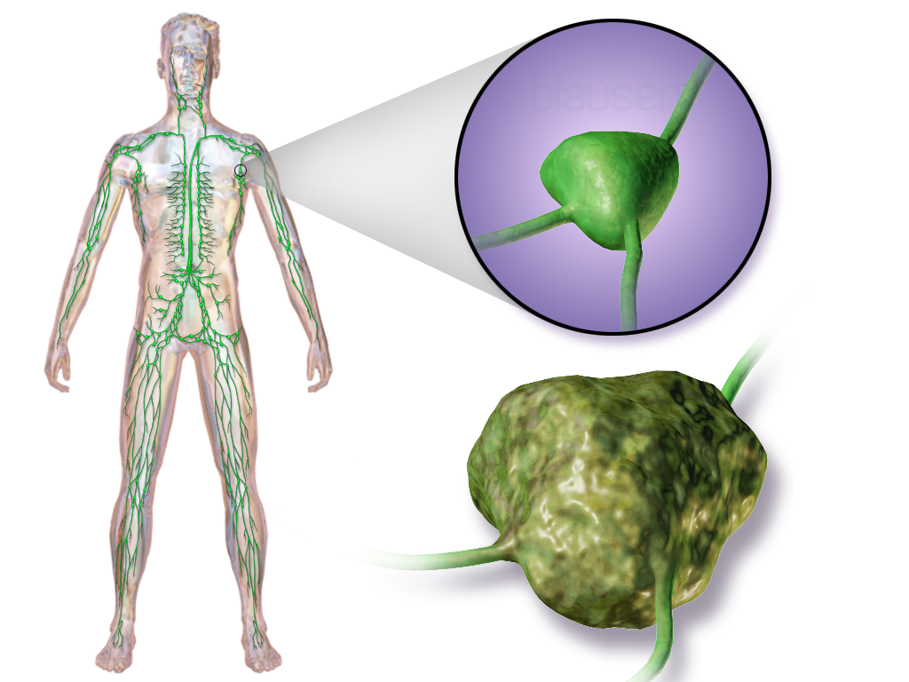
Illustration depicting lymphoma developing in the lymphatic system
SpecialtyHematology and oncology
SymptomsEnlarged lymph nodes, fever, sweats, unintended weight loss, itching, feeling tired[1][2]
Risk factorsEpstein–Barr virus, autoimmune diseases, HIV/AIDS, tobacco smoking[2][3]
Diagnostic methodLymph node biopsy[1][2]
TreatmentChemotherapy, radiation therapy, proton therapy, targeted therapy, surgery[1][2]
PrognosisAverage five year survival 85% (USA)[4]
Frequency4.9 million (2015)[5]
Deaths204,700 (2015)[6]

Lymphoma is a group of blood and lymph tumors that develop from lymphocytes (a type of white blood cell).[7] The name typically refers to just the cancerous versions rather than all such tumors.[7] Signs and symptoms may include enlarged lymph nodes, fever, drenching sweats, unintended weight loss, itching, and constantly feeling tired.[1][2] The enlarged lymph nodes are usually painless.[1] The sweats are most common at night.[1][2]

Many subtypes of lymphomas are known.[8] The two main categories of lymphomas are the non-Hodgkin lymphoma (NHL) (90% of cases)[9][10] and Hodgkin lymphoma (HL) (10%).[9] Lymphomas, leukemias and myelomas are a part of the broader group of tumors of the hematopoietic and lymphoid tissues.[11]

Risk factors for Hodgkin lymphoma include infection with Epstein–Barr virus and a history of the disease in the family.[1] Risk factors for common types of non-Hodgkin lymphomas include autoimmune diseases, HIV/AIDS, infection with human T-lymphotropic virus, immunosuppressant medications, and some pesticides.[2][12] Eating large amounts of red meat and tobacco smoking may also increase the risk.[3][13][14] Diagnosis, if enlarged lymph nodes are present, is usually by lymph node biopsy.[1][2] Blood, urine, and bone marrow testing may also be useful in the diagnosis.[2] Medical imaging may then be done to determine if and where the cancer has spread.[1][2] Lymphoma most often spreads to the lungs, liver, and brain.[1][2]

Treatment may involve one or more of the following: chemotherapy, radiation therapy, proton therapy, targeted therapy, and surgery.[1][2] In some non-Hodgkin lymphomas, an increased amount of protein produced by the lymphoma cells causes the blood to become so thick that plasmapheresis is performed to remove the protein.[2] Watchful waiting may be appropriate for certain types.[2] The outcome depends on the subtype, with some being curable and treatment prolonging survival in most.[9] The five-year survival rate in the United States for all Hodgkin lymphoma subtypes is 85%,[4] while that for non-Hodgkin lymphomas is 69%.[15] Worldwide, lymphomas developed in 566,000 people in 2012 and caused 305,000 deaths.[16] They make up 3–4% of all cancers, making them as a group the seventh-most-common form.[16][17] In children, they are the third-most-common cancer.[18] They occur more often in the developed world than in the developing world.[16]

Signs and symptoms

[edit]
The lymph nodes where lymphoma most commonly develops

Lymphoma may present with certain nonspecific symptoms; if the symptoms are persistent, an evaluation to determine their cause, including possible lymphoma, should be undertaken.

Diagnosis

[edit]
An initial evaluation of a suspected lymphoma is to make a "touch prep" wherein a glass slide is lightly pressed against excised lymphoid tissue, and subsequently stained (usually H&E stain) for evaluation under light microscopy.

Lymphoma is definitively diagnosed by a lymph-node biopsy, meaning a partial or total excision of a lymph node examined under the microscope.[22] This examination reveals histopathological features that may indicate lymphoma. After lymphoma is diagnosed, a variety of tests may be carried out to look for specific features characteristic of different types of lymphoma. These include:

Classification

[edit]
Lymph node with mantle cell lymphoma (low-power view, H&E)

According to the World Health Organization (WHO), lymphoma classification should reflect in which lymphocyte population the neoplasm arises.[23] Thus, neoplasms that arise from precursor lymphoid cells are distinguished from those that arise from mature lymphoid cells.[23] Most mature lymphoid neoplasms comprise the non-Hodgkin lymphomas.[23] Historically, mature histiocytic and dendritic cell (HDC) neoplasms have been considered mature lymphoid neoplasms, since these often involve lymphoid tissue.[23]

Lymphoma can also spread to the central nervous system, often around the brain in the meninges, known as lymphomatous meningitis (LM).[24]

Hodgkin lymphoma

[edit]

Hodgkin lymphoma accounts for about 15% of lymphomas.[25] It differs from other forms of lymphomas in its prognosis and several pathological characteristics. A division into Hodgkin and non-Hodgkin lymphomas is used in several of the older classification systems. A Hodgkin lymphoma is marked by the presence of a type of cell called the Reed–Sternberg cell.[26][27]

Non-Hodgkin lymphomas

[edit]

Non-Hodgkin lymphomas, which are defined as being all lymphomas except Hodgkin lymphoma, are more common than Hodgkin lymphoma. A wide variety of lymphomas are in this class, and the causes, the types of cells involved, and the prognoses vary by type. The number of cases per year of non-Hodgkin lymphoma increases with age. It is further divided into several subtypes.[28]

Epstein–Barr virus-associated lymphoproliferative diseases

[edit]
Follicular lymphoma replacing a lymph node

Epstein–Barr virus-associated lymphoproliferative diseases are a group of benign, premalignant, and malignant diseases of lymphoid cells (i.e., B cells, T cells, NK cells, and histiocytic-dendritic cells) in which one or more of these cell types is infected with the Epstein–Barr virus (EBV). The virus may be responsible for the development and/or progression of these diseases. In addition to EBV-positive Hodgkin lymphomas, the World Health Organization (2016) includes the following lymphomas, when associated with EBV infection, in this group of diseases: Burkitt lymphoma; large B cell lymphoma, not otherwise specified; diffuse large B cell lymphoma associated with chronic inflammation; fibrin-associated diffuse large B cell lymphoma; primary effusion lymphoma; plasmablastic lymphoma; extranodal NK/T cell lymphoma, nasal type; peripheral T cell lymphoma, not otherwise specified; angioimmunoblastic T-cell lymphoma; follicular T cell lymphoma; and systemic T cell lymphoma of childhood.[29]

WHO classification

[edit]

The WHO classification, published in 2001 and updated in 2008, 2017, and 2022,[30] is based upon the foundations laid within the "revised European–American lymphoma classification" (REAL). This system groups lymphomas by cell type (i.e., the normal cell type that most resembles the tumor) and defining phenotypic, molecular, or cytogenetic characteristics. The five groups are shown in the table. Hodgkin lymphoma is considered separately within the WHO and preceding classifications, although it is recognized as being a tumor, albeit markedly abnormal, of lymphocytes of mature B cell lineage.[31]

Of the many forms of lymphoma, some are categorized as indolent (e.g. small lymphocytic lymphoma), compatible with a long life even without treatment, whereas other forms are aggressive (e.g. Burkitt's lymphoma), causing rapid deterioration and death. However, most of the aggressive lymphomas respond well to treatment and are curable. The prognosis, therefore, depends on the correct diagnosis and classification of the disease, which is established after examination of a biopsy by a pathologist (usually a hematopathologist).[32]

Lymphoma subtypes (WHO 2008)
Mature B cell neoplasms
DNA-microarray analysis of Burkitt's lymphoma and diffuse large B-cell lymphoma (DLBCL) showing differences in gene expression patterns. Colors indicate levels of expression; green indicates genes that are underexpressed in lymphoma cells (as compared to normal cells), whereas red indicates genes that are overexpressed in lymphoma cells.
3–4% of lymphomas in adults
Small resting lymphocytes mixed with variable numbers of large activated cells, lymph nodes diffusely effaced
CD5, surface immunoglobulin
5-year survival rate 50%.[33]
Occurs in older adults, usually involves lymph nodes, bone marrow and spleen, most patients have peripheral blood involvement, indolent
About 5% of lymphomas in adults
Variable cell size and differentiation, 40% show plasma cell differentiation, homing of B cells to epithelium creates lymphoepithelial lesions.
CD5, CD10, surface Ig
Frequently occurs outside lymph nodes, very indolent, may be cured by local excision
About 40% of lymphomas in adults
Small "cleaved" [cleft] cells (centrocytes) mixed with large activated cells (centroblasts), usually nodular ("follicular") growth pattern
CD10, surface Ig
About 72–77%[34]
Occurs in older adults, usually involves lymph nodes, bone marrow and spleen, associated with t(14;18) translocation overexpressing Bcl-2, indolent
About 3–4% of lymphomas in adults
Lymphocytes of small to intermediate size growing in diffuse pattern
CD5
About 50[35] to 70%[35]
Occurs mainly in adult males, usually involves lymph nodes, bone marrow, spleen and GI tract, associated with t(11;14) translocation overexpressing cyclin D1, moderately aggressive
About 40–50% of lymphomas in adults
Variable, most resemble B cells of large germinal centers, diffuse growth pattern
Variable expression of CD10 and surface Ig
Five-year survival rate 60%[36]
Occurs in all ages, but most commonly in older adults, may occur outside lymph nodes, aggressive
< 1% of lymphomas in the United States
Round lymphoid cells of intermediate size with several nucleoli, starry-sky appearance by diffuse spread with interspersed apoptosis
CD10, surface Ig
Five-year survival rate 50%[37]
Endemic in Africa, sporadic elsewhere, more common in immunocompromised and children, often visceral involvement, highly aggressive
Mature T cell and natural killer (NK) cell neoplasms
Most common cutaneous lymphoid malignancy
Usually small lymphoid cells with convoluted nuclei that often infiltrate the epidermis, creating Pautrier microabscesseses
CD4
5-year survival 75%[38]
Localized or more generalized skin symptoms, generally indolent, in a more aggressive variant, Sézary's disease, skin erythema and peripheral blood involvement
Most common T cell lymphoma
Variable, usually a mix small to large lymphoid cells with irregular nuclear contours
CD3
Probably consists of several rare tumor types, often disseminated and generally aggressive
Precursor lymphoid neoplasms
15% of childhood acute lymphoblastic leukemia and 90% of lymphoblastic lymphoma.[39]: 635 
Lymphoblasts with irregular nuclear contours, condensed chromatin, small nucleoli and scant cytoplasm without granules
TdT, CD2, CD7
It often presents as a mediastinal mass because of involvement of the thymus. It is highly associated with NOTCH1 mutations, and is most common in adolescent males.
Hodgkin lymphoma
Most common type of Hodgkin lymphoma
Reed–Sternberg cell variants and inflammation, usually broad sclerotic bands that consist of collagen
CD15, CD30
Most common in young adults, often arises in the mediastinum or cervical lymph nodes
    • Mixed cellularity Hodgkin lymphoma
Second-most common form of Hodgkin lymphoma
Many classic Reed–Sternberg cells and inflammation
CD15, CD30
Most common in men, more likely to be diagnosed at advanced stages than the nodular sclerosis form Epstein–Barr virus involved in 70% of cases
Immunodeficiency-associated lymphoproliferative disorders
  • Associated with a primary immune disorder
  • Associated with the human immunodeficiency virus (HIV)
  • Post-transplant
  • Associated with methotrexate therapy
  • Primary central nervous system lymphoma occurs most often in immunocompromised patients, in particular those with AIDS, but it can occur in the immunocompetent, as well. It has a poor prognosis, particularly in those with AIDS. Treatment can consist of corticosteroids, radiotherapy, and chemotherapy, often with methotrexate.

Previous classifications

[edit]

Several previous classifications have been used, including Rappaport 1956, Lennert/Kiel 1974, BNLI, Working formulation (1982), and REAL (1994).

The Working Formulation of 1982 was a classification of non-Hodgkin lymphoma. It excluded the Hodgkin lymphomas and divided the remaining lymphomas into four grades (low, intermediate, high, and miscellaneous) related to prognosis, with some further subdivisions based on the size and shape of affected cells. This purely histological classification included no information about cell surface markers or genetics and made no distinction between T-cell lymphomas and B-cell lymphomas. It was widely accepted at the time of its publication but by 2004 was obsolete.[40]

In 1994, the Revised European-American Lymphoma (REAL) classification applied immunophenotypic and genetic features in identifying distinct clinicopathologic entities among all the lymphomas except Hodgkin lymphoma.[41] For coding purposes, the ICD-O (codes 9590–9999)[42] and ICD-10 (codes C81-C96)[43] are available.

Staging

[edit]
Diagram showing common sites where lymphoma spreads

After a diagnosis and before treatment, cancer is staged. This refers to determining if the cancer has spread, and if so, whether locally or to distant sites. Staging is reported as a grade between I (confined) and IV (spread). The stage of a lymphoma helps predict a patient's prognosis and is used to help select the appropriate therapy.[44]

The Ann Arbor staging system is routinely used for staging of both HL and NHL. In this staging system, stage I represents localized disease contained within a lymph node group, II represents the presence of lymphoma in two or more lymph nodes groups, III represents spread of the lymphoma to lymph nodes groups on both sides of the diaphragm, and IV indicates spread to tissue outside the lymphatic system. Different suffixes imply the involvement of different organs, for example, S for the spleen and H for the liver. Extra-lymphatic involvement is expressed with the letter E. In addition, the presence of B symptoms (one or more of the following: unintentional loss of 10% body weight in the last 6 months, night sweats, or persistent fever of 38 °C or more) or their absence is expressed with B or A, respectively.[45]

CT scan or PET scan imaging modalities are used to stage cancer. PET scanning is advised for fluorodeoxyglucose-avid lymphomas, such as Hodgkin lymphoma, as a staging tool that can even replace bone marrow biopsy. For other lymphomas, CT scanning is recommended for staging.[44]

Age and poor performance status are other established poor prognostic factors.[46] This means that people who are elderly or too sick to take care of themselves are more likely to be killed by lymphoma than others.
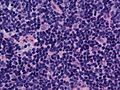
Differential diagnosis

[edit]

Certain lymphomas (extranodal NK/T-cell lymphoma, nasal type and type II enteropathy-associated T-cell lymphoma) can be mimicked by two benign diseases that involve the excessive proliferation of nonmalignant NK cells in the GI tract, natural killer cell enteropathy, a disease wherein NK cell infiltrative lesions occur in the intestine, colon, stomach, or esophagus, and lymphomatoid gastropathy, a disease wherein these cells' infiltrative lesions are limited to the stomach. These diseases do not progress to cancer, may regress spontaneously and do not respond to, and do not require, chemotherapy or other lymphoma treatments.[47]

Treatment

[edit]

Prognoses and treatments are different for HL and between all the different forms of NHL,[48] and also depend on the grade of tumor, referring to how quickly a cancer replicates. Paradoxically, high-grade lymphomas are more readily treated and have better prognoses:[49] Burkitt lymphoma, for example, is a high-grade tumor known to double within days, and is highly responsive to treatment.

Low-grade

[edit]

Many low-grade lymphomas remain indolent (growing slowly or not at all) for many years – sometimes, for the rest of the person's life. With an indolent lymphoma, such as follicular lymphoma, watchful waiting is often the initial course of action, because monitoring is less risky and less harmful than early treatment.[50]

If a low-grade lymphoma becomes symptomatic, radiotherapy or chemotherapy are the treatments of choice. Although these treatments do not permanently cure the lymphoma, they can alleviate the symptoms, particularly painful lymphadenopathy. People with these types of lymphoma can live near-normal lifespans, even though the disease is technically incurable.

Some centers advocate the use of single agent rituximab in the treatment of follicular lymphoma rather than the wait-and-watch approach. Watchful waiting is not a desirable strategy for everyone, as it leads to significant distress and anxiety in some people. It has been called "watch and worry".[51]

High-grade

[edit]

Treatment of some other, more aggressive, forms of lymphoma [which?] can result in a cure in the majority of cases, but the prognosis for people with a poor response to therapy is worse.[52] Treatment for these types of lymphoma typically consists of aggressive chemotherapy, including the CHOP or R-CHOP regimen. A number of people are cured with first-line chemotherapy. Most relapses occur within the first two years, and the relapse risk drops significantly thereafter.[53] For people who relapse, high-dose chemotherapy followed by autologous stem cell transplantation is a proven approach.[54]

The treatment of side effects is also important as they can occur due to the chemotherapy or the stem cell transplantation. It was evaluated whether mesenchymal stromal cells can be used for the treatment and prophylaxis of graft-versus-host diseases. The evidence is very uncertain about the therapeutic effect of mesenchymal stromal cells to treat graft-versus-host diseases on the all-cause mortality and complete disappear of chronic acute graft-versus-host diseases. Mesenchymal stromal cells may result in little to no difference in the all-cause mortality, relapse of malignant disease and incidence of acute and chronic graft-versus-host diseases if they are used for prophylactic reason.[55] Moreover, it was seen that platelet transfusions for people undergoing a chemotherapy or a stem cell transplantation for the prevention of bleeding events had different effects on the number of participants with a bleeding event, the number of days on which a bleeding occurred, the mortality secondary to bleeding and the number of platelet transfusions depending on the way they were used (therapeutic, depending on a threshold, different dose schedules or prophylactic).[56][57]

Four chimeric antigen receptor T cell therapies are FDA-approved for non-Hodgkin lymphoma, including lisocabtagene maraleucel (for relapsed or refractory large B-cell lymphoma with two failed systemic treatments), axicabtagene ciloleucel, tisagenlecleucel (for large B-cell lymphoma), and brexucabtagene autoleucel (for mantle cell lymphoma). These therapies come with certification and other restrictions.[58]

Hodgkin lymphoma

[edit]

Hodgkin lymphoma typically is treated with radiotherapy alone, as long as it is localized.[59]

Advanced Hodgkin disease requires systemic chemotherapy, sometimes combined with radiotherapy.[60] Chemotherapy used includes the ABVD regimen, which is commonly used in the United States. Other regimens used in the management of Hodgkin lymphoma include BEACOPP and Stanford V. Considerable controversy exists regarding the use of ABVD or BEACOPP. Briefly, both regimens are effective, but BEACOPP is associated with more toxicity. Encouragingly, a significant number of people who relapse after ABVD can still be salvaged by stem cell transplant.[61]

Scientists evaluated whether positron emission tomography scans between the chemotherapy cycles can be used to make assumptions about the survival. The evidence is very uncertain about the effect of negative (= good prognosis) or positive (= bad prognosis) interim PET scan results on the progression-free survival. Negative interim PET scan results may result in an increase in progression-free survival compared if the adjusted result was measured. Negative interim PET scan results probably result in a large increase in the overall survival compared to those with a positive interim PET scan result.[62]

Current research evaluated whether Nivolumab can be used for the treatment of a Hodgkin's lymphoma. The evidence is very uncertain about the effect of Nivolumab for patients with a Hodgkin's lymphoma on the overall survival, the quality of life, the survival without a progression, the response rate (=complete disappear) and grade 3 or 4 serious adverse events.[63]

Palliative care

[edit]

Palliative care, a specialized medical care focused on the symptoms, pain, and stress of a serious illness, is recommended by multiple national cancer treatment guidelines as an accompaniment to curative treatments for people with lymphoma.[64][65] It is used to address both the direct symptoms of lymphoma and many unwanted side effects that arise from treatments.[66][67] Palliative care can be especially helpful for children who develop lymphoma, helping both children and their families deal with the physical and emotional symptoms of the disease.[66][68][69][70] For these reasons, palliative care is especially important for people requiring bone marrow transplants.[71][72]

Supportive treatment

[edit]

Adding physical exercises to the standard treatment for adult patients with haematological malignancies like lymphomas may result in little to no difference in the mortality, the quality of life and the physical functioning. These exercises may result in a slight reduction in depression. Furthermore, aerobic physical exercises probably reduce fatigue. The evidence is very uncertain about the effect on anxiety and serious adverse events.[73]

Prognosis

[edit]
Five-year relative survival by stage at diagnosis[74]
Stage at diagnosis Five-year relative
survival (%)
Percentage
of cases (%)
Localized (confined to primary site) 82.3 26
Regional (spread to regional lymph nodes) 78.3 19
Distant (cancer has metastasized) 62.7 47
Unknown (unstaged) 68.6 8

Epidemiology

[edit]
Deaths from lymphomas and multiple myeloma per million persons in 2012
  0–13
  14–18
  19–22
  23–28
  29–34
  35–42
  43–57
  58–88
  89–121
  122–184

Lymphoma is the most common form of hematological malignancy, or "blood cancer", in the developed world.

Taken together, lymphomas represent 5.3% of all cancers (excluding simple basal cell and squamous cell skin cancers) in the United States and 55.6% of all blood cancers.[75]

According to the U.S. National Institutes of Health, lymphomas account for about 5%, and Hodgkin lymphoma in particular accounts for less than 1% of all cases of cancer in the United States.[76]

Because the whole lymphatic system is part of the body's immune system, people with a weakened immune system such as from HIV infection or from certain drugs or medication also have a higher number of cases of lymphoma.[77]

History

[edit]
Thomas Hodgkin

Thomas Hodgkin published the first description of lymphoma in 1832, specifically of the form named after him.[78] Since then, many other forms of lymphoma have been described.

The term "lymphoma" is from Latin lympha ("water") and from Greek -oma ("morbid growth, tumor").[79]

Research

[edit]

The two types of lymphoma research are clinical or translational research and basic research. Clinical/translational research focuses on studying the disease in a defined and generally immediately applicable way, such as testing a new drug in people. Studies may focus on effective means of treatment, better ways of treating the disease, improving the quality of life for people, or appropriate care in remission or after cures. Hundreds of clinical trials are being planned or conducted at any given time.[80]

Basic science research studies the disease process at a distance, such as seeing whether a suspected carcinogen can cause healthy cells to turn into lymphoma cells in the laboratory or how the DNA changes inside lymphoma cells as the disease progresses. The results from basic research studies are generally less immediately useful to people with the disease,[81] but can improve scientists' understanding of lymphoma and form the foundation for future, more effective treatments.

Other animals

[edit]

References

[edit]
[edit]
Revisions and contributorsEdit on WikipediaRead on Wikipedia
from Grokipedia
Lymphoma is a diverse group of cancers that originate in the , specifically from lymphocytes, which are essential for immune function. These malignancies are broadly classified into two main categories: (HL), characterized by the presence of Reed-Sternberg cells and often highly curable, and (NHL), a more heterogeneous group lacking those cells and accounting for the majority of cases. In 2022, there were an estimated 553,000 new cases of NHL and 83,000 of HL worldwide. In the United States, NHL represents about 3.9% of all new cancer cases with an estimated 80,350 diagnoses in 2025, while HL comprises 0.4% with 8,720 cases. Common symptoms of lymphoma include painless swelling of lymph nodes in the , armpits, or , as well as fever, drenching , unexplained , , and itching, particularly in HL. Additional signs may involve if the chest is affected or rashes in cutaneous forms of NHL. These symptoms arise because abnormal lymphocytes proliferate uncontrollably, forming tumors that disrupt normal lymphatic drainage and immune responses. The exact causes of lymphoma remain unclear, but it results from genetic mutations in lymphocyte DNA that lead to rapid, uncontrolled cell growth. Risk factors include a weakened immune system, such as from HIV/AIDS or immunosuppressive drugs post-organ transplant, certain viral infections like Epstein-Barr virus or human T-cell leukemia/lymphoma virus, autoimmune diseases, and older age, with half of NHL cases diagnosed in people over 65. HL peaks in early adulthood (ages 20–39) and later life (over 65), with a slight male predominance. Diagnosis typically involves physical exams, , biopsies to identify cell types, and staging to assess spread, which is crucial for and treatment planning. Treatments vary by type and stage but commonly include , , , targeted drugs, and transplants, with HL often curable in up to 90% of early cases. Overall five-year relative survival rates are 89% for HL and 74% for NHL, reflecting advances in therapies, though outcomes depend on factors like age, stage, and subtype. As of 2022, approximately 1.07 million people in the were living with lymphoma.

Overview and Classification

Definition and General Characteristics

Lymphoma refers to a diverse group of malignancies that arise from the clonal proliferation of within the , primarily involving B-cells, T-cells, or natural killer (NK) cells at various stages of maturation. These cancers account for approximately 3–4% of all malignancies worldwide, according to GLOBOCAN 2022 estimates. They are characterized by abnormal growth that disrupts normal immune function. Unlike leukemias, which predominantly affect the blood and bone marrow with circulating malignant cells, lymphomas typically originate in solid lymphoid tissues and form discrete masses. Lymphomas exhibit a spectrum of behaviors, ranging from indolent forms that grow slowly and may remain asymptomatic for years to aggressive variants that progress rapidly and require immediate intervention. The two primary categories are , marked by the presence of Reed-Sternberg cells, and non-Hodgkin lymphomas, which encompass a broader array of subtypes. The , integral to immune surveillance, comprises a network of lymphatic vessels that transport —a clear fluid containing lymphocytes—throughout the body, filtering it via lymph nodes, the , and . Lymph nodes, small bean-shaped structures clustered in areas like the , armpits, and , serve as primary sites for lymphoma development, while the and represent additional common extranodal locations where malignant cells can accumulate.

Major Types: Hodgkin and Non-Hodgkin

Lymphomas are broadly classified into two major categories: (HL) and (NHL), a distinction rooted in historical and clinical observations. HL is named after Thomas Hodgkin, the English physician who first described the disease in 1832 based on postmortem examinations of affected lymph nodes. NHL, by contrast, encompasses all other lymphomas that do not exhibit the defining features of HL, a categorization that emerged as diagnostic criteria evolved in the 19th and 20th centuries. Hodgkin lymphoma accounts for approximately 10% of all lymphoma cases worldwide and is pathologically defined by the presence of large, multinucleated Reed-Sternberg cells within lymph nodes, often amid a reactive inflammatory background. These cells, derived from B lymphocytes, exhibit characteristic immunophenotypic markers such as CD15 and positivity. HL displays a bimodal age distribution, with incidence peaks among young adults (ages 15–35) and older adults (over 50 years), reflecting potential differences in across age groups. Non-Hodgkin lymphoma represents about 90% of lymphoma cases globally and comprises a heterogeneous group of malignancies arising from B cells, T cells, or natural killer cells, with B-cell origins predominant in 85–90% of instances. Unlike HL, NHL lacks Reed-Sternberg cells and shows greater morphological and biological diversity, often presenting as nodal or extranodal masses. It is more prevalent among older adults, with a age at around 67 years, though subtypes vary in age predilection. The 1:9 prevalence ratio of HL to NHL underscores their differing epidemiological profiles, with HL's relative uniformity contributing to its higher curability—achieving five-year survival rates exceeding 90% in many settings through standardized and approaches—compared to the more variable outcomes in NHL due to its subtype heterogeneity. The classification system provides a framework for further subdividing these major types based on and genetic features.

WHO and Molecular Classification

The fifth edition of the (WHO) classification of haematolymphoid tumours, published in 2022, organizes lymphoid neoplasms, including lymphomas, hierarchically based on (B-cell, T-cell, or NK-cell), maturity stage (precursor versus mature), and underlying genetic features, replacing earlier provisional entities with more definitive categories informed by integrated molecular data. This framework emphasizes essential diagnostic criteria (e.g., morphology, immunophenotype) alongside desirable advanced testing (e.g., ) to refine subtype assignment, facilitating precise prognostication and research. The classification consolidates (HL) and (NHL) under mature lymphoid neoplasms, with updates reflecting genomic insights such as recurrent mutations and translocations. For classical HL, the 2022 edition retains four main subtypes—nodular sclerosis, mixed cellularity, lymphocyte-rich, and lymphocyte-depleted—distinguished primarily by histopathological patterns and the immunophenotype of Reed-Sternberg cells, which express and often CD15, while lacking typical B-cell markers like CD20. Nodular lymphocyte-predominant HL is classified separately as a distinct entity with CD20-positive lymphocyte-predominant cells. Molecularly, Epstein-Barr virus (EBV) association is noted in mixed cellularity and lymphocyte-depleted subtypes, though detailed mechanisms are addressed elsewhere. Non-Hodgkin lymphomas, predominantly mature B-cell neoplasms in the WHO schema, encompass diverse entities such as (DLBCL, not otherwise specified), , , , and , each defined by characteristic genetics and immunophenotypes. DLBCL is subdivided into germinal center B-cell-like (GCB) and activated B-cell-like (ABC) types via , with CD10, , and MUM1 aiding approximation. typically harbors IGH::BCL2 translocations detectable by (FISH), alongside CD20 and CD10 positivity. features overexpression due to t(11;14) translocations; shows rearrangements (e.g., t(8;14)) and high Ki-67 proliferation (>95%), confirmed by next-generation sequencing (NGS); s exhibit indolent features with CD20 expression and occasional 3 or t(11;18). T-cell and NK-cell lymphomas, such as peripheral T-cell lymphoma (NOS) and anaplastic large cell lymphoma, rely on TCR gene rearrangements and CD3 or markers. By 2025, refinements in DLBCL subtyping have advanced through tools like DLBclass, a neural network-based probabilistic classifier that assigns cases to five genetic subtypes (C1–C5) based on multiplatform genomic data, achieving 89–91% accuracy and enabling high-confidence (>97%) categorization for therapeutic guidance. This aligns with and extends the WHO 2022 categories by incorporating biologic heterogeneity, such as EZB (C1/C4) and MCD (C2/C3) clusters defined by mutations in , , or MYD88 and CD79B.

Pathophysiology

Cellular and Genetic Basis

Lymphomas arise from lymphocytes, which are critical to the adaptive . Most B-cell lymphomas originate from mature B cells that have undergone antigen-driven selection in the germinal centers of lymphoid follicles, where processes like and class-switch recombination occur. In contrast, T-cell lymphomas typically derive from T cells that mature in the , with malignant transformation often occurring in post-thymic or thymic precursor stages. Malignant of these lymphocytes generally involves the activation of or inactivation of tumor suppressor genes, disrupting normal control, , and differentiation. For instance, oncogene activation can result from chromosomal translocations that juxtapose proto-oncogenes with immunoglobulin enhancers, leading to their constitutive expression, while tumor suppressor loss often stems from point mutations or deletions that impair and cell surveillance mechanisms. Key genetic events underpin this transformation across lymphoma subtypes. In B-cell lymphomas, the t(14;18)(q32;q21) translocation, found in approximately 85-90% of follicular lymphomas, fuses the gene with the locus (IGH), inhibiting by overexpressing the anti-apoptotic protein BCL2. Somatic in tumor suppressors like TP53, which occurs in 20-30% of diffuse large B-cell lymphomas (DLBCL), compromise genomic stability and promote survival of damaged cells. In T-cell lymphomas, in , present in over 50% of T-cell acute lymphoblastic leukemias/lymphomas, activate the , driving uncontrolled proliferation by altering T-cell development and repressing tumor suppressors. These alterations often accumulate through error-prone DNA editing processes, such as activation-induced deaminase () activity in B cells, which inadvertently targets non-immunoglobulin loci. The plays a crucial role in lymphoma sustenance and progression, with malignant cells recruiting and interacting with non-malignant immune cells to evade host immunity. Lymphoma cells often upregulate , a ligand that binds PD-1 on T cells to suppress cytotoxic responses, as seen in classical where PD-L1 expression on Reed-Sternberg cells and surrounding macrophages fosters an immunosuppressive niche. This interaction not only promotes tumor survival but also facilitates and stromal remodeling through signaling. Progression from benign to overt malignancy typically follows a model of clonal , where initial genetic hits confer a proliferative advantage to a subset of lymphocytes, leading to expansion and acquisition of additional . In , for example, early t(14;18) clones may persist asymptomatically for years before secondary hits, such as TP53 mutations, drive transformation to aggressive DLBCL via branching clonal dynamics. This stepwise highlights the interplay between intrinsic genetic changes and selective pressures within the lymphoid tissue.

Role of Infections and Immune Dysregulation

Infections play a significant role in the pathogenesis of certain lymphomas by providing chronic antigenic stimuli that drive aberrant B- or T-cell proliferation and transformation. Beyond viruses, bacterial infections such as Helicobacter pylori are causally linked to extranodal marginal zone lymphoma (MALT) of the stomach, where chronic gastritis leads to sustained B-cell activation and accumulation of genetic aberrations; eradication of H. pylori can induce regression in early stages. Similarly, hepatitis C virus (HCV) infection is associated with an increased risk of B-cell non-Hodgkin lymphomas, including splenic marginal zone lymphoma and DLBCL, through chronic immune stimulation and direct B-cell tropism promoting lymphoproliferation. Epstein-Barr virus (EBV) is strongly associated with classical (HL), where it is detected in approximately 40% of cases in immunocompetent individuals, particularly in mixed cellularity and lymphocyte-depleted subtypes, through latent infection of B cells that evades immune surveillance. Similarly, EBV contributes to (BL) by immortalizing B cells, cooperating with translocations to promote anti-apoptotic signals and override oncogene-induced cell death. Human T-cell virus type 1 (HTLV-1) is the causative agent of adult T-cell /lymphoma (ATLL), infecting + T cells and inducing clonal expansion over decades via viral proteins that dysregulate host transcription factors like , leading to T-cell transformation in 3-5% of carriers. In primary effusion lymphoma (PEL), a rare effusion-based B-cell , human herpesvirus 8 (HHV-8, also known as Kaposi sarcoma-associated herpesvirus) is universally present, often co-occurring with human virus () infection, where viral latency promotes cytokine-independent growth of infected plasmablastic cells. Immune dysregulation further exacerbates lymphoma risk by impairing tumor surveillance and fostering persistent lymphoid activation. Autoimmune diseases, such as Sjögren's syndrome, are linked to an elevated incidence of (MZL), particularly extranodal MZL of (MALT), through chronic inflammation in salivary glands that sustains B-cell survival signals and genetic instability. Post-transplant , typically involving inhibitors and antimetabolites, markedly increases the risk of post-transplant lymphoproliferative disorders (PTLD), which are predominantly EBV-driven B-cell proliferations arising from impaired T-cell control of viral latency in the early post-transplant period. Key mechanisms underlying these associations include chronic antigenic stimulation, which induces polyclonal B-cell expansion and subsequent monoclonal transformation, as seen in autoimmune-driven MALT lymphomas where persistent autoantigen exposure mimics infectious triggers. Viral oncoproteins further hijack cellular pathways; for instance, EBV's latent membrane protein 1 (LMP1) functions as a constitutively active mimic of the CD40 receptor, aggregating into signaling complexes that activate , PI3K/Akt, and JAK/STAT pathways to promote B-cell survival and proliferation independent of external ligands. In HTLV-1-associated ATLL, the viral Tax protein similarly drives hyperactivation, while HHV-8 in PEL encodes viral homologs and interleukin-6 mimics that sustain progression. Post-2020 research has highlighted potential links between severe acute respiratory syndrome coronavirus 2 () infection and in immunocompromised hosts, with case reports documenting EBV reactivation and rapid PTLD onset following COVID-19 vaccination or infection, possibly due to transient immune shifts favoring latent viral persistence.

Clinical Presentation

Signs and Symptoms

Lymphoma often presents with a range of constitutional and local symptoms that reflect the involvement of the and potential systemic effects. The most characteristic constitutional symptoms, known as , include unexplained fever (typically above 38°C), drenching , and unintentional exceeding 10% of body weight over the previous six months. These symptoms occur in approximately 30% of patients at and are more common in advanced disease stages across both Hodgkin and non-Hodgkin lymphomas. Local signs frequently involve painless swelling of lymph nodes, particularly in the cervical, axillary, and inguinal regions, which may be noticed as firm, rubbery lumps under the skin. Additional local manifestations can include , leading to abdominal fullness or pain on the left side, and , which may cause discomfort in the upper right or early . These findings arise from lymphoid tissue proliferation and are typically the initial clues prompting medical evaluation. Other common symptoms encompass persistent , which affects daily functioning due to the disease's impact on immune and metabolic processes, and generalized pruritus, often severe and unrelieved by standard treatments. In specifically, affected lymph nodes may cause pain shortly after alcohol consumption, a unique feature reported in approximately 1-5% of cases. Less commonly, lymphoma can lead to paraneoplastic syndromes or oncologic emergencies such as , resulting from mediastinal mass compression and manifesting as facial and upper body edema, dyspnea, and venous distension. may also occur due to epidural involvement, presenting with , , sensory changes, or bowel/ dysfunction, particularly in aggressive subtypes. These presentations vary somewhat by lymphoma type but underscore the need for prompt recognition.

Patterns by Lymphoma Type

Hodgkin lymphoma (HL) typically manifests in young adults, often in the second or third decade of life, with a characteristic involvement of mediastinal lymph nodes that can lead to compressive symptoms such as and dyspnea due to mass effect on surrounding structures like the trachea or bronchi. In contrast, (NHL) more commonly affects older adults, with presentations frequently involving extranodal sites such as the , , or , where symptoms arise from local infiltration rather than nodal enlargement alone. For instance, gastrointestinal involvement may cause , , or , while cutaneous manifestations include rashes or plaques, and can present with headaches or neurological deficits. Subtype-specific patterns in NHL further diversify clinical features; (DLBCL), an aggressive form, often shows rapid tumor growth leading to quickly enlarging masses that cause obstructive symptoms or like fever and . Conversely, , an indolent subtype, typically progresses slowly with minimal or waxing-and-waning symptoms, such as painless , and may remain asymptomatic for years. This distinction between indolent and aggressive NHL underscores their differing clinical trajectories: low-grade forms like follicular lymphoma exhibit gradual progression with infrequent acute symptoms, whereas high-grade variants such as DLBCL demand prompt recognition due to their swift advancement and potential for rapid deterioration. Rare presentations in NHL include bone marrow-only involvement in small lymphocytic lymphoma, where patients may lack peripheral lymphadenopathy and instead show cytopenias or fatigue from marrow infiltration without overt nodal disease.

Diagnosis

Initial Evaluation and Laboratory Tests

The initial evaluation of suspected lymphoma begins with a thorough and to identify key clinical features suggestive of the disease. Patients often present with symptoms such as persistent , unexplained fatigue, or constitutional symptoms that warrant further investigation. The history specifically assesses for , defined as unexplained fever greater than 38°C, drenching , and unintentional exceeding 10% of body weight over six months, which are associated with more aggressive disease and influence prognosis. Additionally, the history explores risk factors including immune dysregulation, prior infections, and family history of hematologic malignancies. The physical examination focuses on detecting , typically painless and involving cervical, axillary, or inguinal nodes greater than 1 cm in diameter, as well as assessing for . Evaluation of overall is critical, often using the Eastern Cooperative Oncology Group (ECOG) scale, which ranges from 0 (fully active) to 5 (dead) and gauges the patient's ability to perform daily activities and tolerate potential therapies. An ECOG score of 0-2 generally indicates eligibility for standard treatments, while higher scores predict poorer outcomes and may necessitate supportive measures. Laboratory tests form the cornerstone of non-invasive initial assessment, starting with a complete blood count (CBC) to identify cytopenias such as anemia (hemoglobin <10 g/dL) or thrombocytopenia (platelets <150,000/μL), which occur in up to 40% of cases due to bone marrow infiltration or hypersplenism. Elevated lactate dehydrogenase (LDH) levels, often > upper limit of normal, serve as a prognostic marker reflecting tumor burden and rapid cell turnover, correlating with advanced stage and inferior survival in both Hodgkin and non-Hodgkin lymphomas. No highly specific blood tumor markers exist for diagnosing lymphoma; tests like CA125 or immunoglobulin levels may occasionally be referenced but have low specificity and are not routine screening tools. Serum beta-2 microglobulin, when elevated above 2.5 mg/L, is another key prognostic indicator, particularly in diffuse large B-cell lymphoma and mantle cell lymphoma, associating with higher tumor burden and reduced progression-free survival. Serologic testing for human immunodeficiency virus (HIV), hepatitis B virus (HBV), and hepatitis C virus (HCV) is recommended, especially in patients with risk factors, as these infections increase lymphoma risk and influence treatment choices. Epstein-Barr virus (EBV) serology may be assessed in select cases, such as young adults or immunocompromised patients, given its association with up to 40% of Hodgkin lymphoma cases and certain non-Hodgkin subtypes. If peripheral blood involvement is suspected, such as in leukemic-phase lymphomas with circulating atypical lymphocytes, on peripheral blood can detect abnormal immunophenotypes (e.g., aberrant B-cell markers like CD5+ +), aiding in early suspicion of lymphoma before tissue confirmation. This technique identifies clonal populations with high sensitivity, though it is not diagnostic in isolation. As of 2025, (ctDNA) analysis and circulating tumor cells (CTC) have emerged as promising non-invasive tools primarily for treatment monitoring and minimal residual disease detection in high-risk patients, such as those with classical or aggressive B-cell lymphomas, offering ultrasensitive detection of genetic alterations to guide risk stratification, though not as standard initial diagnostic tests. Studies demonstrate ctDNA's utility in identifying earlier than , with detection rates exceeding 90% in high-burden cases, though it is not yet universally routine pending further validation.

Imaging and Biopsy Procedures

Imaging plays a crucial role in the initial assessment of suspected lymphoma by identifying abnormal enlargement, organ involvement, and guiding site selection. -computed (PET-CT) using 18F-fluorodeoxyglucose (FDG) is a primary modality, as most lymphomas are FDG-avid, allowing detection of metabolically active lesions that may not be apparent on anatomical alone. Computed (CT) scans provide detailed anatomical information about masses in the chest, , and , serving as a foundational tool for evaluating . (MRI) is particularly useful for assessing involvement or masses where is a concern, offering superior contrast compared to CT. Bone scans may be employed in cases of suspected skeletal involvement, though they are less sensitive than PET-CT for detecting lymphoma-related bone lesions. Tissue sampling through is essential for definitive , as imaging alone cannot distinguish lymphoma from other causes of . Excisional biopsy, which removes an entire , is preferred because it preserves tissue necessary for accurate subtyping and ruling out mimics like reactive . Core needle biopsy provides smaller samples and may suffice for accessible superficial nodes but often requires follow-up excisional biopsy if evaluation is inadequate, as it can lead to nondiagnostic or inconclusive results in up to 30% of cases. cytology is minimally invasive but has significant limitations for lymphoma , as it yields dissociated cells without architectural context, resulting in low specificity for subtyping and frequent false negatives. Following , histopathological examination incorporates to characterize malignant cells using panels of antibodies targeting (CD) markers. In classic Hodgkin lymphoma, Reed-Sternberg cells typically express CD15 and , aiding differentiation from non-Hodgkin lymphomas where these markers are less common. Broad IHC panels, including for B-cell lineage, CD3 for T-cell, and others like CD45 and PAX5, enable precise to support classification. Molecular testing on biopsy samples further refines diagnosis by detecting genetic alterations. (FISH) is routinely used to identify recurrent chromosomal translocations, such as t(14;18) in or t(8;14) in , which are hallmark features of specific subtypes. Next-generation sequencing (NGS) panels assess for mutations in genes like MYD88, , or TP53, providing insights into clonal relationships and aiding in cases where morphology and IHC are equivocal.

Staging Systems

Staging systems for lymphoma provide a standardized framework to assess the extent of disease, guiding treatment decisions and prognosis by categorizing involvement of s, extranodal sites, and systemic features. These systems evolved from anatomical assessments to incorporate , reflecting advances in diagnostic technology. The Ann Arbor staging system, developed in 1971, remains foundational for both (HL) and (NHL). It defines four stages based on the number and location of involved sites: stage I involves a single region or lymphoid structure; stage II includes two or more node regions on the same side of the diaphragm; stage III encompasses nodes on both sides of the diaphragm, potentially with splenic involvement; and stage IV indicates disseminated extranodal disease such as or liver infiltration. Subclassifications include "A" for absence of (fever, , >10% in six months) and "B" for their presence; "E" denotes limited extranodal extension contiguous to a nodal mass; and bulky disease, though not formally staged, is noted for its prognostic implications. The modifications, proposed in 1990, refined the Ann Arbor system to integrate computed tomography (CT) for more precise evaluation of nodal and splenic involvement, replacing outdated lymphangiography. Key additions include the "X" designation for bulky disease, defined as a nodal mass greater than 10 cm in diameter or one-third the thoracic diameter at the widest point on chest imaging, which influences intensity. These updates emphasized residual mass assessment post-treatment and enhanced across clinical trials. The Lugano classification, introduced in 2014, modernized staging for FDG-avid lymphomas by prioritizing positron emission tomography-computed tomography (PET-CT) over CT alone for initial evaluation and response assessment in HL and most NHL subtypes. It retains the Ann Arbor framework with elements but mandates PET-CT to define stages, reducing the need for routine in HL and FDG-avid subtypes like , as PET-CT can stage involvement based on metabolic activity; stage assignment incorporates metabolic activity to better delineate viable disease. As of 2025, ongoing revisions by the International Conference on Malignant Lymphomas (ICML) committee consider integrating metabolic tumor volume () from PET-CT for refined risk stratification, though the core system remains unchanged pending validation of and circulating tumor DNA's clinical utility. For advanced-stage HL, the International Prognostic Score (IPS) complements staging by identifying high-risk patients within stages III and IV using seven independent factors: age 45 years or older, male sex, stage IV disease, serum albumin below 4.0 g/dL, hemoglobin less than 10.5 g/dL, white blood cell count at or above 15,000/μL, and lymphocyte count below 600/μL or less than 8% of total white cells. Each factor scores one point, with higher totals (0-7) correlating to poorer 5-year freedom-from-progression rates, from 84% (score 0) to 42% (score ≥5), aiding in therapeutic tailoring without altering the primary staging.

Differential Diagnosis

The differential diagnosis of lymphoma encompasses a range of conditions that present with , constitutional symptoms, or organ involvement, necessitating careful clinical, laboratory, and pathologic evaluation to distinguish malignant lymphoid proliferation from benign or other malignant processes. Key discriminators include the pattern of enlargement (e.g., firm, fixed, and painless in lymphoma versus tender and mobile in reactive processes), presence of (fever, , ), and ancillary tests such as and . Excisional with remains essential for definitive separation, as alone may be inconclusive. Infectious etiologies, particularly reactive from conditions like (due to Epstein-Barr virus) or (TB), often mimic lymphoma through generalized lymphadenopathy and fever. Mononucleosis typically affects younger patients with acute onset, , and heterophile antibody positivity on (e.g., Monospot test), while TB presents with , in a spiking , and granulomatous inflammation on confirmed by acid-fast bacilli cultures or PCR. These are distinguished from lymphoma by the absence of monoclonal lymphoid populations on and resolution with antimicrobial therapy, whereas lymphoma shows persistent clonal proliferation. Other malignancies, including metastatic carcinoma, , and , must be excluded in patients with suggestive of lymphoma. Metastatic carcinoma often involves supraclavicular nodes and is identified by cytologic evidence of epithelial markers (e.g., cytokeratins) on , contrasting with the lymphoid markers (e.g., , CD3) in lymphoma. , particularly , may present with peripheral blood involvement and is differentiated by revealing circulating mature lymphocytes or blasts, unlike the nodal predominance in lymphoma. , such as those in , show spindle cell morphology and mesenchymal markers (e.g., ) on , without the characteristic Reed-Sternberg cells or B-cell clonality of lymphoma. Autoimmune disorders like (RA) and can cause reactive nodal enlargement that overlaps with lymphoma clinically. In RA, is symmetric and associated with joint symptoms; it is distinguished by positive or anti-citrullinated protein antibodies on and rapid improvement with corticosteroids, unlike the steroid-refractory nature of lymphoma. features fibroinflammatory infiltrates with elevated serum IgG4 levels and responds to steroids, with showing IgG4-positive plasma cells (>10/) and storiform , setting it apart from the neoplastic lymphoid aggregates in lymphoma. Benign conditions such as represent another important mimic, particularly the multicentric form, which presents with systemic symptoms and generalized similar to lymphoma. It is differentiated by histologic patterns of or plasmacytosis and testing for human herpesvirus-8 (HHV-8), which is positive in up to 50% of HIV-associated cases but negative in most idiopathic or lymphoma instances; IL-6 elevation may also support the diagnosis.

Treatment

Principles of Therapy

The primary goals of lymphoma therapy vary by disease subtype and stage, aiming for cure in limited-stage or aggressive lymphomas through aggressive multimodal interventions, while focusing on long-term control and symptom palliation for indolent forms. For aggressive B-cell non-Hodgkin lymphomas (B-NHL), such as , rituximab combined with , , , and (R-CHOP) serves as the foundational backbone regimen, achieving rates exceeding 60% in early stages when integrated with other modalities. In contrast, indolent lymphomas like often prioritize or low-intensity therapy to manage progression without immediate curative intent, preserving . Treatment modalities encompass , , , and (HSCT), employed in combination to target lymphoproliferative cells while minimizing toxicity. remains central, often combined with such as anti-CD20 monoclonal antibodies like rituximab to enhance efficacy against B-cell malignancies. is particularly effective for localized disease, providing durable local control in up to 90% of early-stage cases. For high-risk or relapsed disease, autologous or allogeneic HSCT offers a chance for long-term remission, with cure potential in select aggressive subtypes. Risk-adapted strategies guide therapy intensity, de-escalating treatment for low-risk patients to reduce long-term toxicities like secondary malignancies or , while escalating for high-risk cases based on factors such as scores or interim response assessments. This approach, often incorporating early (PET) imaging, allows omission of consolidative in favorable responders, maintaining efficacy with decreased side effects. As of 2025, precision medicine principles increasingly shape lymphoma therapy, leveraging biomarkers like circulating tumor DNA (ctDNA) and minimal residual disease (MRD) monitoring to tailor regimens and predict relapse. MRD assessment via next-generation sequencing enables early detection of residual disease post-therapy, guiding decisions on maintenance therapy or HSCT, with studies showing improved progression-free survival in biomarker-driven adjustments for B-NHL. This shift toward personalized approaches integrates genomic profiling to select targeted agents, enhancing outcomes while avoiding overtreatment.

Treatment for Hodgkin Lymphoma

Treatment for Hodgkin lymphoma (HL) is highly effective and stage-dependent, with cure rates exceeding 80% overall, guided by (PET) imaging for response assessment and risk stratification using factors like International Prognostic Score (IPS) for advanced disease. Standard approaches integrate multi-agent with or without involved-site (ISRT), prioritizing regimens that balance efficacy and toxicity, such as bleomycin omission to reduce pulmonary risks. For early-stage favorable classical HL (stages I-II without risk factors), the standard regimen is 2-4 cycles of (, , , ) followed by 20-30 Gy ISRT to involved sites, achieving 5-year (PFS) rates of 90-95%. PET-directed therapy after 2 cycles allows omission of in responders ( score 1-3), as supported by the HD16 trial showing comparable outcomes with reduced long-term . In early unfavorable disease (with risk factors like bulky mediastinal mass or extranodal involvement), 4 cycles of plus ISRT is preferred, with alternatives including 2 cycles of followed by escalated BEACOPP (, etoposide, , cyclophosphamide, , procarbazine, ) and additional cycles for PET-positive cases, yielding 5-year PFS of 85-90%. Advanced-stage HL (stages III-IV) treatment emphasizes intensive chemotherapy, with preferred regimens per 2025 NCCN guidelines including 6 cycles of nivolumab plus AVD (doxorubicin, vinblastine, dacarbazine; omitting bleomycin), demonstrating 2-year PFS of 92% in the SWOG S1826 trial. For high-risk patients (IPS ≥3), escalated BEACOPP integrated with (as in BrECADD: , etoposide, , cyclophosphamide, procarbazine, , ) plus support is recommended, improving 4-year PFS to 94% over escalated BEACOPP while managing and secondary malignancy risks through dose adjustments. integration with AVD (BV-AVD) serves as an alternative for advanced disease, with 2-year PFS of 83% and reduced bleomycin-related toxicity. ISRT (30 Gy) is reserved for residual PET-positive sites post-chemotherapy. Relapsed or refractory HL management focuses on salvage therapy followed by autologous stem cell transplantation (ASCT) in eligible patients, with second-line regimens such as plus nivolumab achieving overall response rates of 85% and complete responses in 67%, enabling bridge to ASCT with 3-year disease-free survival of 50-60%. Standard salvage options include (ifosfamide, , ) or GVD (gemcitabine, vinorelbine, liposomal ), selected based on prior exposure, prior to high-dose conditioning and ASCT, which remains the curative standard for chemosensitive relapse. PD-1 inhibitors like nivolumab are approved for multiply relapsed cases, with response rates of 65-70% in post-ASCT settings. As of 2025, advances include biologic subtyping based on genomic profiles (e.g., C1 subtype with high load linked to activity), which informs risk stratification and guides use, such as upfront nivolumab for PD-L1-enriched tumors to enhance . Reduced-toxicity regimens like N-AVD and BV-AVD have become first-line standards, minimizing pulmonary toxicity while maintaining efficacy, as validated in phase 3 trials showing noninferiority to historical benchmarks.

Treatment for Non-Hodgkin Lymphoma

Treatment for (NHL) varies significantly by subtype, grade, and patient factors, with indolent forms often managed conservatively and aggressive or high-grade variants requiring intensive regimens. The majority of NHL cases are B-cell derived, and therapies commonly incorporate rituximab, an anti-CD20 , combined with or targeted agents to improve outcomes. For T-cell NHL subtypes, treatments often involve regimens like CHOEP (, , , , ) or targeted therapies such as for CD30-positive cases. For indolent subtypes such as and , initial management frequently involves a watch-and-wait approach for patients with advanced-stage , as median time to requiring is 2-3 years with no impact on cause-specific survival or overall survival. When treatment is indicated, rituximab monotherapy is a standard option, achieving response rates of 40-50% in relapsed cases and 60-80% in untreated lymphoplasmacytic lymphoma, a related indolent entity. in combination with rituximab (R² regimen) represents an effective alternative for relapsed or refractory indolent NHL, including , with a 6-year progression-free survival of 60% in untreated , comparable to rituximab plus . Aggressive NHL, particularly diffuse large B-cell lymphoma (DLBCL), the most common subtype, is typically treated with R-CHOP (rituximab, cyclophosphamide, doxorubicin, vincristine, and prednisone) as first-line therapy, which cures approximately 50% of advanced-stage patients through 4-6 cycles depending on stage and bulk. For high-risk cases (International Prognostic Index score 2-5), polatuzumab vedotin substituted for vincristine in the Pola-R-CHP regimen improves 2-year progression-free survival to 76.7% compared to 70.2% with R-CHOP, based on the phase 3 POLARIX trial, and is preferred for stages III-IV disease. In relapsed or refractory DLBCL, chimeric antigen receptor T-cell (CAR-T) therapy with axicabtagene ciloleucel, targeting CD19, yields a 4-year overall survival of 54.6% and median progression-free survival of 14.7 months in patients after two prior lines, serving as a preferred option post-autologous stem cell transplant failure. High-grade NHL subtypes, including and , demand intensive multiagent chemotherapy due to rapid proliferation. The R-hyper-CVAD regimen (rituximab plus hyperfractionated , , , and dexamethasone, alternating with high-dose and cytarabine) is standard for advanced-stage , achieving a 5-year of 71%. (CNS) prophylaxis is essential, given a 20-30% risk of involvement in , typically involving 4-6 doses of intrathecal , particularly for cases with testicular or renal primary sites. For , similar intensive approaches like R-hyper-CVAD followed by autologous transplant are used in younger fit patients, though outcomes vary by subtype aggressiveness. As of 2025, advances include bispecific antibodies like , a CD20-CD3 engager approved for relapsed or refractory large B-cell lymphomas after two prior lines, demonstrating an overall response rate of 59% as monotherapy and high durable responses in combinations such as with or R-CHOP in frontline settings. Additionally, molecular of DLBCL, such as through genetic into clusters like those defined by Chapuy et al., is increasingly guiding selection, with ongoing trials tailoring regimens based on DLBCL class features to optimize targeted interventions.

Palliative and Supportive Care

Palliative and supportive care in lymphoma focuses on alleviating symptoms, preventing complications, and enhancing throughout the disease course, particularly for experiencing treatment-related side effects or disease progression. This approach integrates multidisciplinary interventions to address physical, emotional, and social needs, often alongside active therapy. Early involvement of teams has been shown to improve outcomes, including reduced symptom burden and better alignment of care goals. Pain management is a of supportive care for lymphoma patients, targeting symptoms from enlargement, organ compression, or skeletal involvement. Opioids, such as or , are commonly used as first-line therapy for moderate to severe caused by nodal compression or tumor infiltration, with careful to balance analgesia and side effects like or . For associated with lymphoma infiltration or secondary metastases, bisphosphonates like are recommended to inhibit activity, reduce , and provide relief, often administered intravenously every 3-4 weeks. Infection prevention is critical in lymphoma due to immunosuppression from the disease or therapies, with prophylactic strategies tailored to risk levels. Antiviral prophylaxis with acyclovir or valacyclovir is standard for patients at risk of or varicella-zoster reactivation, particularly during intensive . For (PCP), trimethoprim-sulfamethoxazole is the preferred agent for prophylaxis in high-risk cases, such as those with prolonged lymphopenia. (G-CSF), like , is employed to mitigate by accelerating recovery, reducing the incidence of by up to 50% in vulnerable patients. Psychosocial support addresses the emotional and practical challenges of lymphoma, including and reproductive concerns. Counseling interventions, such as cognitive-behavioral therapy, help manage cancer-related by promoting techniques and addressing psychological contributors, leading to modest improvements in daily functioning. preservation is discussed early for patients of reproductive age prior to gonadotoxic , with options like or cryopreservation offered to mitigate risks, supported by guidelines emphasizing and multidisciplinary referral. For patients with lymphoma, integrates services to focus on comfort and dignity in the terminal phase. enrollment is recommended when curative intent shifts to symptom control, providing home-based support for , , and existential distress, with evidence showing reduced hospitalizations and improved family satisfaction in hematologic malignancies. Early consultation in relapsed cases facilitates goals-of-care discussions, increasing utilization rates.

Prognosis

Prognostic Factors

Prognostic factors in lymphoma encompass a range of clinical, biologic, and response-based indicators that help predict patient outcomes and guide risk stratification. These factors are crucial for tailoring therapeutic approaches, particularly in distinguishing between favorable and adverse risk groups across (HL) and (NHL) subtypes. Clinical parameters such as age, disease stage, serum (LDH) levels, number of extranodal sites, and are foundational predictors of prognosis in both HL and NHL. Age greater than 60 years is consistently associated with poorer outcomes in lymphoma due to reduced tolerance to intensive therapies and higher burden. Advanced (III or IV) at indicates more widespread disease and correlates with inferior . Elevated LDH levels reflect tumor burden and aggressive biology, serving as an independent adverse factor. Involvement of more than one extranodal site similarly signals disseminated disease and worse . , often measured by the Eastern Cooperative Oncology Group (ECOG) scale, with scores of 2 or higher, predicts reduced survival by indicating frailty and limited functional reserve. Biologic prognostic indices integrate these clinical factors to refine risk assessment in specific NHL subtypes. The (IPI) for (DLBCL), developed in 1993 and revised as the R-IPI in the rituximab era, incorporates five key elements: age over 60, advanced stage, elevated LDH, more than one extranodal site, and ECOG ≥2, stratifying patients into low, low-intermediate, high-intermediate, and high-risk groups with distinct outcomes. For , the Follicular Lymphoma International Prognostic Index (FLIPI) uses five adverse factors—age >60 years, stage III/IV, hemoglobin <12 g/dL, involvement of >4 nodal areas, and elevated LDH—to categorize patients into low (0-1 factors), intermediate (2 factors), or high (≥3 factors) risk, predicting progression-free and overall survival. These indices remain widely adopted for their simplicity and prognostic utility in clinical decision-making. Response-based factors provide dynamic insights into treatment efficacy and long-term prognosis. In HL, interim (PET) imaging after 2 cycles of is a strong predictor of outcome, with a negative scan indicating favorable in over 80% of advanced-stage cases, while positivity identifies high-risk patients for escalation. (MRD) negativity post-therapy, assessed via molecular techniques like PCR or , is associated with superior progression-free and overall survival in both HL and NHL, including DLBCL and , by confirming deep remission. Emerging biologic markers, particularly relevant as of 2025, enhance prognostic precision through liquid biopsy and tissue analysis. (ctDNA) levels, detectable via ultrasensitive sequencing, outperform conventional imaging in predicting remission; undetectable ctDNA after frontline therapy correlates with a 2-year exceeding 95% in large B-cell lymphomas. signatures, derived from or immune profiling, reveal immune cell interactions that influence outcomes; for instance, T-cell inflamed profiles in relapsed HL predict better treatment response, while stromal-rich signatures in DLBCL indicate poorer . These factors, including ctDNA dynamics and microenvironmental features, are increasingly integrated into risk models for personalized monitoring.

Survival Outcomes and Relapse

Survival outcomes for (HL) are generally favorable, with an overall cure rate of approximately 80% following standard frontline regimens such as . For early-stage disease (stages ), the 5-year overall survival (OS) exceeds 85%, reaching 93% for localized and 95% for regional involvement based on recent SEER data. Advanced-stage HL (stages III and IV) has a 5-year OS of around 84%, though modern risk-adapted therapies continue to improve these figures. In contrast, survival for (NHL) varies widely by subtype. (DLBCL), the most common aggressive NHL, achieves cure rates of about 60% with frontline R-CHOP chemoimmunotherapy, with 5-year OS exceeding 60% overall and up to 96% for low-risk cases per NCCN-IPI scoring. Indolent NHL subtypes, such as , are typically incurable but offer prolonged survival, with 10-year OS rates around 70% for low-risk patients and exceeding 90% in select series. Relapse occurs in 20-30% of HL patients after initial treatment, often within the first three years, with second-line salvage therapies achieving response rates of 50-70% and long-term remission in about half of cases. Aggressive NHL, including DLBCL, has higher rates of 30-40%, particularly in advanced disease, where second-line salvage regimens yield 2-year OS of up to 58% when incorporating rituximab. As of 2025, chimeric antigen receptor T-cell (CAR-T) therapy has notably improved outcomes in relapsed DLBCL, with 12-month OS rates reaching 50% in refractory settings, surpassing traditional salvage chemotherapy. This advancement, exemplified by CD19-directed CAR-T products like , has extended 4-year OS to over 50% in select relapsed/refractory cohorts.

Epidemiology

Incidence and Prevalence

Lymphoma, encompassing both (HL) and (NHL), represents a significant global cancer burden, with an estimated 635,000 new cases diagnosed worldwide in 2022 according to GLOBOCAN estimates from the International Agency for Research on Cancer (IARC). Of these, NHL accounted for the majority, with approximately 553,000 cases, while HL contributed about 82,000 cases, comprising roughly 13% of all lymphomas. These figures reflect a slight increase from 2020, when around 627,000 cases were reported, driven primarily by and aging demographics rather than rising age-standardized incidence rates (ASIR). The global ASIR for all lymphomas remains relatively stable at approximately 6.6 per 100,000, but absolute numbers are projected to rise by approximately 40% by 2040 due to these demographic shifts. In 2022, the global 5-year prevalence was approximately 2 million people living with lymphoma, with NHL accounting for about 1.74 million cases and HL for 0.29 million. Age distribution varies markedly between subtypes. HL exhibits a bimodal pattern, with peak incidence in young adults aged 20-30 years and a secondary rise after age 55, making it the most common cancer in adolescents aged 15-19 in many regions. In contrast, NHL predominates in older populations, with over half of cases diagnosed in individuals aged 65 and above, reflecting its association with age-related immune dysregulation. HL constitutes a smaller proportion of lymphomas overall, often less than 15% globally, underscoring NHL's dominance in cancer statistics. Regional variations highlight socioeconomic disparities in lymphoma occurrence. NHL incidence is notably higher in developed countries, with ASIRs exceeding 10 per 100,000 in regions like Western Europe, North America, and Australia/New Zealand, compared to 3-5 per 100,000 in low- and middle-income countries. This pattern aligns with higher human development index (HDI) areas, where lifestyle and environmental factors contribute to elevated rates. HL, however, shows a reverse trend, with higher incidence and a greater proportion of Epstein-Barr virus (EBV)-associated cases in developing countries, where up to 70-90% of pediatric and young adult HL may link to EBV infection, versus 30-50% in high-income settings. Recent trends indicate a post-pandemic rebound in reported cases following an initial decline during the era due to diagnostic delays. In the , HL incidence dipped by approximately 7.5% in 2020, and US data suggest a recovery in 2023-2024 with catch-up diagnoses in some cancers, though global patterns for lymphoma show mixed recovery. This uptick, estimated at 2-4% annually in absolute terms, continues to be influenced by aging populations, with projections for 2025 anticipating over 650,000 new cases worldwide.

Risk Factors and Demographics

Lymphoma incidence exhibits a notable male predominance, with a male-to-female ratio of approximately 1.5:1 across both Hodgkin lymphoma (HL) and non-Hodgkin lymphoma (NHL) subtypes. Age distribution varies by type: HL displays a bimodal pattern with peaks in young adults (ages 20–34) and older individuals (over 55 years), and a median diagnosis age of 39 years, while NHL predominantly affects older adults, with a median age of 68 years and the highest incidence in those aged 65–74. Several established risk factors contribute to lymphoma development, particularly involving dysregulation. Immunosuppression from conditions such as infection or post-organ transplantation significantly elevates risk, with transplant recipients facing up to a 10-fold increase in NHL incidence due to chronic immunosuppressive therapy. Autoimmune diseases, including , Sjögren's syndrome, , and celiac disease, are associated with heightened NHL risk, often through persistent immune stimulation and inflammation, with relative risks ranging from 2- to 4-fold in affected individuals. Prior exposure to or for other cancers also increases susceptibility to secondary lymphomas, with risks persisting for decades post-treatment and linked to cumulative dose. Lifestyle factors show subtype-specific associations with lymphoma. Obesity, defined by a of 30 or higher, correlates with a modestly elevated of NHL, potentially through chronic low-grade inflammation, though the evidence remains inconsistent across studies. Smoking demonstrates links to certain NHL subtypes, such as , where current or prolonged use may increase by 20–50%, while overall associations with NHL or HL are weaker or absent. In the United States, demographic patterns reveal ethnic variations in incidence. HL rates are highest among (3.1 per 100,000), exceeding those in Blacks (2.5), Hispanics (2.8), and Asians/Pacific Islanders (1.2). For NHL, incidence is also elevated in (20.3 per 100,000) compared to Blacks (15.9), Hispanics (17.4), and Asians/Pacific Islanders (13.1), with lower rates in Asian populations potentially influenced by genetic factors despite higher prevalence in some subgroups, which itself raises NHL risk.

History

Early Discoveries

The earliest systematic description of what would later be recognized as Hodgkin lymphoma (HL) came in 1832, when British physician Thomas Hodgkin presented his observations from a series of postmortem examinations at Guy's Hospital in London. In his paper "On Some Morbid Appearances of the Absorbent Glands and Spleen," Hodgkin detailed seven cases—though the published version focused on six—characterized by painless enlargement of lymph nodes, spleen, and sometimes other organs, without evidence of suppuration or typical infectious causes. These findings highlighted a distinct pathological entity involving the lymphatic system, though Hodgkin himself did not propose a specific name or etiology, attributing it possibly to inflammation or degeneration. His work laid the groundwork for recognizing lymphomas as a unique category of disease, distinct from other glandular pathologies. The term "lymphoma" emerged later in the 19th century amid growing efforts to classify lymphoid malignancies. In 1871, Austrian surgeon and pathologist introduced the phrase "malignant lymphoma" to describe a group of aggressive tumors arising from lymphatic tissues, distinguishing them from benign enlargements and sarcomas. Billroth's coinage reflected the era's pathological focus on cellular proliferation and , drawing from his extensive surgical and experience, and it encompassed what are now known as both HL and (NHL) variants. This provided a unified framework for these conditions, shifting attention from mere descriptive to malignant behavior, though early usage often lumped diverse lymphoid neoplasms together without clear subtypes. Advancements in at the turn of the enabled more precise identification of cellular hallmarks. In 1898, Carl Sternberg described large, multinucleated cells in biopsies from patients with HL, initially mistaking the disease for an infectious process akin to . This was expanded in 1902 by Dorothy Reed, who, in her doctoral thesis, characterized these "mirror-image" giant cells—later named Reed-Sternberg (RS) cells—as for HL, based on detailed histological studies of affected tissues. Reed's work, published between 1902 and 1905, emphasized their owl-eye appearance and scarcity amid reactive inflammatory cells, solidifying HL's identity as a neoplastic rather than purely inflammatory disorder and facilitating its separation from other lymphomas. By the mid-20th century, histopathological refinements further delineated HL from NHL. In the 1940s and 1950s, pathologist Robert Lukes, working at institutions like the , developed early classifications that highlighted differences in cellular composition, nodal architecture, and clinical behavior between HL and the broader spectrum of NHLs. Lukes' contributions, including his 1966 collaboration with James Butler on HL subtypes, built on wartime-era biopsies and emphasized RS cells' exclusivity to HL, enabling pathologists to distinguish it from the more heterogeneous NHL group through systematic grading of lymphocyte depletion, nodularity, and mixed cellularity. These efforts marked a pivotal shift toward subtype-specific diagnostics, influencing subsequent international consensus systems.

Development of Modern Therapies

The development of modern therapies for lymphoma began in the 1960s with the introduction of combination regimens that achieved the first cures for advanced (HL). In 1964, Vincent T. DeVita Jr. and colleagues at the developed the MOPP regimen, consisting of (mechlorethamine), , procarbazine, and , which demonstrated durable remissions in patients with previously incurable stages of HL. This marked a , as MOPP achieved complete response rates of over 80% in advanced disease, with long-term survival rates exceeding 50%, establishing as a curative modality rather than merely palliative. Prior efforts with single-agent alkylators had shown only temporary responses, but MOPP's multi-drug approach exploited non-cross-resistant mechanisms to overcome tumor heterogeneity. Building on this foundation in the 1970s, researchers sought to mitigate MOPP's significant toxicities, including sterility and secondary leukemias. In 1973, Gianni Bonadonna and his team at the Istituto Nazionale dei Tumori in Milan introduced the ABVD regimen—adriamycin (doxorubicin), bleomycin, vinblastine, and dacarbazine—as a non-cross-resistant alternative for MOPP failures. Initial trials reported in 1975 showed ABVD yielding complete remission rates comparable to MOPP (around 75%) but with markedly lower rates of gonadal toxicity and myelosuppression, while improving failure-free survival to over 70% at five years. By the late 1970s, ABVD had supplanted MOPP as the standard for advanced HL due to its superior therapeutic index, influencing global treatment protocols and reducing long-term morbidity. The 1990s heralded the era of targeted , transforming (NHL) management, particularly for B-cell subtypes. Rituximab, a chimeric anti-CD20 , received FDA approval on November 26, 1997, for relapsed or refractory CD20-positive low-grade or follicular B-cell NHL. As the first for lymphoma, rituximab induced objective response rates of 50-60% as monotherapy and, when combined with like CHOP, improved overall survival by 30-40% in (DLBCL), revolutionizing B-cell NHL treatment by selectively depleting malignant cells while sparing normal tissues. Its integration into frontline regimens by the early established as a cornerstone, with subsequent approvals expanding its use across indolent and aggressive B-cell lymphomas. The 2010s advanced precision medicine with antibody-drug conjugates and cellular therapies for relapsed/refractory cases. , an anti-CD30 antibody conjugated to , was first approved by the FDA on August 19, 2011, for relapsed HL after autologous transplantation or at least two prior therapies. This agent achieved objective response rates of 75% in heavily pretreated patients, with median durations exceeding 20 months, addressing a critical unmet need in CD30-positive lymphomas like HL and anaplastic large cell lymphoma. Concurrently, chimeric receptor () T-cell therapies emerged; the first approval for lymphoma came on October 18, 2017, with (Yescarta) for relapsed/refractory large after two or more lines of therapy. This anti-CD19 CAR-T product yielded complete remission rates of 50-60% in pivotal trials, offering curative potential for patients ineligible for transplant and spurring approvals for additional CAR-Ts like and lisocabtagene maraleucel by 2021. By 2025, bispecific T-cell engager antibodies had become widespread in clinical practice for relapsed B-cell lymphomas, reflecting rapid adoption following 2020s regulatory milestones. , a /CD3 bispecific, gained FDA approval in May 2023 for third-line relapsed/ DLBCL, achieving complete response rates of 39% in pivotal studies. On , 2025, epcoritamab-bysp received further FDA approval in combination with rituximab and for relapsed or follicular lymphoma after at least one prior line of . Similarly, received approval in 2022 for relapsed or follicular after two or more lines of , while received approval in 2023 for relapsed or diffuse large B-cell or large B-cell lymphoma arising from follicular after two or more lines of . Real-world data from early 2025 showed over 1,100 doses administered across U.S. networks for these bispecific agents, indicating broad integration into salvage regimens. These agents, by simultaneously engaging tumor cells and T-cells, have expanded access to beyond specialized centers, with ongoing expansions to frontline settings underscoring their transformative impact on lymphoma care.

Research Directions

Ongoing Clinical Trials

As of 2025, several phase III clinical trials continue to evaluate novel frontline therapies for (DLBCL), including long-term follow-up from the POLARIX study, which assesses in combination with rituximab, , , and (Pola-R-CHP) versus standard R-CHOP. The trial's five-year outcomes, reported in September 2025, demonstrate sustained benefits, particularly in high-risk subgroups, with ongoing monitoring for overall survival and quality-of-life endpoints. Similarly, the MAGNIFY phase IIIb trial investigates plus rituximab (R²) induction followed by randomized maintenance with R² versus rituximab alone in relapsed/refractory follicular lymphoma (FL) and . Combination immunotherapies represent a major focus in ongoing trials for both indolent and aggressive lymphomas, emphasizing bispecific antibodies and checkpoint inhibitors integrated with . For instance, phase III studies such as NCT04408638 evaluate combined with and in relapsed/refractory large , showing promising efficacy in bridging to transplantation. In frontline settings, trials like those at UCSF and MD Anderson explore nivolumab or added to multi-agent for advanced-stage lymphomas, aiming to reduce toxicity while improving event-free survival. Vaccine-based approaches for (HL) remain in early phases, with exploratory trials investigating personalized neoantigen vaccines post-standard therapy to enhance immune surveillance, though phase III data are pending. For relapsed/refractory settings, T-cell-engaging bispecific antibodies are under active investigation, particularly in high-grade B-cell lymphomas. The EPCORE NHL-2 , expanded in 2025, demonstrates complete responses in patients with relapsed DLBCL using subcutaneous , a CD3xCD20 bispecific, with manageable . Allogeneic CAR-T therapies address manufacturing delays in autologous approaches; phase I/II from Allogene and Caribou Biosciences report durable remissions in 40-50% of relapsed large B-cell lymphoma cases using off-the-shelf CD19-directed products, with November 2025 data highlighting reduced and PD-1 knockout enhancements for improved persistence in second-line large B-cell lymphoma. In 2025, highlights include AI-optimized regimens and ctDNA-guided strategies to personalize . AI tools, as in precision screening trials, enhance patient matching for lymphoma studies by analyzing multimodal data, potentially accelerating enrollment by 30% in phase III protocols. ctDNA monitoring drives efforts, with the SHORTEN-ctDNA (NCT06693830) testing real-time measurable residual assessment to shorten rituximab maintenance in newly diagnosed DLBCL, showing feasibility for reducing treatment duration without compromising outcomes. The PRECISE-HL similarly explores ctDNA to chemotherapy in classical HL, aiming for 85% recurrence-free survival at three years.

Emerging Therapies and Advances

In recent years, antibody-drug conjugates (ADCs) such as have shown promise in regimens for relapsed or (DLBCL), with phase 1b data from 2025 demonstrating an overall response rate of 93.3% and complete response rate of 86.7% when paired with the bispecific antibody , alongside a favorable profile including low rates of . Dual-antigen CAR-T cell therapies, targeting and simultaneously, are emerging to mitigate escape in B-cell lymphomas, with early 2025 phase 1 results from Johnson & Johnson's investigational therapy indicating encouraging efficacy in large B-cell lymphoma patients previously exposed to single-target CAR-T. These approaches aim to enhance tumor penetration and persistence, particularly in cases with bulky or extranodal disease mimicking solid tumor challenges. Precision medicine advancements include -Cas9 editing to target resistant clones in lymphoma, where knockout of PD-1 in CAR-T cells has achieved complete remission rates of 87.5% in relapsed/refractory B-cell by boosting anti-tumor activity and reducing exhaustion. Site-specific integration of CAR transgenes via into the locus improves CAR-T proliferation and efficacy against resistant cells, with efficiencies exceeding 60% in preclinical models, minimizing off-target effects and chromosomal instability. modulation is also gaining traction, as gut influences outcomes in lymphoma; broad-spectrum antibiotics prior to CAR-T therapy correlate with poorer responses, while targeted interventions like fecal transplantation show potential to enhance efficacy by optimizing modulation and reducing toxicity. Diagnostic innovations feature liquid biopsy for (MRD) detection using (ctDNA), which in 2025 prospective studies of large B-cell lymphoma outperformed PET/CT in prognostic accuracy, with MRD-negative patients at end-of-therapy achieving 97% two-year compared to 29% for MRD-positive cases. (AI) enhances PET interpretation by automating segmentation and predicting outcomes, with deep learning models yielding a pooled of 4.11 for and area under the curve of 0.78 across 75 studies, enabling precise risk stratification in . By 2025, the European Society for Medical Oncology (ESMO) guidelines have integrated these advances, recommending non-chemotherapy options like bispecific antibodies and CAR-T for relapsed and , while emphasizing molecular testing such as TP53 analysis to guide precision therapies.00911-1/fulltext) Subtype-specific trials in leverage biologic classifications, identifying two main clusters (C1 with high mutational burden and C2 with chromosomal instability) via genomic profiling, informing adaptive strategies in studies like PRECISE-HL that use ctDNA to adjust chemotherapy intensity. Ongoing clinical trials briefly reference these modalities, testing their integration in high-risk subtypes to improve long-term outcomes.00911-1/fulltext)

Lymphoma in Other Animals

Veterinary Classification

In , lymphoma classification in non-human animals, particularly dogs and cats, adapts the (WHO) system originally developed for human non-Hodgkin lymphomas, emphasizing morphology, immunophenotype, and anatomic distribution while accounting for species-specific features. This adaptation, endorsed by organizations like the World Small Animal Veterinary Association (WSAVA), facilitates consistent diagnosis across veterinary pathology but recognizes limitations such as lower incidence of certain subtypes like in animals compared to humans. The system categorizes lymphomas into B-cell and T-cell types based on with markers like and CD3, with over 30 histopathologic subtypes identified in dogs alone. Lymphoma is one of the most common neoplasms in dogs and cats, with multicentric forms predominating at approximately 80-84% of cases in dogs, involving generalized peripheral . Other anatomic variants include mediastinal lymphoma, affecting the , and gastrointestinal () forms, which comprise 5-7% of canine cases and often present with intestinal involvement in cats. In dogs, B-cell lymphomas are predominant, with (DLBCL) accounting for about 48% of cases, frequently showing plasmacytoid differentiation. Conversely, T-cell lymphomas are more common in cats, comprising subtypes like peripheral not otherwise specified (NOS) and intestinal . Key differences from human classification include the strong association of feline lymphoma with (FeLV), a that increases lymphoma risk up to 60-fold in antigen-positive cats, often leading to multicentric or mediastinal presentations. Unlike humans, no equivalent to exists in domestic animals, with all veterinary cases classified as non-Hodgkin types. Diagnostic approaches parallel human methods but rely more heavily on cytology from fine-needle aspirates for initial confirmation, supplemented by and for , while molecular techniques like PCR for antigen receptor rearrangements (PARR) are used less routinely due to cost and availability constraints.

Comparative Epidemiology and Treatment

Lymphoma represents a significant health concern in , with incidence rates in companion animals providing valuable comparative data to human . In dogs, the estimated incidence is approximately 21.7 cases per 100,000 dogs annually, making it one of the most common hematopoietic malignancies. Certain breeds, such as Golden Retrievers, exhibit higher susceptibility, potentially due to genetic factors. In cats, lymphoma is the most frequently diagnosed cancer, with an annual incidence of about 48 per 100,000 cats, particularly elevated in older males and those infected with (FeLV), which increases risk by promoting viral oncogenesis. These rates underscore lymphoma's in pets, often mirroring environmental exposures shared with humans but influenced by species-specific pathogens like FeLV in cats. Treatment approaches in veterinary draw from protocols but are adapted for shorter expectations and quality-of-life priorities. For multicentric lymphoma in dogs, the CHOP-based multi-agent chemotherapy regimen—combining , , , and —achieves complete remission in 80-90% of cases, with median times of 10-14 months. is commonly used for localized forms, such as nasal lymphoma, offering palliative control with extensions of 6-12 months. In cats, similar CHOP protocols yield lower response rates, with 6-month, 1-year, and 2-year rates of 64%, 57%, and 35%, respectively, due to frequent gastrointestinal involvement and FeLV co-morbidity. Unlike , where cures exceed 70% with intensive therapies, veterinary outcomes emphasize remission over cure, with median survivals of 1-2 years reflecting biological aggressiveness and treatment tolerability limits. Veterinary lymphoma cases offer critical insights into human disease modeling, particularly for immunotherapy development, as dogs spontaneously develop non-Hodgkin-like lymphomas under intact immune systems, facilitating translational research. Canine models have accelerated testing of monoclonal antibodies and checkpoint inhibitors, revealing efficacy patterns applicable to human trials due to shared tumor biology. Zoonotic transmission risks from pet lymphoma remain minimal, with no established direct links to human cases, though shared environments highlight preventive monitoring. As of 2025, veterinary CAR-T cell therapy trials mirror human advancements, with ongoing studies at institutions like the University of Minnesota and University of Pennsylvania evaluating intranodal CD20-targeted CAR-T injections in dogs with B-cell lymphoma, demonstrating early safety and antitumor activity. These efforts underscore animals' role in bridging preclinical and clinical immunotherapy, potentially informing human strategies for refractory lymphomas.

References

Add your contribution
Related Hubs
User Avatar
No comments yet.