Hubbry Logo
search
logo
2146616

Gastrectomy

logo
Community Hub0 Subscribers
Read side by side
from Wikipedia
Gastrectomy
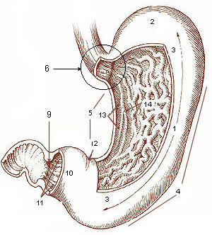
Diagram of the stomach, showing the different regions.
ICD-9-CM43.5-43.9
MeSHD005743
MedlinePlus002945

A gastrectomy is a partial or total surgical removal of the stomach.

Indications

[edit]
A gross anatomy specimen of a total gastrectomy, splenectomy for infiltrative gastric cancer
The same picture, close-up, total gastrectomy, infiltrative gastric cancer
A benign gastric ulcer (from the antrum) of a gastrectomy specimen.

Gastrectomies are performed to treat stomach cancer and perforations of the stomach wall.

For severe duodenal ulcers, it may be necessary to remove the lower portion of the stomach and the upper portion of the small intestine. If there is a sufficient portion of the upper duodenum remaining, a Billroth I procedure is performed, where the remaining portion of the stomach is reattached to the duodenum before the common bile duct. If the stomach cannot be reattached to the duodenum, a Billroth II is performed, wherein the remaining portion of the duodenum is sealed off, a hole is cut into the next section of the small intestine (called the jejunum), and the stomach is reattached at this hole. As the pylorus is used to grind food and slowly release the food into the small intestine, removal of the pylorus can cause food to move into the small intestine faster than normal, leading to gastric dumping syndrome.

Polya's operation

[edit]

Also known as the Reichel–Polya operation, this is a type of posterior gastroenterostomy which is a modification of the Billroth II operation[1] developed by Eugen Pólya and Friedrich Paul Reichel. It involves a resection of 2/3 of the stomach with blind closure of the duodenal stump, and a retrocolic gastrojejunostomy.

Post-operative effects

[edit]

The most obvious effect of the removal of the stomach is the loss of a storage place for food while it is being digested. Since only a small amount of food can be allowed into the small intestine at a time, the patient will have to eat small amounts of food regularly in order to prevent gastric dumping syndrome.

Another major effect is the loss of the intrinsic-factor-secreting parietal cells in the stomach lining. Intrinsic factor is essential for the uptake of vitamin B12 in the terminal ileum, and without it the patient will develop a vitamin B12 deficiency. This can lead to a type of anaemia known as megaloblastic anaemia (can also be caused by folate deficiency, or autoimmune disease where it is specifically known as pernicious anaemia) which severely reduces red-blood cell synthesis (known as erythropoiesis, as well as other haematological cell lineages if severe enough but the red cell is the first to be affected). This can be treated by giving the patient direct injections of vitamin B12. Iron-deficiency anemia can occur as the stomach normally converts iron into its absorbable form.[2]

Another side effect is the loss of ghrelin production, which has been shown to be compensated after a while.[3] Lastly, this procedure is post-operatively associated with decreased bone density and higher incidence of bone fractures. This may be due to the importance of gastric acid in calcium absorption.[4]

Post-operatively, up to 70% of patients undergoing total gastrectomy develop complications such as dumping syndrome and reflux esophagitis.[5] A meta-analysis of 25 studies found that construction of a "pouch", which serves as a "stomach substitute", reduced the incidence of dumping syndrome and reflux esophagitis by 73% and 63% respectively, and led to improvements in quality-of-life, nutritional outcomes, and body mass index.[5]

After Bilroth II surgery, a small amount of residual gastric tissue may remain in the duodenum. The alkaline environment causes the retained gastric tissue to produce acid, which may result in ulcers in a rare complication known as retained antrum syndrome.

All patients lose weight after gastrectomy, although the extent of weight loss is dependent on the extent of surgery (total gastrectomy vs partial gastrectomy) and the pre-operative BMI. Maximum weight loss occurs by 12 months and many patients regain weight afterwards.[6]

History

[edit]

The first successful gastrectomy was performed by Theodor Billroth in 1881 for cancer of the stomach.

Historically, gastrectomies were used to treat peptic ulcers.[7] These are now usually treated with antibiotics, as it was recognized that they are usually due to Helicobacter pylori infection or chemical imbalances in the gastric juices.

In the past a gastrectomy for peptic ulcer disease was often accompanied by a vagotomy, to reduce acid production. This problem is now managed with proton pump inhibitors.

See also

[edit]

References

[edit]
[edit]
Revisions and contributorsEdit on WikipediaRead on Wikipedia
from Grokipedia
Gastrectomy is a surgical procedure involving the partial or complete removal of the stomach, performed to treat various gastric pathologies that do not respond to conservative management.[1] In a partial gastrectomy, only a portion of the stomach is excised, such as the lower part (distal gastrectomy) or upper part (proximal gastrectomy), while a total gastrectomy entails the entire removal of the stomach, with the esophagus then connected directly to the small intestine.[2] These procedures can be conducted via open surgery, laparoscopy, or robotic assistance, aiming to preserve gastrointestinal continuity and function where possible.[3] The primary indication for gastrectomy is gastric cancer, particularly for tumors in the middle or distal stomach requiring distal subtotal resection or extensive disease necessitating total removal to achieve clear margins and lymph node dissection.[2] Other indications include severe peptic ulcer disease with complications like bleeding or perforation, large benign tumors such as gastrointestinal stromal tumors, inflammatory conditions, or polyps that pose a risk of malignancy.[3] In cases of early-stage gastric cancer, less invasive options like endoscopic submucosal dissection may precede or replace gastrectomy, but advanced or multifocal disease often mandates surgical intervention.[2] Contraindications are primarily related to patient fitness, including inability to tolerate general anesthesia or severe comorbidities that increase operative risk.[3] During the procedure, performed under general anesthesia, the surgeon accesses the abdomen through an incision or minimally invasive ports to resect the affected stomach tissue and reconstruct the digestive tract, often using techniques like Billroth I (gastroduodenostomy) or Billroth II (gastrojejunostomy) for partial resections.[3] Lymphadenectomy accompanies oncologic gastrectomies to stage and treat cancer, with D2 dissection recommended for potentially curative cases to improve survival outcomes. In robotic gastrectomy for gastric cancer, mesenteric lymph nodes along the superior mesenteric vein (lymph node station 14v) can be resected as part of D2 or extended lymphadenectomy, particularly in advanced distal gastric cancer, due to improved visualization and dexterity provided by robotic systems.[2][4][5] Postoperative care involves nutritional support, as patients may experience malabsorption, and hospital stays typically last 6-10 days, with full recovery requiring weeks to months.[1] Common risks include anastomotic leaks leading to infection, bleeding, nutritional deficiencies due to reduced stomach capacity, and syndromes like dumping (rapid gastric emptying causing nausea and diarrhea).[2] Morbidity rates range from 17-46%, influenced by surgical approach and patient factors, though minimally invasive techniques often reduce pain and recovery time.[2] Long-term, patients require lifelong dietary modifications, vitamin supplementation (e.g., B12), and monitoring for recurrence in oncologic cases.[1]

Definition and Types

Definition

Gastrectomy is a surgical procedure that involves the partial or total removal of the stomach, a hollow organ in the upper abdomen responsible for storing food and secreting digestive enzymes and acids. The term "gastrectomy" originates from the Greek roots "gaster," meaning stomach, and "ektomē," meaning excision or removal, reflecting its purpose as a targeted resection of gastric tissue.[6][7] This operation alters the digestive process, as the stomach plays a key role in breaking down food and absorbing nutrients, necessitating reconstructive techniques to reconnect the esophagus and small intestine for continued gastrointestinal function.[8] The procedure is classified based on the extent of resection: partial gastrectomy removes a portion of the stomach, such as the lower two-thirds in a distal subtotal approach, while total gastrectomy entails complete excision of the organ, often including nearby lymph nodes in oncologic cases. Partial procedures aim to preserve as much healthy tissue as possible, whereas total removal is typically reserved for more extensive diseases. Gastrectomy can be performed via open surgery, involving a large abdominal incision, or minimally invasively using laparoscopy, which employs small incisions and a camera for precision.[1][8] Primarily indicated for gastric malignancies, gastrectomy serves curative intent by excising tumors and affected margins, but it also addresses benign conditions like intractable peptic ulcers, severe gastritis, or perforations unresponsive to conservative management. In bariatric contexts, variants like sleeve gastrectomy reduce stomach volume to promote weight loss by limiting food intake and altering hunger hormones. Postoperatively, patients adapt to life without a full stomach through dietary modifications, as the surgery impacts nutrient absorption, particularly vitamin B12 and iron, requiring lifelong supplementation in many cases.[8][1]

Types

Gastrectomy procedures are classified primarily by the extent of stomach removal, with the choice depending on the underlying condition, such as gastric cancer, peptic ulcers, or severe obesity. The main types include partial (or subtotal), total, proximal, distal, and pylorus-preserving variants, each tailored to preserve as much function as possible while achieving therapeutic goals.[2][8][9] Partial gastrectomy, also known as subtotal gastrectomy, involves the removal of a portion of the stomach, typically up to two-thirds or about 80%, leaving the remainder intact for reconstruction. This type is commonly performed for cancers located in the lower or middle stomach, allowing preservation of the cardiac sphincter to maintain some normal digestive function. Reconstruction often connects the remaining stomach to the small intestine via methods like Billroth II or Roux-en-Y.[2][9][8] Total gastrectomy entails complete removal of the stomach, including the cardia, body, and pylorus, along with surrounding lymph nodes and omentum in oncologic cases. It is indicated for diffuse or proximal gastric cancers, such as those with signet ring cell histology or CDH1 mutations, requiring wide margins (4-6 cm) for complete resection. The esophagus is then anastomosed to the jejunum, usually in a Roux-en-Y configuration, to restore gastrointestinal continuity.[2][9][10] Proximal gastrectomy removes the upper third of the stomach, preserving the distal portion and pylorus to minimize postoperative nutritional deficiencies. This approach is suitable for early-stage (cT1N0M0) cancers confined to the proximal stomach, with reconstruction options like esophagogastrostomy to reduce reflux complications.[2] Distal gastrectomy targets the middle and lower stomach, sparing the proximal portion while ensuring adequate margins (4-6 cm proximally). It is used for tumors in the distal two-thirds, with reconstruction typically involving Billroth II or Roux-en-Y gastrojejunostomy; preservation of short gastric vessels aids in maintaining blood supply to the remnant.[2] Pylorus-preserving distal gastrectomy is a specialized form of distal resection that spares the pylorus and infrapyloric vessels, aiming to maintain gastric emptying and reduce bile reflux. Indicated for early middle-stomach cancers (cT1N0M0) with at least 4 cm distal margins, it may limit certain lymphadenectomy steps but improves quality of life by preserving pyloric function.[2] In addition to these oncologic-focused types, sleeve gastrectomy represents a partial gastrectomy used primarily in bariatric surgery, removing about 80% of the stomach along the greater curvature to create a sleeve-shaped remnant. This reduces stomach capacity and alters hunger hormones, aiding significant weight loss in patients with class III obesity.[8] Oesophagogastrectomy, sometimes considered a variant, involves partial stomach removal combined with distal esophagectomy for tumors at the gastroesophageal junction, reshaping the remaining stomach into a conduit for esophageal replacement.[9]

Indications and Contraindications

Indications

Gastrectomy is primarily indicated for the treatment of gastric malignancies, particularly adenocarcinoma, where surgical resection aims to achieve curative outcomes by removing the tumor with adequate margins while addressing lymph node involvement. For early-stage gastric cancer confined to the mucosa or submucosa without lymph node metastasis, less extensive procedures like endoscopic submucosal dissection may suffice, but gastrectomy—such as distal, proximal, or total—is recommended for more advanced or unfavorably located tumors to ensure negative margins of 4-6 cm and perform lymphadenectomy (D1 or D2 extent based on tumor location).[2] Proximal gastrectomy is suitable for upper-third early gastric cancers (cT1N0M0), while total gastrectomy is indicated for tumors involving the proximal stomach, signet-ring cell histology, or diffuse involvement.[2] In cases of locally advanced disease invading adjacent organs, multivisceral resection may be performed following neoadjuvant therapy to achieve R0 resection.[2] For benign conditions, gastrectomy is reserved for complications of peptic ulcer disease (PUD) that are refractory to medical management, including proton pump inhibitors and endoscopic interventions. Indications include life-threatening issues such as free perforation, uncontrollable bleeding (especially from giant ulcers >3 cm), or gastric outlet obstruction unresponsive to therapy, where procedures like antrectomy with vagotomy and reconstruction (e.g., Roux-en-Y) are employed to control hemorrhage and prevent recurrence. Other benign indications include resection of large gastrointestinal stromal tumors (GISTs), extensive gastric polyposis with malignant potential, or severe inflammatory conditions such as gastric involvement in Crohn's disease, when endoscopic or medical management fails.[3][11] Historically more common, surgical intervention for PUD has declined with effective pharmacotherapy, but remains essential in select refractory or emergent cases.[12] In bariatric surgery, sleeve gastrectomy—a partial gastrectomy removing about 80% of the stomach—is indicated for severe obesity to promote weight loss and resolve comorbidities. Eligibility typically requires a body mass index (BMI) of 40 or higher, or 35-39.9 with serious weight-related conditions such as type 2 diabetes, hypertension, or severe sleep apnea, following failed attempts at nonsurgical weight loss and commitment to lifestyle changes.[13] Prophylactic total gastrectomy is recommended for individuals with germline CDH1 mutations associated with hereditary diffuse gastric cancer, as endoscopic surveillance often fails to detect multifocal signet-ring cell carcinomas at an early stage. Guidelines suggest the procedure between ages 20-30 for mutation carriers with a family history of gastric cancer, offering near-complete risk reduction despite nutritional challenges.[14][15]

Contraindications

Gastrectomy, whether partial or total, carries significant risks, and its performance is contraindicated in certain clinical scenarios to avoid excessive morbidity or mortality. Absolute contraindications include conditions that preclude safe administration of general anesthesia, such as severe cardiovascular instability or uncontrolled respiratory failure, as these render the patient unable to tolerate the procedure.[2][3] In the context of gastric cancer, extensive distant metastases detected via imaging or PET/CT, such as to the liver, lungs, or other organs, represent an absolute contraindication to curative-intent gastrectomy, shifting management toward systemic therapy or palliative care.[16] Relative contraindications encompass patient factors that increase perioperative risks but may not entirely preclude surgery if benefits outweigh harms, particularly after optimization. These include advanced age, severe cardiopulmonary dysfunction, malnutrition manifesting as anemia or hypoproteinemia, significant ascites, and disseminated malignancy with diffuse peritoneal metastases, all of which can impair wound healing and anastomotic integrity.[2][3] Coagulation disorders, pregnancy, and poor overall performance status—such as vital organ failure or diminished life expectancy due to comorbidities—also fall into this category, necessitating multidisciplinary evaluation.[16] For tumors fixed to adjacent structures like the liver, pancreas, or posterior abdominal wall, resection may be relatively contraindicated for curative purposes, though palliative gastrectomy could be considered for complications like bleeding or obstruction.[3] In oncologic settings, unresectable local disease or stage IV gastric cancer without feasible cytoreduction further contraindicates radical gastrectomy, as guidelines emphasize surgery only for potentially curable stages I-III without distant spread.[16] For minimally invasive approaches like laparoscopic or robotic gastrectomy, a history of upper abdominal surgery is a relative contraindication due to potential adhesions increasing injury risk.[3] Preoperative assessment, including nutritional support and comorbidity management, is essential to mitigate these risks and determine surgical candidacy.[2]

Surgical Techniques

Open Gastrectomy

Open gastrectomy represents the conventional surgical method for partial or total removal of the stomach, typically employed in cases of gastric malignancy or severe benign conditions where extensive access is required. This approach involves a single large incision to provide direct visualization and manipulation of abdominal structures, distinguishing it from minimally invasive techniques. It is particularly indicated for advanced tumors, when laparoscopic access is inadequate, or in patients with prior abdominal surgeries that complicate minimally invasive entry.[2] Preoperative preparation for open gastrectomy includes comprehensive staging via endoscopy, endoscopic ultrasound, and computed tomography to assess tumor extent and resectability. Patients receive prophylactic antibiotics, and a nasogastric tube is placed for decompression. General anesthesia is administered, with the patient positioned supine and the abdomen prepped sterilely. Smoking cessation is advised weeks prior, and medications such as blood thinners are discontinued to minimize bleeding risks.[2][1] The procedure begins with an incision, usually upper midline from the xiphoid process to the umbilicus or a left subcostal approach for better exposure of the upper abdomen, depending on tumor location. Intraoperative exploration assesses for distant metastases; if none are found, the greater and lesser omenta are mobilized by dividing the gastrocolic and gastrosplenic ligaments. For a distal gastrectomy, the duodenum is transected 2-3 cm distal to the pylorus, and the stomach is divided proximally with a 4-6 cm margin from the tumor. In total gastrectomy, the entire stomach is removed, including the esophagus distally and duodenum proximally. Vascular control is achieved by ligating the right and left gastric arteries, gastroepiploic vessels, and short gastric vessels as needed. The spleen may be preserved unless involved, though iatrogenic injury occurs in approximately 7-8% of cases, particularly in open procedures.[2][17] Lymphadenectomy is integral, with D2 dissection standard for curative intent in gastric cancer, involving removal of at least 15 lymph nodes from stations 1 through 12, including perigastric, celiac axis, and hepatoduodenal nodes. This enhances staging accuracy and oncologic outcomes compared to D1 dissection. Reconstruction follows resection: partial gastrectomy often uses Billroth I (gastroduodenostomy) or Billroth II (gastrojejunostomy), while total gastrectomy requires Roux-en-Y esophagojejunostomy to restore continuity. The procedure duration typically ranges from 3 to 6 hours, with closure in layers using absorbable sutures for the peritoneum and fascia.[2][10] Postoperatively, patients remain intubated briefly if extensive, transitioning to nasogastric decompression and parenteral nutrition. Drains are placed to monitor for anastomotic leaks, a key early complication with rates of 2-10% in open approaches due to direct tissue handling. While open gastrectomy allows thorough dissection in complex cases, it is associated with higher morbidity (13-46%) from wound infections, prolonged ileus, and longer hospital stays (5-10 days) compared to laparoscopic methods.[2][18]

Minimally Invasive Gastrectomy

Minimally invasive gastrectomy refers to surgical procedures for partial or total removal of the stomach that utilize small incisions and specialized instruments, primarily laparoscopic or robotic-assisted approaches, to minimize trauma to surrounding tissues compared to traditional open surgery. These techniques have gained prominence for treating gastric cancer, offering enhanced precision and visualization while aiming to preserve oncologic efficacy.[19] Laparoscopic gastrectomy (LG) involves the use of a laparoscope and thin instruments inserted through 4-6 small ports in the abdomen, allowing for dissection, lymph node retrieval, and reconstruction without a large midline incision. Robotic-assisted minimally invasive gastrectomy (RAMIG) builds on this by employing robotic systems, such as the da Vinci platform, which provide three-dimensional visualization, tremor filtration, and articulated instruments for improved dexterity in complex maneuvers such as D2 lymphadenectomy, including resection of mesenteric lymph nodes (specifically station 14v along the superior mesenteric vein) as part of D2 or extended lymphadenectomy, particularly in advanced distal gastric cancer due to enhanced precision, visualization, and dexterity in complex anatomical areas. Both approaches typically follow similar steps to open surgery, including mobilization of the stomach, vascular ligation, and reconstruction via Billroth I/II or Roux-en-Y methods, but with reduced postoperative pain and scarring.[19][20][21] Indications for minimally invasive gastrectomy are primarily early-stage gastric adenocarcinoma (T1-T3, N0-N2), as endorsed by guidelines from the National Comprehensive Cancer Network and Japanese Gastric Cancer Association, though evidence supports extension to locally advanced disease (T4a) in experienced centers. Contraindications include bulky T4b tumors, extensive prior abdominal surgery causing adhesions, or hemodynamic instability precluding pneumoperitoneum. Patient selection emphasizes body mass index under 30 and surgeon expertise, with over 50 cases recommended for proficiency in LG.[19] Short-term outcomes demonstrate clear benefits over open gastrectomy, including reduced intraoperative blood loss (typically 100-200 mL versus 300-500 mL), shorter hospital length of stay (7-8 days versus 9-11 days), and faster return to oral intake. A network meta-analysis of randomized controlled trials (RCTs) confirmed lower overall complication rates with minimally invasive approaches (15-20% versus 25-30% for open), encompassing anastomotic leaks, wound infections, and pulmonary issues, without increased mortality (0-1% across groups). Robotic assistance further mitigates certain risks, such as pancreatic fistula (2-5% versus 5-10% in LG), due to enhanced precision.[22][20][23] Oncologic outcomes are comparable to open surgery, with adequate lymph node harvests (≥15-20 nodes) and R0 resection rates exceeding 90% in landmark RCTs like CLASS-01 and KLASS-02, which involved over 1,000 patients each. Five-year overall survival rates range from 73-97% for stage I-III disease, showing non-inferiority (hazard ratio 0.86-1.03 for survival and disease-free survival). Long-term data from cohort studies indicate similar recurrence patterns, though robotic approaches may yield slightly higher lymph node counts (40 versus 38 in LG). Limitations include prolonged operative times (180-300 minutes versus 120-200 minutes for open) and higher costs for RAMIG ($13,000-15,000 versus $10,000 for LG), necessitating cost-effectiveness analyses in resource-limited settings.[19][23][20] Ongoing trials, such as those under the IDEAL framework (stages 3-4), continue to refine these techniques, with evidence levels strongest for LG in distal gastrectomy and emerging for RAMIG in total procedures. Adoption has surged, with minimally invasive methods comprising over 70% of gastrectomies in high-volume Asian centers by 2023.[20]

Reconstruction Methods

Billroth Procedures

The Billroth procedures, developed by Austrian surgeon Theodor Billroth in the late 19th century, are foundational reconstruction techniques following distal gastrectomy for conditions such as gastric cancer or peptic ulcers. Billroth I, first performed on January 29, 1881, involves an end-to-end gastroduodenostomy, reconnecting the remnant stomach directly to the duodenum after removal of the distal stomach and pylorus.[24] This method aims to restore a more physiological pathway for food transit, preserving the natural route through the duodenum.[25] In contrast, Billroth II, introduced on January 14, 1885, employs a gastrojejunostomy, anastomosing the gastric remnant to a loop of jejunum (typically 20-40 cm distal to the ligament of Treitz) while closing the duodenal stump.[24] This bypasses the duodenum, allowing food to enter the jejunum directly.[25] Billroth I is considered simpler and more anatomically physiological, potentially reducing the risk of certain postoperative issues like afferent loop syndrome, but it requires adequate mobilization of the duodenum to avoid tension at the anastomosis site.[26] Studies indicate lower overall complication rates with Billroth I compared to Billroth II, with a meta-analysis of 15 randomized controlled trials reporting a 20.1% complication rate for Billroth I versus 37.2% for Billroth II (P=0.000), including reduced incidences of anastomotic leaks (1.3% vs. 3.3%) and deep infections (2.7% vs. 10.4%).[26] Additionally, Billroth I is associated with shorter hospital stays.[26] However, it can lead to duodenogastric reflux of bile and pancreatic secretions, promoting remnant gastritis and increasing risks of esophagitis.[27] Billroth II offers technical feasibility when duodenal mobilization is challenging, such as in cases of inflammation or tumor invasion, and it may lower the risk of duodenal stump leakage in select patients.[25] Despite this, it is linked to higher rates of early postoperative complications, including severe infections and prolonged recovery, as evidenced by risk-adjusted observed-to-expected ratios favoring Billroth I (0.74 vs. 1.03).[26] Long-term, Billroth II also contributes to significant reflux, potentially leading to alkaline reflux gastritis and nutritional deficiencies due to bacterial overgrowth in the blind duodenal loop.[27] Comparisons between the two procedures highlight Billroth I as the preferred choice when oncological margins and anatomy permit, due to its superior safety profile and physiological benefits, though both are associated with reflux-related morbidity that has driven the adoption of alternatives like Roux-en-Y reconstruction.[26][25] Historical success with Billroth II, applied in 32 patients by 1885 with improved survival rates over Billroth I's initial high mortality, underscores its role in advancing gastric surgery despite modern refinements.[24]

Roux-en-Y Reconstruction

Roux-en-Y reconstruction is a widely used method to restore gastrointestinal continuity following partial or total gastrectomy, involving the creation of a Y-shaped anastomosis between the remnant stomach (or esophagus in total gastrectomy) and the jejunum.[2] This technique divides the jejunum approximately 10-15 cm distal to the ligament of Treitz, forming a Roux limb of 40-60 cm that is anastomosed end-to-end to the gastric remnant via gastrojejunostomy or to the esophagus via esophagojejunostomy in total gastrectomy cases.[2] The distal jejunum is then reconnected to the Roux limb through a jejunojejunostomy, typically performed intracorporeally with linear or circular staplers during laparoscopic procedures, and can be configured antecolic or retrocolic to minimize tension.[28] This configuration diverts biliary and pancreatic secretions away from the gastrojejunal anastomosis, reducing exposure of the upper gastrointestinal tract to alkaline reflux.[29] The primary indications for Roux-en-Y reconstruction include distal gastrectomy for tumors near the pylorus or when a small remnant stomach is anticipated, as well as total gastrectomy for proximal gastric malignancies, where it serves as a standard to maintain intestinal and biliary continuity while optimizing oncologic margins.[2] It is particularly favored in cases of gastric cancer to prevent reflux-related complications that could compromise quality of life or long-term surveillance.[28] Compared to Billroth I (which reconnects the duodenum directly to the stomach) or Billroth II (which uses a gastrojejunostomy with duodenal loop), Roux-en-Y is preferred for its lower risk of bile reflux gastritis and esophagitis, with studies showing reduced endoscopic evidence of remnant gastritis.[29][28] Advantages of Roux-en-Y include improved postoperative quality of life through decreased dumping syndrome, reflux esophagitis, and marginal ulcers, as evidenced by randomized controlled trials demonstrating superior functional outcomes and nutritional status over Billroth procedures.[29] For instance, it supports better long-term endoscopic findings and patient-reported outcomes in distal gastrectomy, with shorter emptying times for solid foods compared to pouch variants in total gastrectomy.[2] However, the procedure is more technically demanding, often extending operative time (e.g., 185-257 minutes versus 173-203 minutes for Billroth I) and slightly increasing blood loss (e.g., 27.5 mL versus 10 mL).[28] Potential complications specific to Roux-en-Y include anastomotic leakage (reported in 1-5% of cases), Roux stasis syndrome (affecting 10-30% with symptoms like nausea and vomiting due to delayed motility), and internal hernias such as Peterson's space hernia.[2][28] Despite these risks, overall morbidity is comparable to other reconstructions when performed laparoscopically, and long-term outcomes favor Roux-en-Y for reduced alkaline reflux and enhanced survivorship in malignancy cases.[29] Variants like uncut Roux-en-Y, which preserve vagal innervation by avoiding full jejunal division, further mitigate stasis but maintain similar reflux benefits.[28] As of 2025, emerging techniques such as double-tract reconstruction (DTR) after proximal gastrectomy have gained attention for further reducing reflux and improving nutritional outcomes, often combining esophagojejunostomy with a jejunal pouch or additional tract.[30] Other options, like jejunal interposition or pouch reconstructions (e.g., Hunt-Lawrence pouch) for total gastrectomy, aim to enhance reservoir function and minimize long-term deficiencies.[2]

Postoperative Management

Immediate Postoperative Care

Immediate postoperative care following gastrectomy emphasizes close monitoring in a recovery unit to detect and manage potential complications such as anastomotic leaks, bleeding, or infection, while promoting early recovery through enhanced recovery after surgery (ERAS) protocols.[31] Patients are typically transferred to the post-anesthesia care unit (PACU) or intensive care unit if high-risk, where vital signs, including blood pressure, heart rate, oxygen saturation, and urine output, are continuously assessed to ensure hemodynamic stability.[32] Fluid status is optimized using restrictive intravenous strategies, such as lactated Ringer's at 1 mL/kg/hour, to prevent overload and associated complications like pulmonary edema.[33] Pain management is multimodal and opioid-sparing to minimize respiratory depression and ileus, incorporating scheduled acetaminophen (1 g every 8 hours intravenously, transitioning to oral), nonsteroidal anti-inflammatory drugs if renal function permits, and regional techniques like thoracic epidural analgesia or transversus abdominis plane blocks.[31] Patient-controlled analgesia may be used initially for breakthrough pain, with a goal of transitioning to oral analgesics by postoperative day 4.[32] Prophylaxis against postoperative nausea and vomiting (PONV) is standard, using agents like ondansetron based on risk stratification (e.g., Apfel score), as PONV can delay oral intake and increase aspiration risk.[33] Nutritional support begins early to reduce catabolism and support wound healing, with nasogastric tubes removed on postoperative day 1 unless prolonged ileus is present.[31] For subtotal gastrectomy, clear liquids may be introduced on postoperative day 1 if tolerated, advancing to full liquids and soft diet as able; total gastrectomy patients may require jejunostomy tube feeds starting at 10 mL/hour on day 1.[32] Early oral nutrition on day 1 is safe and feasible, reducing hospital length of stay by approximately 1.5 days without increasing anastomotic leak rates (0% in the early group).[34] though elderly patients (>70 years) need vigilant monitoring for compliance and symptoms like nausea.[34] Early mobilization is initiated within 6-8 hours postoperatively, progressing from sitting in a chair to hallway walking three times daily by day 1, to prevent deep vein thrombosis (DVT), pneumonia, and muscle loss.[33] Thromboembolic prophylaxis with low-molecular-weight heparin is administered for 7-10 days (extended to 4 weeks in high-risk cases), combined with incentive spirometry and leg exercises every 2 hours while awake to enhance pulmonary function.[31] Routine peritoneal drains and urinary catheters are avoided to reduce infection risk and facilitate mobility, with blood glucose monitored every 6 hours in diabetics and mornings in others to prevent hyperglycemia-related complications.[32] Wound care involves keeping incisions clean and dry, with sterile dressings changed as needed, and monitoring for signs of surgical site infection such as erythema or drainage.[33] Laboratory monitoring includes complete blood count, electrolytes, and inflammatory markers like C-reactive protein on postoperative days 1, 3, and 5 to detect early leaks or infections, guiding interventions like imaging if suspected.[31] These ERAS elements collectively reduce complication rates and hospital length of stay (by approximately 1.5 days in studies).[34] Hospital stays typically last 6-14 days, or up to two weeks for complex or total gastrectomies, with full recovery requiring weeks to months.

Long-term Follow-up

Long-term follow-up after gastrectomy focuses on surveillance for disease recurrence, management of nutritional deficiencies, and assessment of quality of life, with protocols varying based on whether the procedure was performed for curative intent in gastric cancer or prophylactically for hereditary conditions such as CDH1 mutations.[35][36] For patients undergoing gastrectomy for gastric cancer, surveillance includes periodic cross-sectional imaging tailored to disease stage and recurrence risk, with routine screening potentially discontinued after 5 years due to the rarity of late recurrences.[35] Upper gastrointestinal endoscopy is recommended annually for up to 20 years post-operation to detect remnant gastric cancer early, particularly in the first 5 years when curative resection rates are higher and overall survival is better.[37] Western guidelines, such as those from ESMO and NCCN, emphasize symptom-driven follow-up over routine tumor markers or imaging, as no randomized trials demonstrate survival benefits from intensive surveillance.[38] Nutritional monitoring is lifelong, addressing common sequelae like anemia from iron or vitamin B12 deficiency and osteoporosis from calcium and vitamin D malabsorption.[35][38] After total gastrectomy, the stomach's role in initiating protein digestion via pepsin and hydrochloric acid is eliminated, shifting primary protein breakdown to pancreatic proteases (trypsin, chymotrypsin) and intestinal peptidases in the small intestine. This, combined with rapid intestinal transit, reduced mixing, and altered anatomy, leads to mild malabsorption of proteins (nitrogen) and fats, often contributing to malnutrition. However, with adequate intake, positive nitrogen balance and weight gain are achievable. Patients require higher protein consumption through small, frequent meals and nutritional supplements to support healing and prevent muscle loss.[39][40] Annual blood tests for full blood count, ferritin, calcium, vitamin B12, and other micronutrients are standard, alongside supplementation with a daily multivitamin, calcium (at least 1.5 g/day), vitamin D (target serum level >30 ng/ml), and vitamin B12 as per local protocols.[36] Weight stabilization typically occurs within 2 years, but ongoing dietitian reviews help manage gastrointestinal symptoms such as dumping syndrome or reflux, as well as to optimize protein intake and overall nutritional status.[38] In prophylactic total gastrectomy, consensus guidelines recommend annual in-person or remote assessments involving multidisciplinary teams, including surgeons, gastroenterologists, dietitians, and nurse specialists, starting 3-6 months postoperatively and continuing lifelong after initial recovery (1-2 years).[36] These include evaluations of weight, symptoms, and micronutrient levels, with support from psychologists for psychological adjustment and geneticists for ongoing cancer risk screening.[36] Quality of life generally recovers to baseline within 1 year for most patients, though limited resections facilitate faster improvement compared to total gastrectomy.[41] Long-term follow-up thus integrates oncologic vigilance with supportive care to mitigate chronic impacts on physical and emotional well-being.[38]

Complications and Risks

Early Complications

Early complications following gastrectomy encompass adverse events occurring within 30 days of surgery, including surgical site issues, infections, and systemic responses that can prolong recovery, increase reintervention needs, and elevate mortality risk. These complications arise due to the procedure's complexity, involving resection of part or all of the stomach and reconstruction, often in patients with underlying comorbidities like gastric cancer. Overall incidence rates range from 12% to 28.5% in large cohorts, with higher rates observed in total versus partial gastrectomy.[42] The most clinically significant early complications include anastomotic leakage, pulmonary issues, intra-abdominal abscesses, and wound infections. Anastomotic leakage, a disruption at the surgical join sites, occurs in approximately 7% of cases and is a leading cause of postoperative mortality (contributing 29.2% of deaths) and reoperations, often requiring drainage or revision surgery. Pulmonary complications, such as pneumonia and atelectasis, affect about 15% of patients and account for 21.6% of mortality, exacerbated by postoperative pain, immobility, and diaphragmatic irritation. Intra-abdominal abscesses and wound infections are common, typically managed with antibiotics and drainage. Other notable issues include bleeding (from staple lines or vessels), duodenal stump leakage, and pancreatic fistula.[43] Risk factors for these early complications are multifactorial, encompassing patient-related elements like age over 65 years, diabetes, hypertension, and prior abdominal surgery, as well as procedural aspects such as operative duration, blood loss, and Roux-en-Y reconstruction. These factors highlight the importance of preoperative optimization, including nutritional assessment (e.g., prealbumin levels) and careful intraoperative techniques like minimally invasive approaches to mitigate risks. Complications not only extend hospital stays and healthcare costs but also impair long-term outcomes, with affected patients showing reduced survival compared to uncomplicated cases.[43]

Late Complications

Late complications following gastrectomy, often referred to as postgastrectomy syndromes, encompass a range of chronic symptoms and physiological disturbances that arise months to years after surgery due to alterations in gastric anatomy, motility, and absorption. These syndromes affect approximately 20-30% of patients to varying degrees, with 2-5% experiencing severe, disabling symptoms that impact quality of life.[44] The incidence and severity depend on the extent of resection (partial versus total), reconstruction method (e.g., Billroth I, Billroth II, or Roux-en-Y), and patient factors such as age and sex, with females and older patients at higher risk for persistent issues.[45] Common late complications include dumping syndrome, nutritional deficiencies, bile reflux gastritis, and Roux stasis syndrome, each stemming from disrupted reservoir function, rapid transit of food, or impaired nutrient uptake.[46] Dumping syndrome is one of the most prevalent late complications, occurring in up to 40% of patients after gastrectomy, particularly those with Roux-en-Y reconstruction. It manifests in two phases: early dumping (within 30 minutes of eating) caused by rapid osmotic fluid shifts into the small intestine leading to gastrointestinal (bloating, cramps) and vasomotor symptoms (palpitations, flushing); and late dumping (1-3 hours post-meal) due to reactive hypoglycemia from exaggerated insulin release. Diagnosis typically involves a modified oral glucose tolerance test monitoring hematocrit, pulse, and glucose levels. Management focuses on dietary modifications such as small, frequent meals low in simple carbohydrates, with pharmacological options like acarbose for late symptoms or octreotide for refractory cases; surgical interventions, such as conversion to Roux-en-Y, are reserved for severe instances.[44] Nutritional deficiencies represent another major category of late complications, arising from reduced intrinsic factor production, bypassed duodenum, or altered absorption sites, affecting 20-80% of patients within 4-5 years post-surgery. Iron deficiency anemia is common (up to 40% incidence), resulting from achlorhydria and loss of acidic environment needed for iron solubilization, while vitamin B12 deficiency (prevalence 20-80% at 48 months) stems from intrinsic factor absence after total or proximal gastrectomy. Other deficiencies include calcium, vitamin D, and folate, potentially leading to osteoporosis or megaloblastic anemia. In patients undergoing total gastrectomy, the elimination of the stomach's role in initiating protein digestion via pepsin and hydrochloric acid shifts primary protein breakdown to pancreatic proteases (trypsin, chymotrypsin) and intestinal peptidases. Combined with rapid intestinal transit, reduced food mixing, and altered anatomy, this results in mild malabsorption of proteins (nitrogen) and fats, which can contribute to malnutrition. However, with adequate intake—including higher protein consumption through small, frequent meals and supplements—positive nitrogen balance, weight stabilization, and prevention of muscle loss are achievable. Routine monitoring via serum ferritin, B12 levels, and endoscopy is recommended, with treatment involving oral or parenteral supplementation (e.g., 1-2 mg daily vitamin B12 or 100-200 mg elemental iron). Prophylactic multivitamins and mineral supplements are standard in long-term follow-up to mitigate risks.[44][46][39][47] Bile reflux gastritis, affecting up to 70% of patients after Billroth II reconstruction, occurs due to loss of the pyloric sphincter and duodenal passage, allowing alkaline bile to reflux into the gastric remnant and cause chronic inflammation, erosions, and symptoms like epigastric pain or vomiting. Endoscopic biopsy confirms diagnosis by showing foveolar hyperplasia and inflammation. Initial management includes ursodeoxycholic acid (UDCA) to alter bile composition or sucralfate for mucosal protection; persistent cases may require surgical diversion via Roux-en-Y gastrojejunostomy. Postvagotomy diarrhea, seen in about 30% of patients (clinically significant in 10%), results from vagal nerve disruption accelerating small bowel transit and bacterial overgrowth, managed with dietary fiber, loperamide, or cholestyramine.[44] Roux stasis syndrome, specific to Roux-en-Y reconstructions, affects 10-30% of patients and involves nausea, vomiting, and bloating from impaired motility in the Roux limb due to retrograde peristalsis and phase III migrating motor complex disruption. It is diagnosed by exclusion via scintigraphy or manometry after ruling out mechanical obstruction. Treatment emphasizes small frequent meals and prokinetic agents like metoclopramide; severe cases may necessitate jejunal feeding or rarely, reconstructive surgery. Additionally, gallstone formation risk increases post-gastrectomy due to bile stasis from vagotomy or rapid weight loss, with prophylactic UDCA recommended in high-risk patients to reduce incidence by up to 30%. Long-term surveillance, including annual nutritional assessments and endoscopy, is crucial to detect and address these complications early, improving outcomes in survivors of gastric cancer or other indications for gastrectomy.[44][45]

History and Developments

Early History

The earliest attempts at gastrectomy occurred in the late 19th century amid high surgical risks and limited antisepsis. In April 1879, French surgeon Jules Péan performed a partial resection of the pyloric stomach for cancer in a cachectic patient, but the individual succumbed on the fifth postoperative day due to complications.[24] This was followed in November 1880 by Polish surgeon Ludwik Rydygier, who conducted the first recorded partial gastrectomy for gastric cancer; however, the patient died just 12 hours after the procedure.[24] These pioneering efforts highlighted the formidable challenges of gastric surgery, including infection and hemorrhage, yet they laid foundational groundwork for subsequent innovations. The breakthrough in gastrectomy came with Austrian surgeon Theodor Billroth, widely regarded as the pioneer of the procedure. On January 29, 1881, Billroth performed the first successful partial gastrectomy—known as Billroth I—for pyloric carcinoma in 43-year-old Viennese woman Therese Heller; the operation involved resecting the distal stomach and creating an end-to-end anastomosis between the gastric remnant and duodenum, lasting approximately 1.5 hours, with the patient surviving four months before succumbing to metastases.[24] Building on this, Billroth and his collaborator Anton Wölfler refined techniques through canine experiments, leading to the inaugural Billroth II procedure on January 14, 1885, which connected the gastric remnant to the jejunum via gastrojejunostomy to address anastomotic tensions observed in the earlier method.[24] These operations marked a shift toward viable gastric resections, though early mortality rates remained elevated due to delayed diagnoses and postoperative infections. Further early advancements emerged in the 1880s, expanding gastrectomy's applications beyond malignancy. In 1882, Rydygier reported the first partial gastrectomy for benign gastric ulcer, a procedure met with initial skepticism in surgical circles but demonstrating potential for non-cancerous conditions.[48] Across the Atlantic, in 1886, Belfast surgeon Thomas Sinclair executed the region's inaugural gastrectomy for cancer, contributing to the procedure's gradual adoption in Europe despite persistent high risks.[48] By the close of the century, these developments had established partial gastrectomy as a feasible, albeit hazardous, intervention, influencing the evolution of gastric surgery toward improved outcomes in the 20th century.

Modern Advancements

In recent years, gastrectomy procedures have evolved significantly toward minimally invasive techniques, driven by advancements in laparoscopy and robotics, which aim to reduce perioperative morbidity while maintaining oncologic efficacy for gastric cancer treatment. Laparoscopic gastrectomy has become the standard for early-stage disease, offering shorter hospital stays and faster recovery compared to open surgery, as evidenced by long-term data showing comparable overall survival rates despite slightly lower lymph node yields. Robotic-assisted gastrectomy, utilizing systems like the da Vinci Xi, further enhances precision through three-dimensional visualization and articulated instruments, particularly beneficial for complex lymph node dissections in advanced cases. A 2025 meta-analysis of 15 studies involving 2,240 patients demonstrated that robotic total gastrectomy (RTG) reduced intraoperative blood loss by 18.1 mL (P < 0.001), increased retrieved lymph nodes by 3.4 (P < 0.001), lowered severe complications (relative risk 0.65, P = 0.024), and shortened hospital stays by 0.8 days (P = 0.032) compared to laparoscopic approaches, though operative times were longer by 33 minutes (P < 0.001); oncologic outcomes, including 3-year overall and disease-free survival, remained equivalent.[49] Functional-preserving gastrectomies represent another key advancement, focusing on maintaining postoperative quality of life by minimizing resection extent in select early gastric cancers. Proximal gastrectomy (PG) with innovative reconstructions, such as double-tract or Kamikawa anastomoses, has reduced reflux esophagitis incidence to as low as 4.6-10.6%, improving nutritional outcomes and anemia rates versus total gastrectomy. Pylorus-preserving gastrectomy (PPG) preserves the pyloric sphincter to mitigate dumping syndrome and bile reflux, with studies reporting a 96.3% 5-year survival rate and lower gallstone formation (2.3% vs. 8.7% in distal gastrectomy). Segmental gastrectomy (SG), often guided by sentinel lymph node navigation, allows targeted resection for middle-third tumors, achieving 96.6% 5-year survival without increased recurrence in recent cohorts. The 2024 SENORITA trial, involving over 500 patients, validated stomach-preserving surgery via sentinel node biopsy with a 98.5% detection rate, enabling organ preservation in clinically node-negative early gastric cancer while ensuring oncologic safety.[50][51] Emerging technologies further refine these procedures, including reduced-port robotic gastrectomy (REPROG), which uses fewer incisions for enhanced cosmesis and recovery. In the KLASS-13 study of 1,060 REPROG patients propensity-matched to 1,965 conventional laparoscopic cases, REPROG yielded a shorter hospital stay (6.1 vs. 7.8 days, P < 0.001), comparable complication rates (2.0% vs. 2.4%, P = 0.424), and a low conversion rate of 0.19%, positioning it as a feasible option for experienced centers. Intraoperative innovations like indocyanine green near-infrared fluorescence imaging improve lymph node visualization during robotic distal gastrectomy, enhancing dissection accuracy. Laparoscopic-endoscopic cooperative surgery (LECS) integrates endoscopy for precise tumor excision in submucosal lesions, with multicenter data from 2024 showing low morbidity and preserved function. Future directions include AI-assisted planning and telesurgery to broaden access, though ongoing trials like JCOG1907 are evaluating long-term robotic efficacy in advanced disease.[52][53][54][55]

References

User Avatar
No comments yet.