



Community hub
Inguinal hernia
View on Wikipedia
| Inguinal hernia | |
|---|---|
 | |
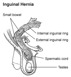
| Diagram of an indirect, scrotal inguinal hernia (median view from the left). | |
| Pronunciation | |
| Specialty | General surgery |
| Symptoms | Pain, bulging in the groin[1] |
| Complications | Strangulation[1] |
| Usual onset | < 1 year old, > 50 years old[2] |
| Risk factors | Family history, smoking, chronic obstructive pulmonary disease, obesity, pregnancy, peritoneal dialysis, collagen vascular disease, connective tissue disease, previous open appendectomy[1][2][3] |
| Diagnostic method | Based on symptoms, medical imaging[1] |
| Treatment | Conservative, surgery[1] |
| Frequency | 27% (males), 3% (females)[1] |
| Deaths | 59,800 (2015)[4] |
An inguinal hernia or groin hernia is a hernia (protrusion) of abdominal cavity contents through the inguinal canal. Symptoms, which may include pain or discomfort, especially with or following coughing, exercise, or bowel movements, are absent in about a third of patients. Symptoms often get worse throughout the day and improve when lying down. A bulging area may occur that becomes larger when bearing down. Inguinal hernias occur more often on the right than the left side. The main concern is strangulation, where the blood supply to part of the intestine is blocked. This usually produces severe pain and tenderness in the area.[1]
Risk factors for the development of a hernia include: smoking, chronic obstructive pulmonary disease, obesity, pregnancy, peritoneal dialysis, collagen vascular disease, and previous open appendectomy, among others.[1][2] Predisposition to hernias is genetic[5] and they occur more often in certain families.[6][7][8][1] Deleterious mutations causing predisposition to hernias seem to have dominant inheritance (especially for men). It is unclear if inguinal hernias are associated with heavy lifting. Hernias can often be diagnosed based on signs and symptoms. Occasionally, medical imaging is used to confirm the diagnosis or rule out other possible causes.[1]
Groin hernias that do not cause symptoms in males do not need repair. Repair, however, is generally recommended in females due to the higher rate of femoral hernias (also a type of groin hernia), which have more complications. If strangulation occurs, immediate surgery is required. Repair may be done by open surgery or by laparoscopic surgery. Open surgery has the benefit of possibly being done under local anesthesia rather than general anesthesia. Laparoscopic surgery generally has less pain following the procedure.[1][9]
In 2015, inguinal, femoral, and abdominal hernias affected about 18.5 million people.[10] About 27% of males and 3% of females develop a groin hernia at some time in their life.[1] Groin hernias occur most often before the age of one and after the age of fifty.[2] Globally, inguinal, femoral, and abdominal hernias resulted in 60,000 deaths in 2015 and 55,000 in 1990.[4][11]
Signs and symptoms
[edit]
Hernias usually present as bulges in the groin area that can become more prominent when coughing, straining, or standing up. The bulge commonly disappears on lying down. Mild discomfort can develop over time. The inability to "reduce", or place the bulge back into the abdomen, usually means the hernia is 'incarcerated' which requires emergency surgery.

As the hernia progresses, contents of the abdominal cavity, such as the intestines, can descend into the hernia and run the risk of being pinched within the hernia, causing an intestinal obstruction. Significant pain at the hernia site is suggestive of a more severe course, such as incarceration (the hernia cannot be reduced back into the abdomen) and subsequent ischemia and strangulation (when the hernia becomes deprived of blood supply).[12] If the blood supply of the portion of the intestine caught in the hernia is compromised, the hernia is deemed "strangulated" and gut ischemia and gangrene can result, with potentially fatal consequences. The timing of complications is not predictable.
Pathophysiology
[edit]In males, indirect hernias follow the same route as the descending testes, which migrate from the abdomen into the scrotum during the development of the urinary and reproductive organs. The larger size of their inguinal canal, which transmits the testicle and accommodates the structures of the spermatic cord, might be one reason why men are 25 times more likely to have an inguinal hernia than women. Although several mechanisms, such as the strength of the posterior wall of the inguinal canal and shutter mechanisms compensating for raised intra-abdominal pressure, prevent hernia formation in normal individuals, the exact importance of each factor remains under debate. The physiological school of thought thinks that the risk of hernia is due to a physiological difference between patients who develop a hernia and those who do not, namely the presence of aponeurotic extensions from the transversus abdominis aponeurotic arch.[13]
Inguinal hernias mostly contain the omentum or a part of the small intestines, however, some unusual contents may be an appendicitis, diverticulitis, colon cancer, urinary bladder, ovaries, and rarely malignant lesions.[14]
-
 Illustration of an inguinal hernia.
 -
 Different types of inguinal hernias.
 -
 Inguinal fossae


Diagnosis
[edit]


There are two types of inguinal hernia, direct and indirect, which are defined by their relationship to the inferior epigastric vessels. Direct inguinal hernias occur medial to the inferior epigastric vessels when abdominal contents herniate through a weak spot in the fascia of the posterior wall of the inguinal canal, which is formed by the transversalis fascia. Indirect inguinal hernias occur when abdominal contents protrude through the deep inguinal ring, lateral to the inferior epigastric vessels; this may be caused by failure of embryonic closure of the processus vaginalis.
In the case of the female, the opening of the superficial inguinal ring is smaller than that of the male. As a result, the possibility for hernias through the inguinal canal in males is much greater because they have a larger opening and therefore a much weaker wall through which the intestines may protrude.
| Type | Description | Relationship to inferior epigastric vessels | Covered by internal spermatic fascia? | Usual onset |
|---|---|---|---|---|
| Direct inguinal hernia | Enters through a weak point in the fascia of the abdominal wall (Hesselbach triangle) | Medial | No | Adult |
| Indirect inguinal hernia | Protrudes through the inguinal ring and is ultimately the result of the processus vaginalis failing to close after the testicle's passage during the embryonic stage | Lateral | Yes | Congenital / Adult |
Inguinal hernias, in turn, belong to groin hernias, which also includes femoral hernias. A femoral hernia is not via the inguinal canal, but via the femoral canal, which normally allows passage of the common femoral artery and vein from the pelvis to the leg.
In Amyand's hernia, the content of the hernial sac is the appendix.

In Littre's hernia, the content of the hernial sac contains a Meckel's diverticulum.
Clinical classification of hernia is also important, according to which the hernia is classified into
- Reducible hernia: can be pushed back into the abdomen by putting manual pressure on it.
- Irreducible/Incarcerated hernia: cannot be pushed back into the abdomen by applying manual pressure.
Irreducible hernias are further classified into
- Obstructed hernia: is one in which the lumen of the herniated part of the intestine is obstructed.
- Strangulated hernia: is one in which the blood supply of the hernia contents is cut off, thus leading to ischemia. The lumen of the intestine may be patent or not.
Direct inguinal hernia
[edit]The direct inguinal hernia enters through a weak point in the fascia of the abdominal wall, and its sac is noted to be medial to the inferior epigastric vessels. Direct inguinal hernias may occur in males or females, but males are ten times more likely to get a direct inguinal hernia.[15]
A direct inguinal hernia protrudes through a weakened area in the transversalis fascia near the medial inguinal fossa within an anatomic region known as the inguinal or Hesselbach's triangle, an area defined by the edge of the rectus abdominis muscle, the inguinal ligament and the inferior epigastric artery. These hernias are capable of exiting via the superficial inguinal ring and are unable to extend into the scrotum.
When a patient develops a simultaneous direct and indirect hernia on the same side, it is called a pantaloon hernia or saddlebag hernia because it resembles a pair of pants with the epigastric vessels in the crotch, and the defects can be repaired separately or together. Another term for pantaloon hernia is Romberg's hernia.
Since the abdominal walls weaken with age, direct hernias tend to occur in the middle-aged and elderly. This is in contrast to indirect hernias, which can occur at any age, including the young, since their etiology includes a congenital component where the inguinal canal is left more patent (compared to individuals less susceptible to indirect hernias).[16][17] Additional risk factors include chronic constipation, being overweight or obese, chronic cough, family history and prior episodes of direct inguinal hernias.[15]
Indirect inguinal hernia
[edit].jpg)
.jpg)
An indirect inguinal hernia results from the failure of embryonic closure of the deep inguinal ring. In the male, it can occur after the testicle has passed through the deep inguinal ring. It is the most common cause of groin hernia. A double indirect inguinal hernia has two sacs.
In the male fetus, the peritoneum gives a coat to the testicle as it passes through this ring, forming a temporary connection called the processus vaginalis. In normal development, the processus is obliterated once the testicle is completely descended. The permanent coat of peritoneum that remains around the testicle is called the tunica vaginalis. The testicle remains connected to its blood vessels and the vas deferens, which make up the spermatic cord and descend through the inguinal canal to the scrotum.
The deep inguinal ring, which is the beginning of the inguinal canal, remains as an opening in the fascia transversalis, which forms the fascial inner wall of the spermatic cord. When the opening is larger than necessary for passage of the spermatic cord, the stage is set for an indirect inguinal hernia. The protrusion of the peritoneum through the internal inguinal ring can be considered an incomplete obliteration of the processus.
In an indirect inguinal hernia, the protrusion passes through the deep inguinal ring and is located lateral to the inferior epigastric artery. Hence, the conjoint tendon is not weakened.
There are three main types
- Bubonocele: In this case, the hernia is limited to the inguinal canal.
- Funicular: here, the processus vaginalis is closed at its lower end just above the epididymis. The content of the hernial sac can be felt separately from the testis, which lies below the hernia.
- Complete (or scrotal): here, the processus vaginalis is patent throughout. The hernial sac is continuous with the tunica vaginalis of the testis. The hernia descends to the bottom of the scrotum, and it is difficult to differentiate the testis from the hernia.
In females, groin hernias are only 4% as common as in males. Indirect inguinal hernia is still the most common groin hernia for females. If a woman has an indirect inguinal hernia, her internal inguinal ring is patent, which is abnormal for females. The protrusion of the peritoneum is not called "processus vaginalis" in women, as this structure is related to the migration of the testicle to the scrotum. It is simply a hernia sac. The eventual destination of the hernia contents for a woman is the labium majus on the same side, and hernias can enlarge one labium dramatically if they are allowed to progress. [citation needed]
Medical imaging
[edit]A physician may diagnose an inguinal hernia, as well as the type, from medical history and physical examination.[20] For confirmation or in uncertain cases, medical ultrasonography is the first choice of imaging, because it can both detect the hernia and evaluate its changes with for example pressure, standing and Valsalva maneuver.[21]
When assessed by ultrasound or cross sectional imaging with CT or MRI, the major differential in diagnosing indirect inguinal hernias is differentiation from spermatic cord lipomas, as both can contain only fat and extend along the inguinal canal into the scrotum.[22]
On axial CT, lipomas originate inferior or lateral to the cord, and are located inside the cremaster muscle, while inguinal hernias lie anteromedial to the cord and are not intramuscular. Large lipomas may appear nearly indistinguishable as the fat engulfs anatomic boundaries, but they do not change position with coughing or straining.[22]
Differential diagnosis
[edit]Differential diagnosis of the symptoms of inguinal hernia mainly includes the following potential conditions:[23]
- Femoral hernia
- Epididymitis
- Testicular torsion
- Lipomas
- Inguinal adenopathy (lymph node swelling)
- Groin abscess
- Saphenous vein dilation, called saphena varix
- Vascular aneurysm or pseudoaneurysm
- Hydrocele
- Varicocele
- Cryptorchidism (undescended testes)
Management
[edit]Conservative
[edit]There is currently no medical recommendation about how to manage an inguinal hernia condition in adults, due to the fact that, until the early 2010s,[24][25] elective surgery used to be recommended. The hernia truss (or hernia belt) is intended to contain a reducible inguinal hernia within the abdomen. It is not considered to provide a cure, and if the pads are hard and intrude into the hernia aperture, they may cause scarring and enlargement of the aperture. In addition, most trusses with older designs are unable to effectively contain the hernia at all times, because their pads do not remain permanently in contact with the hernia. The more modern variety of truss is made with non-intrusive flat pads and comes with a guarantee to hold the hernia securely during all activities. They have been described by users as providing greater confidence and comfort when carrying out physically demanding tasks.[citation needed] However, their use is controversial, as data to determine whether they help prevent hernia complications is lacking.[1] A truss also increases the probability of complications, which include strangulation of the hernia, atrophy of the spermatic cord, and atrophy of the fascial margins. This allows the defect to enlarge and makes subsequent repair more difficult.[26] Their popularity is nonetheless likely to increase, as many individuals with small, painless hernias are now delaying hernia surgery due to the risk of post-herniorrhaphy pain syndrome.[27] Elasticated pants[specify] used by athletes may also provide useful support for the smaller hernia.[citation needed]
Surgical
[edit]
Surgical correction of inguinal hernias is called a hernia repair. It is not recommended in minimally symptomatic hernias, for which watchful waiting is advised, due to the risk of post-herniorraphy pain syndrome. Surgery is commonly performed as outpatient surgery. Various surgical strategies may be considered in the planning of inguinal hernia repair. These include the consideration of mesh use (e.g., synthetic or biologic), open repair, use of laparoscopy, type of anesthesia (general or local), appropriateness of bilateral repair, etc. Mesh or non-mesh repairs have both benefits in different areas, but mesh repairs may reduce the rate of hernia reappearance, visceral or neurovascular injuries, length of hospital stay, and time to return to activities of daily living.[28] In emergency surgery, it is currently uncertain if mesh or non-mesh repair works best.[29] Laparoscopy is most commonly used for non-emergency cases; however, a minimally invasive open repair may have a lower incidence of post-operative nausea and mesh associated pain. During surgery conducted under local anaesthesia, the patient will be asked to cough and strain during the procedure to help in demonstrating that the repair is without tension and sound.[30]

(photo: United States Military Medical Archives)
The photograph is blurry as the patient was shaking too much.
Constipation after hernia repair results in strain to evacuate the bowel, causing pain, and fear that the sutures may rupture. Opioid analgesia makes constipation worse. Promoting an easy bowel motion is important post-operatively.
Surgical correction is always recommended for inguinal hernias in children.[31]
Emergency surgery for incarceration and strangulation carries much higher risk than planned, "elective" procedures.[32] However, the risk of incarceration is low, estimated at 0.2% per year.[33] On the other hand, surgery has a risk of inguinodynia (10-12%), and this is why males with minimal symptoms are advised to watchful waiting.[33][34] However, if they experience discomfort while doing physical activities or they routinely avoid them for fear of pain, they should seek surgical evaluation.[35] For female patients, surgery is recommended even for asymptomatic patients.[36]
Epidemiology
[edit]A direct inguinal hernia is less common (~25–30% of inguinal hernias) and usually occurs in men over 40 years of age.
Men have an 8 times higher incidence of inguinal hernia than women.[37]
See also
[edit]References
[edit]- ^ a b c d e f g h i j k l m Fitzgibbons RJ J, Forse RA (19 February 2015). "Clinical practice. Groin hernias in adults" (PDF). The New England Journal of Medicine. 372 (8): 756–63. doi:10.1056/NEJMcp1404068. PMID 25693015. Archived from the original (PDF) on 18 November 2021. Retrieved 18 November 2021.
- ^ a b c d Domino FJ (2014). The 5-minute clinical consult 2014 (22nd ed.). Philadelphia, Pa.: Wolters Kluwer Health/Lippincott Williams & Wilkins. p. 562. ISBN 978-1-4511-8850-9.
- ^ Burcharth J, Pommergaard HC, Rosenberg J (2013). "The inheritance of groin hernia: a systematic review". Hernia. 17 (2): 183–9. doi:10.1007/s10029-013-1060-4. PMID 23423330. S2CID 27799467.
- ^ a b GBD 2015 Mortality and Causes of Death Collaborators (8 October 2016). "Global, regional, and national life expectancy, all-cause mortality, and cause-specific mortality for 249 causes of death, 1980-2015: a systematic analysis for the Global Burden of Disease Study 2015". Lancet. 388 (10053): 1459–1544. doi:10.1016/S0140-6736(16)31012-1. PMC 5388903. PMID 27733281.
- ^ Öberg S, Andresen K, Rosenberg J (2017). "Etiology of Inguinal Hernias: A Comprehensive Review". Frontiers in Surgery. 4: 52. doi:10.3389/fsurg.2017.00052. PMC 5614933. PMID 29018803.
- ^ Mihailov E, Nikopensius T, Reigo A, Nikkolo C, Kals M, Aruaas K, et al. (2017). "Whole-exome Sequencing Identifies a Potential TTN Mutation in a Multiplex Family With Inguinal Hernia - PubMed". Hernia: The Journal of Hernias and Abdominal Wall Surgery. 21 (1): 95–100. doi:10.1007/s10029-016-1491-9. PMC 5281683. PMID 27115767.
- ^ Sezer S, Şimşek N, Celik HT, Erden G, Ozturk G, Düzgün AP, et al. (2014). "Association of Collagen Type I Alpha 1 Gene Polymorphism With Inguinal Hernia - PubMed". Hernia: The Journal of Hernias and Abdominal Wall Surgery. 18 (4): 507–12. doi:10.1007/s10029-013-1147-y. PMID 23925543. S2CID 22999363.
- ^ Gong Y, Shao C, Sun Q, Chen B, Jiang Y, Guo C, et al. (1994). "Genetic Study of Indirect Inguinal Hernia - PubMed". Journal of Medical Genetics. 31 (3): 187–92. doi:10.1136/jmg.31.3.187. PMC 1049739. PMID 8014965.
- ^ Simons MP, Aufenacker T, Bay-Nielsen M, et al. (August 2009). "European Hernia Society guidelines on the treatment of inguinal hernia in adult patients". Hernia. 13 (4): 343–403. doi:10.1007/s10029-009-0529-7. PMC 2719730. PMID 19636493.
- ^ GBD 2015 Disease and Injury Incidence and Prevalence Collaborators (8 October 2016). "Global, regional, and national incidence, prevalence, and years lived with disability for 310 diseases and injuries, 1990-2015: a systematic analysis for the Global Burden of Disease Study 2015". Lancet. 388 (10053): 1545–1602. doi:10.1016/S0140-6736(16)31678-6. PMC 5055577. PMID 27733282.
- ^ GBD 2013 Mortality and Causes of Death Collaborators (17 December 2014). "Global, regional, and national age-sex specific all-cause and cause-specific mortality for 240 causes of death, 1990-2013: a systematic analysis for the Global Burden of Disease Study 2013". Lancet. 385 (9963): 117–71. doi:10.1016/S0140-6736(14)61682-2. PMC 4340604. PMID 25530442.
- ^ Neutra R, Velez A, Ferrada R, Galan R (January 1981). "Risk of incarceration of inguinal hernia in Cali, Colombia". Journal of Chronic Diseases. 34 (11): 561–564. doi:10.1016/0021-9681(81)90018-7. PMID 7287860.
- ^ Desarda MP (16 April 2003). "Surgical physiology of inguinal hernia repair - a study of 200 cases". BMC Surgery. 3 (1) 2. doi:10.1186/1471-2482-3-2. PMC 155644. PMID 12697071.
- ^ Yoell JH (September 1959). "SURPRISES IN HERNIAL SACS—Diagnosis of Tumors by Microscopic Examination". California Medicine. 91 (3): 146–148. ISSN 0008-1264. PMC 1577810. PMID 13846556.
- ^ a b "Direct Inguinal Hernia". University of Connecticut. Archived from the original on April 27, 2012. Retrieved May 6, 2012.
- ^ James Harmon M.D. Lecture 13. Human Gross Anatomy. University of Minnesota. September 4, 2008.
- ^ "Hernia: Treatment, Types, Symptoms (Pain) & Surgery".
- ^ "UOTW #16 - Ultrasound of the Week". Ultrasound of the Week. 2 September 2014. Retrieved 27 May 2017.
- ^ "UOTW #40 - Ultrasound of the Week". Ultrasound of the Week. 9 March 2015.
- ^ LeBlanc KE, LeBlanc LL, LeBlanc KA (15 June 2013). "Inguinal hernias: diagnosis and management". American Family Physician. 87 (12): 844–8. PMID 23939566.
- ^ Stavros AT, Rapp C (September 2010). "Dynamic Ultrasound of Hernias of the Groin and Anterior Abdominal Wall". Ultrasound Quarterly. 26 (3): 135–169. doi:10.1097/RUQ.0b013e3181f0b23f. PMID 20823750. S2CID 31835133.
- ^ a b Burkhardt JH, Arshanskiy Y, Munson JL, Scholz FJ (March 2011). "Diagnosis of Inguinal Region Hernias with Axial CT: The Lateral Crescent Sign and Other Key Findings". RadioGraphics. 31 (2): E1 – E12. doi:10.1148/rg.312105129. PMID 21415178.
- ^ Klingensmith ME, Chen LE, Glasgow SC, Goers TA, Melby SJ (2008). The Washington manual of surgery. Philadelphia: Wolters Kluwer Health/Lippincott Williams & Wilkins. ISBN 978-0-7817-7447-5.
- ^ Simons MP, Aufenacker T, Bay-Nielsen M, Bouillot JL, Campanelli G, Conze J, et al. (August 2009). "European Hernia Society guidelines on the treatment of inguinal hernia in adult patients". Hernia. 13 (4): 343–403. doi:10.1007/s10029-009-0529-7. PMC 2719730. PMID 19636493.
- ^ Rosenberg J, Bisgaard T, Kehlet H, Wara P, Asmussen T, Juul P, et al. (February 2011). "Danish Hernia Database recommendations for the management of inguinal and femoral hernia in adults". Dan Med Bull. 58 (2): C4243. PMID 21299930.
- ^ Purkayastha S, Chow A, Athanasiou T, Tekkis P, Darzi A (July 2008). "Inguinal hernia". BMJ Clin Evid. 2008. PMC 2908002. PMID 19445744.
- ^ Aasvang E, Kehlet H (July 2005). "Chronic postoperative pain: the case of inguinal herniorrhaphy". Br J Anaesth. 95 (1): 69–76. doi:10.1093/bja/aei019. PMID 15531621.
- ^ Lockhart K, Dunn D, Teo S, Ng JY, Dhillon M, Teo E, et al. (2018). "Mesh versus non-mesh for inguinal and femoral hernia repair". Cochrane Database of Systematic Reviews. 2018 (9) CD011517. doi:10.1002/14651858.CD011517.pub2. PMC 6513260. PMID 30209805.
- ^ Sæter AH, Fonnes S, Li S, Rosenberg J, Andresen K (2023-11-27). Cochrane Colorectal Group (ed.). "Mesh versus non-mesh for emergency groin hernia repair". Cochrane Database of Systematic Reviews. 2023 (11). doi:10.1002/14651858.CD015160.pub2. PMC 10680123.
- ^ Inguinal Hernia
- ^ "Inguinal Hernia". UCSF Pediatric Surgery. Archived from the original on 2020-10-26. Retrieved 2018-11-15.
- ^ Sæter AH, Fonnes S, Rosenberg J, Andresen K (November 2022). "Mortality after emergency versus elective groin hernia repair: a systematic review and meta-analysis". Surgical Endoscopy. 36 (11): 7961–7973. doi:10.1007/s00464-022-09327-2. ISSN 0930-2794.
- ^ a b Fitzgibbons RJ, Giobbie-Hurder A, Gibbs JO, Dunlop DD, Reda DJ, McCarthy M, et al. (18 January 2006). "Watchful Waiting vs Repair of Inguinal Hernia in Minimally Symptomatic Men". JAMA. 295 (3): 285–92. doi:10.1001/jama.295.3.285. PMID 16418463.
- ^ Simons MP, Aufenacker TJ, Berrevoet F, Bingener J, Bisgaard T, Bittner R, et al. (2017). World guidelines for groin hernia management (PDF).
- ^ Brooks D. "Overview of treatment for inguinal and femoral hernia in adults". www.uptodate.com. Retrieved 2017-11-19.
- ^ Rosenberg J, Bisgaard T, Kehlet H, Wara P, Asmussen T, Juul P, et al. (February 2011). "Danish Hernia Database recommendations for the management of inguinal and femoral hernia in adults". Danish Medical Bulletin. 58 (2): C4243. ISSN 1603-9629. PMID 21299930.
- ^ "Inguinal hernia". Mayo Clinic. 2017-08-11.
External links
[edit]- Indirect Inguinal Hernia - University of Connecticut Health Center
 Media related to Inguinal hernia at Wikimedia Commons

| International | |
|---|---|
| National | |
| Other | |

Inguinal hernia
View on GrokipediaBackground
Definition and Types
An inguinal hernia is the protrusion of abdominal contents, such as intra-abdominal fat or a loop of intestine, through a weakened area in the lower abdominal wall into the inguinal canal.[7] This condition represents the most common form of hernia, accounting for approximately 75% of all abdominal wall hernias.[8] Inguinal hernias are primarily classified into indirect and direct types based on their anatomical path relative to the inferior epigastric vessels. Indirect inguinal hernias develop lateral to the inferior epigastric vessels, entering the inguinal canal through the deep (internal) inguinal ring, and are generally congenital, resulting from a patent processus vaginalis.[9] Direct inguinal hernias emerge medial to the inferior epigastric vessels, protruding through a weakened posterior wall of the inguinal canal (Hesselbach's triangle), and are typically acquired due to progressive tissue attenuation.[10] A rare variant, known as pantaloon hernia, involves simultaneous direct and indirect defects, producing a characteristic dual-sac or saddlebag configuration.[7] These classifications hold clinical significance, as indirect hernias predominate in younger individuals (especially males) and often manifest as a reducible bulge that may extend into the scrotum, whereas direct hernias are more frequent in middle-aged and older men, presenting as a broader, more localized groin swelling with potentially lower rates of incarceration.[1]Relevant Anatomy
The inguinal canal is an oblique passage through the lower anterior abdominal wall, measuring approximately 4 cm in length in males and slightly shorter in females, extending superomedially from the deep (internal) inguinal ring to the superficial (external) inguinal ring.[11] This canal serves as a conduit for structures passing between the abdomen and the external genitalia or perineum, with its formation resulting from eversion and fusion of layers of the abdominal musculature during embryonic development.[11] The canal's obliquity provides a structural safeguard against intra-abdominal pressure transmission.[12] The boundaries of the inguinal canal are well-defined and contribute to its stability. The anterior wall is formed by the aponeurosis of the external oblique muscle, reinforced laterally by fibers of the internal oblique muscle in its lateral third.[11] The posterior wall consists of the transversalis fascia throughout its length, strengthened medially by the conjoint tendon.[11] The roof is composed of the arched aponeurotic fibers of the internal oblique and transversus abdominis muscles, which converge medially to form the conjoint tendon.[11] The floor is created by the inguinal ligament, the rolled inferior edge of the external oblique aponeurosis, with medial reinforcement from the lacunar ligament.[11] These layered boundaries maintain the canal's integrity under normal physiological conditions.[12] Key components within and around the inguinal canal include the internal and external inguinal rings, which mark its entrances. The internal inguinal ring is an oval defect in the transversalis fascia, located midway between the anterior superior iliac spine and the pubic symphysis, approximately 1.25 cm above the inguinal ligament and lateral to the inferior epigastric vessels.[11] The external inguinal ring is a triangular slit in the external oblique aponeurosis, positioned superior and slightly lateral to the pubic tubercle, with its margins formed by medial and lateral crura.[11] The inferior epigastric vessels serve as a critical anatomical landmark, ascending along the posterior abdominal wall and lying lateral to the internal ring, thereby distinguishing potential pathways through the region.[11] Hesselbach's triangle, an area of clinical relevance, is bounded inferiorly by the inguinal ligament, medially by the lateral border of the rectus abdominis muscle, and superolaterally by the inferior epigastric vessels.[13] Gender differences in inguinal canal anatomy arise primarily from reproductive development. In males, the canal is wider and longer to accommodate the descent of the testes, transmitting the spermatic cord, which includes the vas deferens, gonadal vessels, and nerves.[11] In females, the canal is narrower and shorter, transmitting the round ligament of the uterus along with associated vessels and lymphatics, reflecting the absence of testicular migration.[11] These variations influence the overall dimensions and contents of the canal.[14] Supporting structures include the conjoint tendon and the ilioinguinal nerve. The conjoint tendon, also known as the falx inguinalis, is a fibrous structure formed by the fused aponeuroses of the internal oblique and transversus abdominis muscles, inserting onto the pubic crest and pectineal line to reinforce the medial posterior wall of the inguinal canal.[15] The ilioinguinal nerve, a branch of the L1 spinal nerve, enters the canal through the internal ring, courses along its length within the spermatic cord or round ligament, and exits via the external ring to provide sensory innervation to the skin of the upper medial thigh, mons pubis, and either the scrotum in males or the labia majora in females.[11]Causes and Risk Factors
Etiology
Inguinal hernias arise from distinct etiological pathways, primarily categorized as congenital or acquired, with underlying tissue weaknesses contributing in both cases. Congenital inguinal hernias, predominantly indirect in type, result from the failure of the embryonic processus vaginalis to close completely after testicular descent, leaving a persistent peritoneal sac that allows abdominal contents to protrude through the internal inguinal ring.[16] This developmental anomaly occurs in approximately 1-5% of full-term infants, with higher rates observed in preterm neonates due to incomplete maturation.[17] Acquired inguinal hernias, often direct, develop later in life due to chronic elevations in intra-abdominal pressure that exceed the tensile strength of the abdominal wall, leading to fascial disruptions at the inguinal canal. Common precipitating activities include heavy lifting, persistent coughing from respiratory conditions, and straining associated with chronic constipation, which repeatedly stress the myopectineal orifice and Hesselbach's triangle.[18] These mechanical forces cause gradual enlargement of pre-existing weaknesses, facilitating herniation without a patent processus vaginalis.[18] Connective tissue abnormalities further predispose individuals to both congenital and acquired forms by compromising the structural integrity of the inguinal region. Deficiencies in collagen synthesis, particularly an imbalance in type I and type III collagen ratios, weaken the transversalis fascia and aponeurotic layers, while elevated activity of matrix metalloproteinases (MMPs)—enzymes that degrade extracellular matrix—accelerates tissue remodeling and breakdown.[18] These molecular disruptions are more pronounced in direct hernias and correlate with aging-related collagen loss.[18] The distinction between congenital and acquired etiologies was first elucidated in the 18th century through anatomical dissections, notably by Dutch physician Petrus Camper, who identified the role of the processus vaginalis in indirect hernias via studies of pediatric and adult cadavers.[19] This foundational work laid the groundwork for later 19th-century contributions, such as those by Astley Cooper, who detailed the transversalis fascia's vulnerability in acquired cases.[20]Predisposing Factors
Predisposing factors for inguinal hernia can be categorized as non-modifiable or modifiable, with the former including inherent biological traits and the latter encompassing lifestyle and environmental influences that heighten susceptibility through increased intra-abdominal pressure or tissue weakening.[7][18] Non-modifiable factors prominently feature male sex, which confers an 8- to 10-fold higher risk compared to females due to anatomical differences in the inguinal canal and hormonal influences on connective tissue.[7] Advanced age also plays a key role, as progressive collagen degradation in the abdominal wall reduces tissue strength and elasticity, with studies showing altered collagen metabolism and increased immature collagen types in older patients.[21][22] Family history indicates a genetic predisposition, with first-degree relatives of affected individuals facing elevated risk; this is exemplified by connective tissue disorders such as Ehlers-Danlos syndrome, where defective collagen synthesis leads to tissue fragility and higher hernia incidence.[23][24] Modifiable factors include smoking, which impairs collagen synthesis and promotes chronic cough, thereby elevating intra-abdominal pressure and tissue breakdown, with evidence linking tobacco use to higher hernia development rates.[7] Chronic constipation and associated straining similarly increase risk by generating repeated pressure on the inguinal region, as observed in epidemiological studies associating bowel habit alterations with hernia occurrence.[25][26] Occupational exposure to heavy manual labor further predisposes individuals, as strenuous activities like lifting impose chronic mechanical stress on the abdominal wall, with systematic reviews confirming elevated risk in professions involving high-effort work, such as construction.[27][28] Iatrogenic factors, including prior lower abdominal surgeries, can contribute by disrupting local tissue integrity, though this primarily heightens recurrence risk in subsequent hernia formations.[29] In recreational and athletic contexts, particularly weightlifting and strength training, certain compound exercises that significantly elevate intra-abdominal pressure are frequently associated with inguinal hernia development or exacerbation, especially in men. Deadlifts and squats are most commonly implicated, as they involve heavy axial loading, core bracing, and often the Valsalva maneuver (breath-holding), which spikes abdominal pressure and strains the inguinal region. Overhead presses and heavy bench presses carry moderate risk. Patient-reported data from surveys indicate that gym activity ranks as the second most common perceived precipitating factor for abdominal wall hernias (after general heavy lifting), particularly among those with sedentary occupations who engage in intense training.[30] Recent reports note a surge in inguinal hernia cases among men aged 25-35 linked to improper form, excessive loads, or rapid progression in gym-based strength training, though hernias typically require a pre-existing weakness (congenital or acquired) to manifest. Prevention in these populations emphasizes proper technique, gradual progression, core strengthening, and avoiding excessive breath-holding during lifts.Pathophysiology
Hernia Formation Mechanisms
The formation of an inguinal hernia arises from biomechanical forces that disrupt the structural integrity of the abdominal wall in the inguinal region. Elevated intra-abdominal pressure, often generated by activities such as the Valsalva maneuver (e.g., coughing, straining, or heavy lifting), applies outward force on the peritoneal lining and underlying fascia, promoting protrusion when local tissue strength is compromised.[31] This process is governed by principles of wall tension, as described by Laplace's law, where the tension (T) in the abdominal wall is given by $ T = \frac{P \times r}{h} $, with P representing intra-abdominal pressure, r the radius of the abdominal cavity, and h the wall thickness; increased pressure or thinned walls thus amplify tension, facilitating bulge formation at sites of weakness.[32] Tissue failure is central to hernia development, primarily involving attenuation or weakening of the transversalis fascia, which forms the posterior wall of the inguinal canal and serves as the primary structural support against protrusion.[33] This fascial thinning, often due to chronic mechanical stress or disordered collagen metabolism (e.g., elevated type III collagen and matrix metalloproteinase activity), allows the peritoneum to herniate, forming a sac that may contain omentum, bowel, or preperitoneal fat.[31] In cases of indirect herniation, progressive dilation of the internal inguinal ring further enables abdominal contents to enter the canal, exacerbating the defect.[33] Inguinal hernia development typically progresses through stages beginning with a pre-hernia phase of latent weakness in the abdominal wall, which may be congenital (e.g., incomplete closure of the processus vaginalis) or acquired through aging, repetitive strain, or connective tissue alterations.[33] This subclinical stage can evolve into overt formation under sustained pressure, often gradually as a sliding hernia where the hernia sac incorporates part of an adjacent organ wall, allowing slow protrusion of contents.[34] Alternatively, acute pressure spikes may precipitate sudden formation, potentially leading directly to an incarcerated hernia where contents become trapped and irreducible due to edema or adhesions.[33] Gender influences hernia formation mechanisms, with inguinal hernias occurring approximately eight times more frequently in males than females due to anatomical differences in canal contents and dimensions.[35] In females, the round ligament—traversing the inguinal canal in place of the spermatic cord—may contribute to weakness at the internal ring, though such cases are less common and typically manifest as indirect hernias from congenital patency rather than fascial attenuation.[36]Differences Between Types
Indirect inguinal hernias originate lateral to the inferior epigastric vessels and pass through the deep inguinal ring, following the path of the persistent processus vaginalis, a congenital peritoneal extension that accompanies testicular descent during fetal development.[9][3] In contrast, direct inguinal hernias arise medial to the inferior epigastric vessels and protrude directly through Hesselbach's triangle due to acquired weakness in the posterior abdominal wall, primarily involving attenuation of the transversalis fascia.[33] These distinct anatomical pathways contribute to key pathophysiological differences: indirect hernias can extend along the inguinal canal into the scrotum in males, often presenting as inguinoscrotal hernias with a piriform shape broad at the base, whereas direct hernias rarely extend beyond the external inguinal ring and maintain a more globular configuration confined to the groin.[37] In pediatric cases, nearly all indirect inguinal hernias are associated with a patent processus vaginalis, which fails to obliterate postnatally and allows peritoneal contents to herniate.[38] Direct inguinal hernias, however, are exceedingly rare in children and predominate in older adults, where chronic strain and tissue degeneration play a larger role; prevalence data indicate that direct hernias constitute a significant portion of cases in men over 40 years, with inguinal hernia incidence overall rising markedly after age 50 due to age-related connective tissue weakening.[7] Comparatively, indirect hernias carry a higher risk of incarceration in children, occurring in up to 12% of cases and approaching 30% in infants under 6 months, owing to the narrow deep ring and active peritoneal dynamics.[39] Direct hernias, linked to vascular factors such as atherosclerosis that elevate intra-abdominal pressure through arterial stiffness and impaired compliance, exhibit lower incarceration rates but may complicate management due to comorbid conditions in elderly patients.[40] A pantaloon hernia, a variant combining both direct and indirect components on the same side, exhibits a characteristic saddlebag appearance with separate sacs straddling the inferior epigastric vessels, complicating surgical repair by requiring dual sac management and increasing recurrence risk.[41] These type-specific features influence diagnostic palpation, with an impulse felt at the fingertip for indirect hernias and bulging against the side of the examining finger for direct hernias.[33]Clinical Presentation
Signs and Symptoms
Inguinal hernias often present with a noticeable bulge in the groin area, which becomes more prominent when the individual is standing, coughing, or straining, and may reduce or disappear when lying down.[7] Patients commonly report a dull ache, burning sensation, or feeling of heaviness or pressure in the groin, particularly exacerbated by activities such as bending, lifting, prolonged standing, or prolonged sitting. Although an inguinal hernia can cause pain or discomfort during prolonged sitting, such pain is more commonly triggered by activities that increase intra-abdominal pressure, such as prolonged standing, coughing, straining, or lifting. This pain may occur suddenly after exercise or straining and can include a "popping" sensation; it is often triggered or worsened by increased abdominal pressure, such as laughing, coughing, or lifting, and typically relieves with rest but recurs with activity; symptoms often improve or disappear when lying down. Symptoms may also be prominent during high-pressure exercises like pull-ups.[7][1][6] In males, the hernia may extend into the scrotum, causing additional discomfort or swelling around the testicles if the protruding tissue descends there.[3] In some cases, the hernia or associated swelling can irritate or compress nearby sensory nerves in the inguinal region, such as the ilioinguinal, iliohypogastric, or genitofemoral nerves. This may cause referred or radiating pain, burning, aching, or electric sensations extending into the inner thigh, hip, pelvis, or occasionally down the leg toward the knee. However, unlike classic sciatica (which typically radiates down the back of the leg to the foot), hernia-related pain rarely extends below the knee or follows the posterior leg distribution. This referred pain is often activity-related and may accompany the typical groin discomfort. Self-checking for possible signs of an inguinal hernia is not a reliable substitute for professional medical diagnosis, as it can miss small or internal hernias, lead to misinterpretation, and does not detect complications. Authoritative sources strongly recommend consulting a physician if a hernia is suspected, due to the risk of serious complications such as strangulation.[1][7] A general method described in medical resources for observing potential signs includes these steps:- Stand upright in front of a mirror with good lighting, preferably naked or with underwear removed for clear view of the groin area.
- Look for any visible bulge or swelling in the groin (the area between the lower abdomen and upper thigh) or scrotum (in men).
- Cough repeatedly or bear down (as if having a bowel movement) to increase abdominal pressure and check if a bulge appears, enlarges, or becomes more noticeable.
- Gently place your hand over the groin area to feel for any protrusion or impulse during coughing.