
Community hub
0 subscribers8 pages, 0 posts
Recent from talks
Be the first to start a discussion here.
Be the first to start a discussion here.
Be the first to start a discussion here.
Be the first to start a discussion here.
Contribute something
Welcome to the community hub built to collect knowledge and have discussions related to Inner plexiform layer.
Nothing was collected or created yet.
Inner plexiform layer
View on Wikipediafrom Wikipedia
| Inner plexiform layer | |
|---|---|
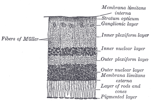
 Section of retina. (Inner plexiform layer labeled at right, fourth from the top.) | |
 Plan of retinal neurons. (Inner plexiform layer labeled at left, fifth from the top.) | |
| Details | |
| Identifiers | |
| Latin | stratum plexiforme internum retinae |
| TA98 | A15.2.04.015 |
| FMA | 58704 |
| Anatomical terminology | |
The inner plexiform layer is an area of the retina that is made up of a dense reticulum of fibrils formed by interlaced dendrites of retinal ganglion cells and cells of the inner nuclear layer. Within this reticulum a few branched spongioblasts are sometimes embedded.[1]
References
[edit]- ^ Nolte, John (2002). The Human Brain: An Introduction to Its Functional Anatomy. 5th ed. St. Louis: Mosby. pp. 416–7. ISBN 0-323-01320-1.
External links
[edit]- Overview Archived 2010-07-01 at the Wayback Machine at utah.edu
- Histology image: 07902loa – Histology Learning System at Boston University
| Fibrous tunic (outer) |
|  | |||||
|---|---|---|---|---|---|---|---|
| Uvea / vascular tunic (middle) |
| ||||||
| Retina (inner) |
| ||||||
| Anatomical regions of the eye |
| ||||||
| Other | |||||||
This article about the eye is a stub. You can help Wikipedia by expanding it. |