
Muscular dystrophy
View on Wikipedia| Muscular dystrophy | |
|---|---|
| Other names | Muscle dysfunction |
 | |
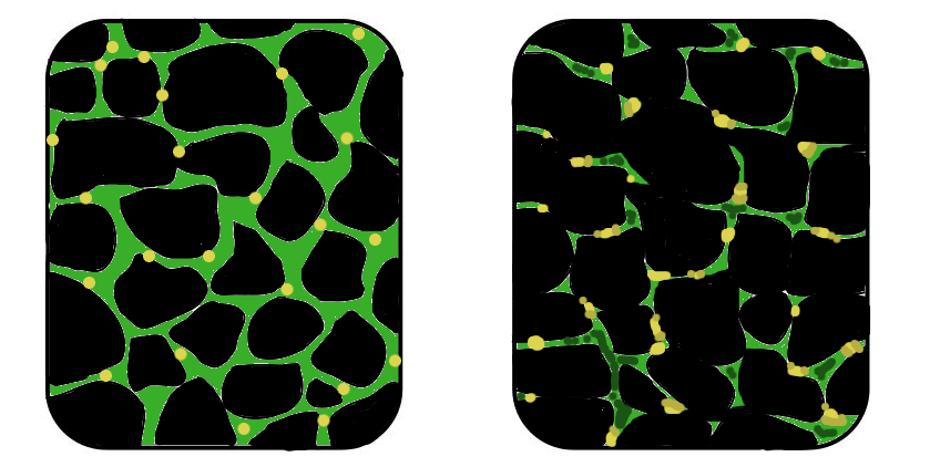
| In affected muscle (right), the tissue has become disorganized and the concentration of dystrophin (green) is greatly reduced, compared to normal muscle (left). | |
| Specialty | Neuromuscular medicine |
| Symptoms | Increasing weakening, breakdown of skeletal muscles, trouble walking[1][2] |
| Duration | Chronic[1] |
| Types | > 30, including Duchenne muscular dystrophy, Becker muscular dystrophy, facioscapulohumeral muscular dystrophy, limb–girdle muscular dystrophy, myotonic dystrophy[1][2] |
| Causes | Genetic (X-linked recessive, autosomal recessive, or autosomal dominant)[2] |
| Diagnostic method | Genetic testing[2] |
| Treatment | Pharmacotherapy, physical therapy, braces, corrective surgery, assisted ventilation[1][2] |
| Prognosis | Depends on the particular disorder[1] |
Muscular dystrophies (MD) are a genetically and clinically heterogeneous group of rare neuromuscular diseases that cause progressive weakness and breakdown of skeletal muscles over time.[1] The disorders differ as to which muscles are primarily affected, the degree of weakness, how fast they worsen, and when symptoms begin.[1] Some types are also associated with problems in other organs.[2]
Over 30 different disorders are classified as muscular dystrophies.[1][2] Of those, Duchenne muscular dystrophy (DMD) accounts for approximately 50% of cases and affects males beginning around the age of four.[1] Other relatively common muscular dystrophies include Becker muscular dystrophy, facioscapulohumeral muscular dystrophy, and myotonic dystrophy,[1] whereas limb–girdle muscular dystrophy and congenital muscular dystrophy are themselves groups of several – usually extremely rare – genetic disorders.
Muscular dystrophies are caused by mutations in genes, usually those involved in making muscle proteins.[2] The muscle protein, dystrophin, is in most muscle cells and works to strengthen the muscle fibers and protect them from injury as muscles contract and relax.[3] It links the muscle membrane to the thin muscular filaments within the cell. Dystrophin is an integral part of the muscular structure. An absence of dystrophin can cause impairments: healthy muscle tissue can be replaced by fibrous tissue and fat, causing an inability to generate force.[4] Respiratory and cardiac complications can occur as well. These mutations are either inherited from parents or may occur spontaneously during early development.[2] Muscular dystrophies may be X-linked recessive, autosomal recessive, or autosomal dominant.[2] Diagnosis often involves blood tests and genetic testing.[2]
There is no cure for any disorder from the muscular dystrophy group.[1] Several drugs designed to address the root cause are currently available including gene therapy (Elevidys), and antisense drugs (Ataluren, Eteplirsen etc.).[2] Other medications used include glucocorticoids (Deflazacort, Vamorolone); calcium channel blockers (Diltiazem); to slow skeletal and cardiac muscle degeneration, anticonvulsants to control seizures and some muscle activity, and Histone deacetylase inhibitors (Givinostat) to delay damage to dying muscle cells.[1] Physical therapy, braces, and corrective surgery may help with some symptoms[1] while assisted ventilation may be required in those with weakness of breathing muscles.[2]
Outcomes depend on the specific type of disorder.[1] Many affected people will eventually become unable to walk[2] and Duchenne muscular dystrophy in particular is associated with shortened life expectancy.
Muscular dystrophy was first described in the 1830s by Charles Bell.[2] The word "dystrophy" comes from the Greek dys, meaning "no, un-" and troph- meaning "nourish".[2]
Signs and symptoms
[edit]
The signs and symptoms consistent with muscular dystrophy are:[5]
- Progressive muscular wasting
- Poor balance
- Scoliosis (abnormal curvature of the spine or the back)[6]
- Muscle contracture leads to limited range of movement of joints.[6]
- Muscle spasms
- Progressive inability to walk
- Waddling gait
- Gowers' sign as in Duchenne muscular dystrophy.
- Calf deformation as in Duchenne muscular dystrophy.
- Respiratory difficulty [7]
- Cardiomyopathy[8]
Causes
[edit]The majority of muscular dystrophies are inherited; the different muscular dystrophies follow various inheritance patterns (X-linked, autosomal recessive or autosomal dominant). In a small percentage of patients, the disorder may have been caused by a de novo (spontaneous) mutation.[9][10]
Diagnosis
[edit]The diagnosis of muscular dystrophy is based on the results of muscle biopsy, increased creatine phosphokinase (CpK3), electromyography, and genetic testing. A physical examination and the patient's medical history will help the doctor determine the type of muscular dystrophy. Specific muscle groups are affected by different types of muscular dystrophy.[11]
An MRI can be used to assess the white matter of the nervous system and measure the merosin levels in young boys. An absence of merosin in young boys will result with neurological deficits and changes in the white matter.[12]
Classification
[edit]| Disorder name | OMIM | Gene | Inheritance pattern | Age of onset | Muscles affected | Description |
|---|---|---|---|---|---|---|
| Becker muscular dystrophy | 300376 | DMD | XR | Childhood | Distal limbs progressing to generalised weakness | A less severe variant of Duchenne muscular dystrophy,[13] affects predominantly boys. |
| Congenital muscular dystrophy | Multiple | Multiple | AD, AR | At birth | Generalised weakness | Symptoms include general muscle weakness and possible joint deformities. Disease progresses slowly, and lifespan is shortened.
Congenital muscular dystrophy includes several disorders with a range of symptoms. Muscle degeneration may be mild or severe. Problems may be restricted to skeletal muscle, or muscle degeneration may be paired with effects on the brain and other organ systems.[14] Several forms of the congenital muscular dystrophies are caused by defects in proteins thought to have some relationship to the dystrophin-glycoprotein complex and to the connections between muscle cells and their surrounding cellular structure. Some forms of congenital muscular dystrophy show severe brain malformations, such as lissencephaly and hydrocephalus.[13] |
| Duchenne muscular dystrophy | 310200 | DMD | XR | Childhood | Distal limbs progressing to generalised weakness, involving respiratory muscles | The most common childhood form of muscular dystrophy, affects predominantly boys (mild symptoms may occur in female carriers). Characterised by progressive muscle wasting. Clinical symptoms become evident when the child begins walking. By age 10, the child may need braces and by age 12, most patients are unable to walk.[15] Typical lifespans range from 15 to 45.[15] Sporadic mutations in this gene occur frequently.[16] |
| Distal muscular dystrophy | 254130 | DYSF | AD, AR | 20–60 years | Distal muscles in hands, forearms and lower legs | Progress is slow and not life-threatening.[17]
Miyoshi myopathy, one of the distal muscular dystrophies, causes initial weakness in the calf muscles, and is caused by defects in the same gene responsible for one form of limb–girdle muscular dystrophy.[13] |
| Emery–Dreifuss muscular dystrophy | Multiple | Multiple | XR, AD, AR | Childhood, early teenage years | Distal limb muscles, limb-girdle, heart | Symptoms include muscle weakness and wasting, starting in the distal limb muscles and progressing to involve the limb–girdle muscles. Most patients also have cardiac conduction defects and arrhythmias.[18][19] |
| Facioscapulohumeral muscular dystrophy | 158900 | DUX4 | AD | Adolescence | Face, shoulders, upper arms, progressing to other muscles | Causes progressive weakness, initially in the muscles of the face, shoulders, and upper arms. Additional muscles are often affected.[20] Affected individuals can become severely disabled, with 20% requiring a wheelchair by age 50.[21] 30% of cases involve spontaneous mutations.[21] Penetrance and severity seem to be lower in females compared to males.[21][22] |
| Limb–girdle muscular dystrophy | Multiple | Multiple | AD, AR | Any | Upper arms and legs | The person normally leads a normal life with some assistance. Rare cardiopulmonary complications can be life-threatening.[23] |
| Myotonic muscular dystrophy | 160900 602668 |
DMPK CNBP |
AD | Adulthood | Skeletal muscles, heart, other muscle groups | Presents with myotonia (delayed relaxation of muscles), as well as muscle wasting and weakness.[24] Varies in severity and manifestations and affects many body systems in addition to skeletal muscles, including the heart, endocrine organs, and eyes.[25] |
| Oculopharyngeal muscular dystrophy | 164300 | PABPN1 | AD, rarely AR | 40–50 years | Eye muscles, face, throat, pelvis, shoulders |
Management
[edit]
There is no cure for muscular dystrophy. In terms of management, physical therapy, occupational therapy, orthotic intervention (e.g., ankle-foot orthosis),[26][27] speech therapy, and respiratory therapy may be helpful.[26] Low-intensity corticosteroids such as prednisone, and deflazacort may help to maintain muscle tone.[28] Orthoses (orthopedic appliances used for support) and corrective orthopedic surgery may be needed to improve the quality of life in some cases.[2] The cardiac problems that occur with Emery–Dreifuss muscular dystrophy (EDMD) and myotonic muscular dystrophy may require a pacemaker.[29] The myotonia (delayed relaxation of a muscle after a strong contraction) occurring in myotonic muscular dystrophy may be treated with medications such as quinine.[30]
Low-intensity assisted exercises, dynamic exercise training, or assisted bicycle training of the arms and legs during a 24-week trial were shown to significantly delay the functional loss of muscular dystrophy. These therapies may be performed in a safe and feasible manner, even for children in their ambulation stages. However, eccentric or intense exercises causing soreness should not be used, as they can cause further damage.[31]
Occupational therapy assists the patient to engage in activities of daily living (such as self-feeding and self-care activities) and leisure activities at the most independent level possible. This may be achieved with use of adaptive equipment or the use of energy-conservation techniques. Occupational therapy may implement changes to a person's environment, both at home or work, to increase the individual's function and accessibility. Furthermore, it addresses psychosocial changes and cognitive decline that may accompany MD, and provides support and education about the disease to the family and patient.[32]
Prognosis
[edit]Prognosis depends on the individual form of muscular dystrophy. Some dystrophies cause progressive weakness and loss of muscle function, which may result in severe physical disability and a life-threatening deterioration of respiratory muscles or heart. Other dystrophies do not affect life expectancy and only cause relatively mild impairment.[2]
History
[edit]In the 1860s, descriptions of boys who grew progressively weaker, lost the ability to walk, and died at an early age became more prominent in medical journals. In the following decade,[33] French neurologist Guillaume Duchenne published a comprehensive account of the most common and severe form of the disease, which now carries his name.[34]
Society and culture
[edit]In 1966 in the U.S. and Canada, Jerry Lewis and the Muscular Dystrophy Association (MDA) began the annual Labor Day telecast The Jerry Lewis Telethon, significant in raising awareness of muscular dystrophy in North America. Some disability-rights advocates have criticized the telethon for portraying victims of the disease as deserving pity rather than respect.[35]
On December 18, 2001, the MD CARE Act was signed into law in the United States. The law amends the Public Health Service Act to provide research for the various muscular dystrophies and established the Muscular Dystrophy Coordinating Committee to help focus research efforts through a coherent research strategy.[36][37]
Research and advocacy
[edit]The Muscular Dystrophy Association (MDA) is involved in research, advocacy, and services for individuals affected by muscular dystrophy. The organization provides resources that contribute to understanding and addressing this condition.
See also
[edit]References
[edit]- ^ a b c d e f g h i j k l m n "NINDS Muscular Dystrophy Information Page". NINDS. March 4, 2016. Archived from the original on 30 July 2016. Retrieved 12 September 2016.
- ^ a b c d e f g h i j k l m n o p q r "Muscular Dystrophy: Hope Through Research". NINDS. March 4, 2016. Archived from the original on 30 September 2016. Retrieved 12 September 2016.
- ^ Gao, Q. Q.; McNally, E. M. (2011-01-17). Terjung, Ronald (ed.). Comprehensive Physiology. Vol. 5 (1 ed.). Wiley. pp. 1223–1239. doi:10.1002/cphy.c140048. ISBN 978-0-470-65071-4. PMC 4767260. PMID 26140716.
- ^ Gao, Quan Q.; McNally, Elizabeth M. (2015-06-24). "The Dystrophin Complex: Structure, Function, and Implications for Therapy". Comprehensive Physiology. 5 (3): 1223–1239. doi:10.1002/cphy.c140048. ISBN 9780470650714. PMC 4767260. PMID 26140716.
- ^ Muscular Dystrophy Clinical Presentation at eMedicine
- ^ a b El-Sobky, Tamer A.; Abdulhady, Hala; Mahmoud, Shady; Amen, John (31 January 2024). "Orthopedic manifestations of congenital muscular dystrophy subtypes in children: Emerging signatures need consolidation: a scoping review". Journal of Musculoskeletal Surgery and Research. 8: 11–23. doi:10.25259/JMSR_229_2023.
- ^ Birnkrant, DJ; Bushby, K; Bann, CM; Alman, BA; Apkon, SD; Blackwell, A; Case, LE; Cripe, L; Hadjiyannakis, S; Olson, AK; Sheehan, DW; Bolen, J; Weber, DR; Ward, LM (April 2018). "Diagnosis and management of Duchenne muscular dystrophy, part 2: respiratory, cardiac, bone health, and orthopaedic management". The Lancet. Neurology. 17 (4): 347–361. doi:10.1016/S1474-4422(18)30025-5. PMC 5889091. PMID 29395990.
- ^ Bădilă, E; Lungu, II; Grumezescu, AM; Scafa Udriște, A; Rosenthal, David; Duboc, Denis; Melacini, Paola (12 May 2021). "Diagnosis of Cardiac Abnormalities in Muscular Dystrophies". Medicina (Kaunas, Lithuania). 57 (5): 488. doi:10.3390/medicina57050488. PMC 8151418. PMID 34066119.
- ^ Choices, NHS. "Muscular dystrophy - Causes - NHS Choices". www.nhs.uk. Archived from the original on 2016-04-02. Retrieved 2016-04-10.
- ^ Griffiths, Anthony JF; Miller, Jeffrey H.; Suzuki, David T.; Lewontin, Richard C.; Gelbart, William M. (2000). Spontaneous mutations. Archived from the original on January 26, 2012.[page needed]
- ^ "NIH /How is muscular dystrophy diagnosed?". NIH.gov. NIH. 2015. Archived from the original on 7 April 2016. Retrieved 10 April 2016.
- ^ Emery, Alan EH (2002-02-23). "The muscular dystrophies". The Lancet. 359 (9307): 687–695. doi:10.1016/S0140-6736(02)07815-7. ISSN 0140-6736. PMID 11879882. S2CID 31578361.
- ^ a b c May 2006 report to Congress Archived 2014-04-05 at the Wayback Machine on Implementation of the MD CARE Act, as submitted by Department of Health and Human Service's National Institutes of Health
- ^ Congenital Muscular Dystrophy~clinical at eMedicine
- ^ a b "Duchenne muscular dystrophy: MedlinePlus Medical Encyclopedia". medlineplus.gov. Archived from the original on 2017-04-05. Retrieved 2017-03-14.
- ^ "Duchenne Muscular Dystrophy. What is muscular dystrophy? | Patient". Patient.info. 2016-04-15. Archived from the original on 2016-12-02. Retrieved 2017-03-14.
- ^ Udd, Bjarne (2011). "Distal muscular dystrophies". Handbook of Clinical Neurology. Vol. 101. pp. 239–62. doi:10.1016/B978-0-08-045031-5.00016-5. ISBN 978-0-08-045031-5. PMID 21496636.
- ^ "OMIM Entry - # 310300 - EMERY-DREIFUSS MUSCULAR DYSTROPHY 1, X-LINKED; EDMD1". Omim.org. Archived from the original on 2017-03-10. Retrieved 2017-03-14.
- ^ "Emery-Dreifuss muscular dystrophy - Genetics Home Reference". Ghr.nlm.nih.gov. 2017-03-07. Archived from the original on 2017-03-12. Retrieved 2017-03-14.
- ^ "facioscapulohumeral muscular dystrophy - Genetics Home Reference". Ghr.nlm.nih.gov. Archived from the original on 2017-03-24. Retrieved 2017-03-14.
- ^ a b c Statland, JM; Tawil, R (December 2016). "Facioscapulohumeral Muscular Dystrophy". Continuum (Minneapolis, Minn.). 22 (6, Muscle and Neuromuscular Junction Disorders): 1916–1931. doi:10.1212/CON.0000000000000399. PMC 5898965. PMID 27922500.
- ^ "Facioscapulohumeral muscular dystrophy: MedlinePlus Medical Encyclopedia". Nlm.nih.gov. 2017-03-09. Archived from the original on 2016-07-04. Retrieved 2017-03-14.
- ^ Jenkins, Simon P.R. (2005). Sports Science Handbook:I - Z. Brentwood, Essex: Multi-Science Publ. Co. p. 121. ISBN 978-0906522-37-0.
- ^ Turner, C.; Hilton-Jones, D. (2010). "The myotonic dystrophies: diagnosis and management" (PDF). Journal of Neurology, Neurosurgery & Psychiatry. 81 (4): 358–67. doi:10.1136/jnnp.2008.158261. PMID 20176601. S2CID 2453622.
- ^ Bird, T. D.; Adam, M. P.; Everman, D. B.; Mirzaa, G. M.; Pagon, R. A.; Wallace, S. E.; Bean LJH; Gripp, K. W.; Amemiya, A. (1993). "Myotonic Dystrophy Type 1". Myotonic Dystrophy Type 1 - GeneReviews® - NCBI Bookshelf. University of Washington, Seattle. PMID 20301344. Archived from the original on 2017-01-18. Retrieved 2017-03-14.
{{cite book}}:|website=ignored (help) - ^ a b "What are the treatments for muscular dystrophy?". NIH.gov. NIH. 2015. Archived from the original on 7 April 2016. Retrieved 10 April 2016.
- ^ "Muscular Dystrophy-OrthoInfo - AAOS". orthoinfo.aaos.org. Archived from the original on 2016-04-12. Retrieved 2016-04-10.
- ^ McAdam, Laura C.; Mayo, Amanda L.; Alman, Benjamin A.; Biggar, W. Douglas (2012). "The Canadian experience with long term deflazacort treatment in Duchenne muscular dystrophy". Acta Myologica. 31 (1): 16–20. PMC 3440807. PMID 22655512.
- ^ Verhaert, David; Richards, Kathryn; Rafael-Fortney, Jill A.; Raman, Subha V. (January 2011). "Cardiac Involvement in Patients With Muscular Dystrophies". Circulation: Cardiovascular Imaging. 4 (1): 67–76. doi:10.1161/CIRCIMAGING.110.960740. PMC 3057042. PMID 21245364.
- ^ Eddy, Linda L. (2013). Caring for Children with Special Healthcare Needs and Their Families: A Handbook for Healthcare Professionals. John Wiley & Sons. ISBN 978-1-118-51797-0.[page needed]
- ^ Jansen, Merel; Van Alfen, Nens; Geurts, Alexander C. H.; De Groot, Imelda J. M. (2013). "Assisted Bicycle Training Delays Functional Deterioration in Boys with Duchenne Muscular Dystrophy". Neurorehabilitation and Neural Repair. 27 (9): 816–827. doi:10.1177/1545968313496326. PMID 23884013. S2CID 9990910.
- ^ Lehman, R. M.; McCormack, G. L. (2001). "Neurogenic and Myopathic Dysfunction". In Pedretti, Lorraine Williams; Early, Mary Beth (eds.). Occupational Therapy: Practice Skills for Physical Dysfunction (5th ed.). Mosby. pp. 802–3. ISBN 978-0-323-00765-8.
- ^ Laing, Nigel G; Davis, Mark R; Bayley, Klair; Fletcher, Sue; Wilton, Steve D (2011). "Molecular Diagnosis of Duchenne Muscular Dystrophy: Past, Present and Future in Relation to Implementing Therapies". The Clinical Biochemist Reviews. 32 (3): 129–134. PMC 3157948. PMID 21912442.
- ^ "Muscular Dystrophy: Hope Through Research". National Institute of Neurological Disorders and Stroke. 23 March 2020. Retrieved 7 April 2020.
- ^ Berman, Ari (2011-09-02). "The End of the Jerry Lewis Telethon—It's About Time". The Nation. Retrieved 2017-03-14.
- ^ H.R. 717--107th Congress (2001) Archived 2012-02-19 at the Wayback Machine: MD-CARE Act, GovTrack.us (database of federal legislation), (accessed Jul 29, 2007)
- ^ Public Law 107-84 Archived 2012-11-07 at the Wayback Machine, PDF as retrieved from NIH website
Further reading
[edit]- De Los Angeles Beytía, Maria; Vry, Julia; Kirschner, Janbernd (2012). "Drug treatment of Duchenne musculardystrophy: available evidence and perspectives". Acta Myologica. 31 (1): 4–8. PMC 3440798. PMID 22655510.
- Bertini, Enrico; D'Amico, Adele; Gualandi, Francesca; Petrini, Stefania (December 2011). "Congenital Muscular Dystrophies: A Brief Review". Seminars in Pediatric Neurology. 18 (4): 277–288. doi:10.1016/j.spen.2011.10.010. PMC 3332154. PMID 22172424.
External links
[edit]

| Types | |
|---|---|
| National/International Organizations | |
| National/International Events |
|
| Clinical trials | |


Muscular dystrophy
View on GrokipediaDefinition and Classification
Core Definition
Muscular dystrophy encompasses a heterogeneous group of more than 30 inherited genetic disorders characterized by progressive weakness and degeneration of skeletal muscles, resulting from mutations in genes that encode proteins essential for muscle structure, stability, and function.[1][3] These conditions primarily affect voluntary muscles, leading to muscle fiber breakdown, replacement by fibrous or fatty tissue, and eventual loss of mobility, though the heart and respiratory muscles may also be involved in certain forms.[11] Unlike acquired muscle disorders, muscular dystrophies stem from germline mutations rather than environmental or infectious causes, with onset varying from infancy to adulthood depending on the specific type.[12] At the molecular level, the core pathology arises from disruptions in the dystrophin-glycoprotein complex or other muscle-associated proteins, which normally link the cytoskeleton to the extracellular matrix, providing mechanical reinforcement during contraction.[3] For instance, in the most common form, Duchenne muscular dystrophy, mutations in the DMD gene abolish dystrophin production, rendering muscle cells vulnerable to damage from everyday mechanical stress and triggering chronic inflammation and necrosis.[2] Inheritance patterns are predominantly X-linked recessive, autosomal recessive, or autosomal dominant, with de novo mutations accounting for up to one-third of cases in some subtypes, underscoring the genetic etiology without reliance on external triggers.[13] Diagnosis relies on clinical presentation corroborated by genetic testing, as histopathological findings like fiber size variation and necrosis are supportive but not specific.[3] While muscular dystrophies share progressive muscle wasting as a hallmark, they differ in affected muscle groups, rate of progression, and extramuscular manifestations, such as cardiomyopathy or cognitive involvement, necessitating subtype-specific classification for management.[14] No curative therapies exist as of 2025, with interventions focused on symptom palliation, including corticosteroids to delay weakness, physical therapy, and emerging gene therapies targeting specific mutations, though efficacy varies and long-term data remain limited.[15] Prevalence estimates indicate Duchenne and Becker forms affect approximately 1 in 3,500 to 5,000 male births worldwide, highlighting the disorders' significant public health burden despite their rarity in aggregate.[16]Major Types and Subtypes
Major Types of Muscular Dystrophy| Disorder name | OMIM | Gene | Inheritance pattern | Age of onset | Muscles affected | Description |
|---|---|---|---|---|---|---|
| Duchenne muscular dystrophy (DMD) | 310200 | DMD | X-linked recessive | Ages 2-5 years | Proximal muscles (hip girdle) | Symptoms emerge around ages 2-3 years, featuring waddling gait, toe walking, Gowers' maneuver due to hip girdle weakness; calf pseudohypertrophy with replacement by fat and connective tissue; elevated serum creatine kinase levels may precede weakness. |
| Becker muscular dystrophy (BMD) | 300276 | DMD | X-linked recessive | Ages 5 to early adulthood | Proximal muscles (quadriceps, hip girdle) | Presents similarly but milder, with waddling gait, lumbar lordosis, quadriceps weakness; ambulation often retained into adulthood. |
| Myotonic dystrophy type 1 (DM1) | 160900 | DMPK | Autosomal dominant | Variable (congenital to adult onset) | Distal muscles, face, hands | Features myotonia (prolonged muscle contraction after effort), distal weakness, ptosis, temporalis wasting; systemic signs include cataracts and hypersomnia. |
| Facioscapulohumeral muscular dystrophy (FSHD) | 158900 | DUX4 | Autosomal dominant | Teens to 20s | Facial, scapular, humeral muscles | Primarily affects facial and shoulder girdle muscles, causing difficulty whistling, incomplete eye closure, inability to raise arms overhead, winged scapulae. |
| Type | Inheritance | Typical Onset | Key Features | Prevalence Estimate |
|---|---|---|---|---|
| Duchenne (DMD) | X-linked recessive | Ages 2-5 | Proximal weakness, pseudohypertrophy, early wheelchair | 1 in 3,500-5,000 male births[5] |
| Becker (BMD) | X-linked recessive | Adolescence/adulthood | Slower progression, cardiac focus | 1 in 18,000-30,000 male births[23] |
| Myotonic DM1 | Autosomal dominant | Variable (congenital to adult) | Myotonia, distal weakness, multisystem | 1 in 8,000[26] |
| FSHD | Autosomal dominant | Teens/20s | Facial/scapulohumeral weakness, asymmetric | 1 in 8,000-15,000[30] |
| LGMD (various) | Mostly autosomal recessive | Childhood-adulthood | Girdle weakness, heterogeneous | Varies by subtype; overall rare[33] |
Genetic and Pathophysiological Foundations
Genetic Mutations and Inheritance Patterns
Muscular dystrophies encompass a heterogeneous group of disorders primarily resulting from mutations in genes encoding proteins critical for muscle structure and function, such as dystrophin, myotonic dystrophy protein kinase, and others involved in the dystrophin-glycoprotein complex. These mutations disrupt sarcolemmal integrity, leading to progressive muscle degeneration. Inheritance patterns vary across subtypes, including X-linked recessive, autosomal dominant, and autosomal recessive modes, with over 30 distinct forms identified. Approximately 70% of cases arise from de novo mutations or carrier transmission, underscoring the genetic heterogeneity.[5] The most prevalent form, Duchenne muscular dystrophy (DMD), affects about 1 in 3,500 to 5,000 male births and is caused by out-of-frame mutations—predominantly large deletions (60-70%), duplications (10%), or point mutations—in the DMD gene at locus Xp21, which spans 2.4 megabases and contains 79 exons. These alterations abolish dystrophin protein expression, essential for linking the cytoskeleton to the extracellular matrix. DMD follows an X-linked recessive pattern, wherein hemizygous males manifest the disease while females are typically asymptomatic carriers due to X-inactivation; carrier females have a 50% risk of transmitting the mutation to offspring, with affected sons comprising half of male progeny.[36][5][37] Becker muscular dystrophy (BMD), a milder allelic variant occurring in 1 in 18,000 to 30,000 males, involves in-frame mutations in the same DMD gene, yielding truncated but partially functional dystrophin levels (typically 10-40% of normal). Inheritance mirrors DMD's X-linked recessive mode, with symptoms onset often delayed until adolescence or adulthood.[37][23] Myotonic dystrophy type 1 (DM1), the most common adult-onset muscular dystrophy with prevalence of 1 in 8,000, stems from pathogenic CTG trinucleotide repeat expansions (typically >50 repeats, up to thousands) in the 3' untranslated region of the DMPK gene on chromosome 19q13.3. This autosomal dominant disorder exhibits anticipation, where repeat instability during meiosis increases expansion size across generations, correlating with earlier onset and severity. A single expanded allele suffices for disease manifestation, with 50% transmission risk to offspring regardless of sex.[38][39] Facioscapulohumeral muscular dystrophy (FSHD) type 1, accounting for 95% of cases and affecting 1 in 8,000 to 15,000 individuals, arises from contraction of the D4Z4 macrosatellite repeat array on chromosome 4q35 from 11-100 units to 1-10 units, coupled with a permissive 4qA haplotype. This epigenetic derepression permits toxic expression of the DUX4 transcription factor from the distal D4Z4 unit, which is normally silenced in somatic tissues. FSHD follows autosomal dominant inheritance, with variable penetrance and a 50% risk per offspring; de novo contractions occur in 10-30% of sporadic cases.[40][41] Limb-girdle muscular dystrophies (LGMDs) comprise diverse subtypes primarily affecting proximal muscles, with over 30 genes implicated in the dystrophin-associated complex, sarcomere, or membrane repair pathways. Most (LGMD2/R types, ~90%) are autosomal recessive, requiring biallelic mutations (e.g., in CAPN3 for LGMD2A or SGCA for LGMD2D), thus necessitating carrier parents and a 25% recurrence risk in siblings; autosomal dominant forms (LGMD1/D types, ~10%) involve heterozygous mutations (e.g., in MYOT) with 50% offspring risk. Prevalence varies, with LGMD2A being common in certain populations at 1 in 15,000.[42][43]| Major Type | Gene/Locus | Common Mutation Types | Inheritance Pattern | Approximate Prevalence |
|---|---|---|---|---|
| Duchenne (DMD) | DMD (Xp21) | Out-of-frame deletions/duplications/point mutations | X-linked recessive | 1:3,500-5,000 males[5] |
| Becker (BMD) | DMD (Xp21) | In-frame deletions/duplications | X-linked recessive | 1:18,000-30,000 males[23] |
| Myotonic Type 1 (DM1) | DMPK (19q13.3) | CTG repeat expansion (>50) | Autosomal dominant | 1:8,000[38] |
| FSHD Type 1 | D4Z4/DUX4 (4q35) | Repeat contraction (1-10 units) | Autosomal dominant | 1:8,000-15,000[40] |
| Limb-Girdle (most) | Various (e.g., CAPN3, SGCA) | Biallelic loss-of-function | Autosomal recessive | Varies; e.g., LGMD2A 1:15,000 in some regions[43] |
Molecular and Cellular Mechanisms
The molecular and cellular mechanisms of muscular dystrophies center on genetic defects that compromise muscle fiber stability, repair, and homeostasis, leading to progressive degeneration. In dystrophinopathies like Duchenne muscular dystrophy (DMD), frameshift or nonsense mutations in the DMD gene abolish functional dystrophin, a 427-kDa cytoskeletal protein that anchors actin filaments to the sarcolemma via the dystrophin-glycoprotein complex (DGC). The DGC, comprising dystroglycans, sarcoglycans, sarcospan, and syntrophins/dystrobrevins, links the intracellular cytoskeleton to the extracellular matrix, buffering mechanical stress during contraction and modulating signaling pathways such as nitric oxide production via neuronal nitric oxide synthase (nNOS).[44][45][46] Absence of dystrophin destabilizes the sarcolemma, causing microtears, elevated extracellular calcium influx through stretch-activated channels, and subsequent activation of calcium-dependent proteases like calpains, which degrade myofibrillar proteins and exacerbate necrosis. This initiates a vicious cycle: necrotic fibers release damage-associated molecular patterns, recruiting inflammatory cells including macrophages and T-lymphocytes, which produce cytokines (e.g., TNF-α, IL-6) that amplify oxidative stress via reactive oxygen species from dysfunctional mitochondria and NADPH oxidase. Satellite cells, muscle stem cells essential for regeneration, exhibit impaired self-renewal and differentiation due to dystrophin deficiency, leading to exhaustion, while persistent inflammation drives fibrotic replacement by extracellular matrix from activated myofibroblasts.[47][48][49] Limb-girdle muscular dystrophies (LGMD) often involve mutations in DGC-associated proteins, such as sarcoglycans in LGMD-R2 subtypes, yielding analogous sarcolemmal fragility and secondary cascades of calcium dysregulation, proteolysis, and fibrosis, though severity varies with residual complex function. In LGMD-R1 calpainopathy (LGMD-R1), capn3 mutations disrupt calpain-3's role in sarcomere remodeling and IκBα degradation for NF-κB signaling, impairing myofiber integrity and repair without primary DGC loss.[44][50][51] Distinct mechanisms characterize non-DGC dystrophies; facioscapulohumeral muscular dystrophy (FSHD) arises from D4Z4 repeat contractions on chromosome 4q35, causing epigenetic derepression and ectopic expression of the transcription factor DUX4 in myofibers. DUX4 induces a fetal-like gene program, generates double-stranded RNA that activates innate immune responses via PKR and MAVS, stabilizes MYC mRNA to promote apoptosis, and disrupts RNA quality control through nonsense-mediated decay interference, culminating in oxidative stress and cell death.[40][52][53] Myotonic dystrophy type 1 features CTG repeat expansions in DMPK, yielding mutant RNA that sequesters splicing factors like MBNL1 into nuclear foci, mis-splicing chloride channel (CLCN1) and other transcripts to cause myotonia, insulin resistance, and gradual myofiber atrophy via impaired excitation-contraction coupling and regenerative deficits.[44]Clinical Presentation
Primary Signs and Symptoms
Muscular dystrophies manifest primarily through progressive weakness and degeneration of skeletal muscles, with symptoms varying by subtype in onset age, severity, and distribution.[11] Common signs and symptoms:- Symmetric proximal muscle weakness, often beginning in the lower limbs and pelvis, leading to impaired mobility.[54]
- Delayed achievement of motor milestones, such as walking beyond 18 months, frequent falls, and difficulty climbing stairs or rising from a seated position.[55]
- Fatigue, myalgias, and contractures as muscles weaken.[54] [56]
- Symptoms emerge around ages 2-3 years, featuring a waddling gait, toe walking, and Gower's maneuver—using hands to "climb" the body when standing from the floor due to hip girdle weakness.[57] [55]
- Calf pseudohypertrophy, where muscles appear enlarged but are replaced by fat and connective tissue, coexists with atrophy elsewhere.[1]
- Muscle cramps and elevated serum creatine kinase levels may precede overt weakness.[5]
- Presents similarly but milder, with onset from age 5 to early adulthood, including lumbar lordosis, waddling gait, and quadriceps weakness.[58] [59]
- Affected individuals often retain ambulation into adulthood.[58] [59]
- Features myotonia—prolonged muscle contraction after voluntary effort, especially in hands and face—alongside distal weakness, ptosis, and temporalis wasting.[26]
- Systemic signs like cataracts and hypersomnia often accompany.[26]
- Primarily affects facial and shoulder girdle muscles, causing difficulty whistling, closing eyes fully, or raising arms overhead, with winged scapulae.[60]
Disease Progression and Complications
Disease progression in muscular dystrophy varies significantly by subtype, with Duchenne muscular dystrophy (DMD) exhibiting the most rapid deterioration, typically leading to loss of ambulation by age 12 and premature death in the third or fourth decade due to respiratory or cardiac failure.[61] In DMD, initial proximal muscle weakness emerges between ages 2 and 5, progressing to pseudohypertrophy of calves, Gowers' maneuver for rising, and eventual wheelchair dependence; respiratory complications, including diaphragmatic weakness and hypoventilation, manifest in the late teens, while cardiomyopathy develops in nearly all cases by age 18, contributing to 20% of mortality even with interventions.[3] Orthopedic issues such as scoliosis (affecting 90-95% of non-ambulatory patients) and joint contractures further exacerbate mobility loss, often requiring surgical correction.[62] Becker muscular dystrophy (BMD), a milder allelic variant of DMD, shows delayed onset around age 5-15 and slower progression, with many patients retaining ambulation into their 40s or beyond; however, cardiac involvement remains prevalent, manifesting as dilated cardiomyopathy in up to 70% by age 40, which is the leading cause of death.[23] Complications include arrhythmias, fractures from falls due to weakness, and less frequent respiratory failure compared to DMD, though growth impairment and cognitive challenges can occur.[23] Facioscapulohumeral muscular dystrophy (FSHD) progresses gradually over decades, often beginning with facial and shoulder girdle weakness in adolescence or early adulthood, with lower limb involvement in 80% of cases but loss of ambulation in only 5-10% lifetime; the disease course is variable and asymmetric, with exacerbations following periods of stability.[32] Complications are primarily musculoskeletal, including winged scapulae, foot drop requiring orthoses, and rare extramuscular issues like hearing loss or retinal vasculopathy in severe cases, though life expectancy approaches normal absent respiratory compromise.[32] Across subtypes, common complications stem from chronic muscle degeneration and immobility, encompassing recurrent infections from weakened cough mechanisms, gastrointestinal dysmotility leading to constipation or aspiration risk, and renal dysfunction in advanced DMD due to immobility and dehydration.[61][62] Monitoring for these via serial echocardiography, spirometry, and functional assessments is essential, as early intervention can mitigate some risks despite inexorable advancement.[3]Diagnostic Approaches
Clinical Evaluation and Testing
Clinical evaluation of muscular dystrophy begins with a comprehensive patient history, emphasizing the age of symptom onset, pattern and progression of muscle weakness (typically proximal and symmetric), presence of delayed motor milestones in children, and family history suggestive of X-linked, autosomal recessive, or dominant inheritance patterns.[15] Physical examination focuses on assessing muscle strength using standardized scales such as the Medical Research Council scale, evaluating gait abnormalities (e.g., waddling gait or Gowers' sign in Duchenne muscular dystrophy), tendon reflexes (often preserved early but diminished later), and signs of pseudohypertrophy, particularly in calves for dystrophinopathies.[3] [63] Initial laboratory testing includes measurement of serum creatine kinase (CK) levels, which are characteristically elevated—often 10 to 100 times the upper limit of normal in early stages due to muscle fiber leakage—serving as a sensitive but nonspecific indicator of ongoing muscle damage across dystrophy subtypes.[15] [3] Electromyography (EMG) and nerve conduction studies are employed to confirm a myopathic process, revealing short-duration, low-amplitude motor unit potentials with early recruitment, while distinguishing from neurogenic disorders by normal or near-normal nerve conduction velocities.[15] [63] Muscle biopsy, though increasingly superseded by genetic testing, provides histopathological confirmation through evidence of dystrophic changes such as fiber size variation, necrosis, inflammatory infiltrates, regeneration, fibrosis, and fatty infiltration; immunohistochemistry or Western blot may assess protein expression (e.g., dystrophin absence in Duchenne cases).[3] [15] Ancillary imaging, including muscle MRI, can delineate patterns of involvement (e.g., selective muscle sparing or replacement by fat and fibrosis) to guide biopsy site selection or subtype differentiation, with T1-weighted sequences highlighting fatty degeneration quantitatively via metrics like the muscle fat fraction.[63]Genetic Confirmation and Differential Diagnosis
Genetic confirmation of muscular dystrophy relies on molecular analysis to detect pathogenic variants in disease-specific genes, establishing a definitive diagnosis in the majority of cases. For Duchenne muscular dystrophy (DMD) and Becker muscular dystrophy (BMD), testing targets the DMD gene on the X chromosome, where deletions or duplications account for 65-80% of mutations, detected via multiplex ligation-dependent probe amplification (MLPA) or comparative genomic hybridization as first-line methods.[64][5] Subsequent next-generation sequencing identifies point mutations, small insertions/deletions, or other variants in up to 30% of cases, with sensitivity exceeding 95% when combined.[65] Samples are typically obtained from peripheral blood leukocytes, though amniotic fluid or chorionic villi enable prenatal diagnosis.[66] For limb-girdle muscular dystrophy (LGMD) subtypes, targeted gene panels or whole-exome sequencing interrogate multiple loci such as CAPN3, DYSF, or SGCA, given the genetic heterogeneity involving over 30 genes.[3] Myotonic dystrophy confirmation involves polymerase chain reaction (PCR) or Southern blot for CTG repeat expansions in DMPK (type 1) or CCTG in CNBP (type 2), with allele sizes correlating to anticipation and severity.[3] Facioscapulohumeral muscular dystrophy (FSHD) is verified by detecting contractions in the DUX4 D4Z4 repeat array on chromosome 4q35, often via Southern blot or methylation-sensitive PCR, distinguishing FSHD1 (95% of cases) from FSHD2.[3] Elevated creatine kinase (CK) levels support suspicion but require genetic corroboration, as historical muscle biopsies assessing dystrophin expression or protein aggregates are now supplementary, reserved for ambiguous cases or to avoid invasive testing in children.[67][5] Differential diagnosis differentiates muscular dystrophies from mimicking neuromuscular conditions through integrated clinical, electrophysiological, and genetic evaluation. Spinal muscular atrophy (SMA), caused by SMN1 deletions, presents with hypotonia and fasciculations but spares CK elevation and shows denervation on electromyography (EMG), confirmed by SMN1 copy number analysis rather than dystrophinopathy panels.[5] Congenital myopathies, such as nemaline or central core disease, exhibit non-progressive weakness with specific biopsy findings (e.g., rod bodies) and mutations in genes like NEB or RYR1, contrasting the progressive fibrosis in dystrophies.[3] Metabolic disorders like Pompe disease (GAA deficiency) feature vacuolar myopathy on biopsy and respond to enzyme replacement, distinguishable by absent family history of X-linked inheritance in DMD.[68] Inflammatory myopathies (e.g., dermatomyositis) show perifascicular atrophy and autoantibodies, with EMG revealing irritative potentials unlike the myopathic changes in dystrophies; corticosteroid responsiveness further aids distinction.[3] Myasthenia gravis involves fatigable weakness and positive acetylcholine receptor antibodies, ruled out by normal muscle histology and response to acetylcholinesterase inhibitors.[68] For intermediate phenotypes, such as between DMD and BMD, genetic variant classification per ACMG guidelines assesses pathogenicity based on reading frame disruption, with in-frame mutations predicting milder courses.[65] Carrier testing for female relatives uses the same assays, informing reproductive risks, while unsolved cases (5-10% in DMD/BMD) may warrant research-grade long-read sequencing.[65] Early genetic delineation optimizes prognosis and trial eligibility, as misdiagnosis delays targeted therapies like exon-skipping for amenable DMD variants.[64]Treatment Strategies
Symptomatic and Supportive Care
Symptomatic and supportive care for muscular dystrophy emphasizes multidisciplinary interventions to alleviate symptoms, preserve function, and avert complications such as contractures, respiratory failure, and scoliosis, particularly in progressive forms like Duchenne muscular dystrophy (DMD).[69] Physical therapy regimens, including stretching and low-resistance exercises, are recommended to maintain joint mobility and muscle strength while minimizing fatigue, with evidence indicating delayed loss of ambulation when initiated early.[15] Occupational therapy supports daily activities through adaptive equipment and training, enhancing independence in self-care tasks.[69] Orthopedic management involves orthotic devices, such as ankle-foot orthoses, to stabilize gait and prevent foot drop, often prescribed before full loss of ambulation in DMD patients around age 10-12 years.[70] Surgical interventions, including spinal fusion for scoliosis correction when curvature exceeds 20-30 degrees, improve posture and pulmonary function, with guidelines advocating preoperative pulmonary optimization.[69] Foot tendon releases address equinovarus deformities, prolonging brace tolerance.[70] Respiratory support is critical as diaphragmatic weakness progresses, with non-invasive ventilation (NIV) initiated when forced vital capacity falls below 50% predicted or nocturnal hypoventilation occurs, extending survival by years through improved gas exchange.[71] Assisted cough techniques, using mechanical insufflation-exsufflation devices, clear secretions and reduce pneumonia risk during intercurrent illnesses.[72] Regular monitoring via spirometry every 6 months guides escalation to daytime NIV or tracheostomy in advanced stages.[73] Nutritional interventions address dysphagia and growth needs, with gastrostomy tubes recommended when oral intake fails to meet caloric requirements, preventing malnutrition that exacerbates respiratory compromise.[74] Assistive technologies, including powered wheelchairs and standing frames, promote mobility and bone health post-ambulation loss.[75] Psychological support and family counseling mitigate emotional burdens, integrated within comprehensive care models.[69]Pharmacological Interventions
Corticosteroids remain the primary pharmacological intervention for Duchenne muscular dystrophy (DMD), the most common and severe form of muscular dystrophy, with moderate-quality evidence from randomized controlled trials (RCTs) indicating improvements in muscle strength, function, and pulmonary metrics over 12 months compared to placebo.[76] Prednisone, typically dosed at 0.75 mg/kg/day, prolongs ambulation by 2-3 years and slows progression of scoliosis and respiratory decline, though long-term use is associated with side effects including weight gain, osteoporosis, and growth suppression.[77] Deflazacort, approved by the FDA in 2016 at 0.9 mg/kg/day, demonstrates comparable efficacy to prednisone in preserving muscle strength over 12 weeks in boys aged 5-15 years, with potentially reduced weight gain but similar risks of cataracts and bone density loss.[77] [78] Histone deacetylase (HDAC) inhibitors like givinostat (Duvyzat), approved by the FDA in March 2024 for ambulatory children aged 6 and older with DMD, target muscle inflammation and fibrosis, showing in phase 3 trials a 1.3-point slower decline in functional motor scores over 72 weeks versus placebo, alongside reductions in fat infiltration on MRI.[79] This approval was based on clinical endpoints rather than surrogates, distinguishing it from prior therapies, though gastrointestinal side effects and neutropenia require monitoring.[80] Antisense oligonucleotide (ASO) therapies for exon skipping, applicable to subsets of DMD patients amenable to specific exon corrections (e.g., up to 29% for exons 45, 51, 53), include eteplirsen (FDA-approved 2016 for exon 51), golodirsen (2019 for exon 53), viltolarsen (2020 for exon 53), and casimersen (2021 for exon 45).[81] These drugs induce partial dystrophin production via intramuscular or intravenous administration, but approvals relied on accelerated pathways using dystrophin levels as surrogates, with limited confirmatory evidence of clinically meaningful improvements in motor function or survival; post-approval studies have shown modest dystrophin increases (0.3-5% of normal) but no consistent benefits in ambulation or cardiac outcomes.[82] [83] For other muscular dystrophies such as limb-girdle (LGMD) or facioscapulohumeral (FSHD), no FDA-approved disease-modifying pharmacological agents exist as of 2025, with management limited to off-label corticosteroids or NSAIDs for symptomatic inflammation, which lack robust efficacy data and may exacerbate weakness in some cases.[84] [85] Ongoing trials, such as p38 MAPK inhibitors like losmapimod for FSHD, explore epigenetic targets but remain unproven.[86] Across types, pharmacological strategies emphasize early initiation to mitigate progression, balanced against adverse effects, with no interventions restoring full dystrophin function or curing the underlying genetic defects.[87]Experimental and Gene-Based Therapies
Gene-based therapies for muscular dystrophy primarily target Duchenne muscular dystrophy (DMD), the most common and severe form, caused by mutations in the DMD gene leading to dystrophin deficiency. These approaches include viral vector-mediated gene replacement, antisense oligonucleotide (ASO)-induced exon skipping to restore partial dystrophin production, and emerging genome editing techniques like CRISPR-Cas9. While some have received regulatory approval, many remain experimental due to limited long-term efficacy data, variable dystrophin expression levels (often 20-80% of normal), and challenges such as immune responses to vectors or off-target edits.[88][89] Adeno-associated virus (AAV) vectors deliver truncated micro-dystrophin genes to muscle cells, aiming to bypass the large size of the full DMD gene. Delandistrogene moxeparvovec (Elevidys), developed by Sarepta Therapeutics, received FDA accelerated approval on June 22, 2023, for ambulatory children aged 4-5 years with confirmed DMD mutations, based on micro-dystrophin expression as a surrogate endpoint rather than definitive clinical benefits.[90] Approval expanded in June 2024 to all ambulatory patients regardless of age, with full approval for this group, but restrictions were imposed in June 2025 for non-ambulatory patients due to safety concerns including acute liver injury and myocarditis risks.[91][92] Confirmatory trials like EMBARK showed transient motor function improvements (e.g., North Star Ambulatory Assessment scores) but failed to meet primary endpoints for sustained benefit, highlighting reliance on biomarkers over functional outcomes.[93] Similar AAV-micro-dystrophin candidates, such as those from RegenxBio (RGX-202) and Solid Biosciences (SGT-003), are in phase 1/2 trials as of 2025, with preclinical data showing muscle strength gains in animal models but human efficacy pending.[94] Exon-skipping therapies use ASOs to mask mutated exons during mRNA splicing, producing truncated but partially functional dystrophin applicable to 13-80% of DMD patients depending on mutation type. Eteplirsen (Exondys 51) for exon 51 skipping was conditionally approved by the FDA in 2016 based on minimal dystrophin increases (0.9% of normal), despite lacking robust clinical efficacy evidence; subsequent drugs like golodirsen and viltolarsen (for exons 53 and 45, respectively) followed similar accelerated paths in 2019 and 2020, with viltolarsen showing stable ambulation in small cohorts over 180 weeks but no significant 6-minute walk test improvements.[89][95] Long-term data indicate modest cardiomyopathy benefits in some models, but overall functional gains remain limited, prompting debates on whether surrogate endpoints justify approvals amid high costs ($300,000+ annually).[96][97] Genome editing with CRISPR-Cas systems offers potential for permanent correction by excising or skipping mutated exons directly in patient cells. As of May 2025, no CRISPR therapies for muscular dystrophy have advanced to late-stage trials, though preclinical studies demonstrate efficient dystrophin restoration in DMD mouse models with minimal off-target effects using Cas9 or Cas12a nucleases.[98] HuidaGene's HG302, a CRISPR-Cas12-based exon-skipping therapy, initiated dosing in its phase 1 MUSCLE trial in December 2024, targeting ambulatory DMD boys with early data from ASGCT 2025 suggesting improved dystrophin expression and motor function.[99][100] Delivery challenges persist, including AAV immunogenicity and scalability for systemic muscle targeting, with ongoing research at centers like the Max Delbrück Center exploring optimized vectors.[101] For rarer dystrophies like limb-girdle, experimental AAV therapies (e.g., Sarepta's SRP-9004 for LGMD type 2D) increased missing protein levels in phase 1 trials as of October 2025, with regulatory submissions planned.[102][103] These approaches underscore causal links between dystrophin restoration and slowed progression but require rigorous validation against placebo-controlled outcomes to confirm disease-modifying effects.Controversies and Critical Evaluations
Debates on Treatment Efficacy and Approvals
The approval of eteplirsen (Exondys 51) by the U.S. Food and Drug Administration (FDA) on September 19, 2016, marked the first antisense oligonucleotide therapy for Duchenne muscular dystrophy (DMD), granted via accelerated approval based on a surrogate endpoint of increased dystrophin production observed in a small open-label study involving 12 boys. Critics, including the FDA's own advisory committee which voted 7-3 against approval citing insufficient evidence of clinical benefit, argued that the surrogate did not reliably predict improvements in muscle function, ambulation, or survival, with dystrophin increases deemed minimal (0.93% of normal levels).[104] The European Medicines Agency (EMA) rejected eteplirsen in 2018, emphasizing the lack of robust phase III data demonstrating meaningful functional outcomes.[105] Subsequent exon-skipping therapies from Sarepta Therapeutics, such as golodirsen (Vyondys 53) approved in December 2019 and viltolarsen (Viltepso) in August 2020, followed similar accelerated pathways relying on dystrophin surrogates from studies of 25 or fewer patients, without placebo-controlled evidence of slowed disease progression.[106] Independent assessments, including a 2019 Institute for Clinical and Economic Review (ICER) report, found no high-quality evidence of clinical benefits for eteplirsen three years post-approval, highlighting risks of over-reliance on unvalidated biomarkers amid annual costs exceeding $300,000 per patient.[107] A 2024 analysis in JAMA noted that U.S. spending on these targeted DMD therapies reached billions despite limited efficacy data, prompting calls for stricter confirmatory trial requirements to verify functional gains like six-minute walk test improvements.[106] Gene-based therapies have intensified debates, exemplified by Sarepta's delandistrogene moxeparvovec (Elevidys), initially granted accelerated approval in June 2023 for ambulatory children aged 4-5 with DMD mutations amenable to exon 2 skipping, based on micro-dystrophin expression rather than clinical endpoints.[108] Traditional approval was extended in 2024 to non-ambulatory patients despite phase III trial failures to meet primary endpoints for motor function, with critics questioning the FDA's decision to prioritize surrogate data over outcomes like North Star Ambulatory Assessment scores showing no significant differences.[109] Safety concerns escalated in 2025 following reports linking Elevidys to acute liver failure and deaths in two teenage trial participants, underscoring risks of immune responses to adeno-associated virus vectors without proportional efficacy gains.[110] Broader critiques target the FDA's accelerated approval framework for rare diseases, where DMD drugs have been greenlit on biomarkers uncorrelated with lifespan extension—DMD patients typically survive into their 20s-30s with supportive care—while confirmatory studies lag or underperform.[111] Proponents, including patient advocacy groups, defend approvals as providing early access in a field lacking curative options, yet empirical data from long-term registries indicate minimal shifts in ventilation-free survival or wheelchair dependency rates attributable to these interventions.[112] As of 2025, regulatory experts advocate reforms mandating adaptive trial designs and real-world evidence to balance innovation against unsubstantiated claims, amid Sarepta's discontinuation of riskier candidates like SRP-5051 due to renal toxicities and unproven benefits.[113][114]Ethical and Risk Considerations in Research
Research into muscular dystrophy, particularly Duchenne muscular dystrophy (DMD), involves ethical challenges stemming from the disease's progressive and fatal nature, which creates pressure for rapid development of innovative therapies despite limited preliminary data.[115] Studies of experimental treatments, such as gene therapies using adeno-associated virus (AAV) vectors, must balance potential benefits against substantial risks, including immune-mediated adverse events that have led to patient deaths.[116] For instance, two fatalities in DMD gene therapy trials were attributed to complement activation and cytokine release triggered by high-dose AAV administration, highlighting the need for rigorous preclinical immune response modeling.[117] A primary risk in these trials is acute immune reactions, such as acute respiratory distress syndrome (ARDS) from innate immune responses or myocarditis, as observed in phase 1/2 studies where participants experienced inflammation of heart tissue.[117] [118] Long-term uncertainties include off-target genetic effects and potential oncogenic risks from viral vectors, which remain uncharacterized due to the novelty of these interventions.[119] Patient and caregiver surveys indicate tolerance for elevated mortality risks—up to 10-20% in some cases—for non-curative gene therapies that might delay progression, reflecting desperation amid absent disease-modifying options.[120] However, ethicists argue that such risk thresholds demand enhanced oversight to prevent undue influence from therapeutic misconception, where participants overestimate benefits based on preclinical hype.[121] Informed consent poses unique hurdles in pediatric DMD research, as trials predominantly enroll young boys incapable of full legal consent, relying on parental proxy decisions amid emotional distress and cognitive impairments from the disease.[122] Parents often weigh side effect burdens, trial eligibility conflicts, and hopes for altruism, yet studies show underappreciation of placebo risks or randomization ethics, complicating true voluntariness.[123] Ethical frameworks emphasize assent from capable children and ongoing re-consent, but neuromuscular progression can erode capacity over time, raising questions about mid-trial withdrawal rights.[124] Controversies illustrate tensions between regulatory flexibility and evidence standards, as seen in the 2016 FDA accelerated approval of eteplirsen, an exon-skipping therapy for DMD, despite advisory committee rejection for insufficient efficacy data beyond surrogate biomarkers like dystrophin production.[125] Critics contended this set a precedent prioritizing patient advocacy over randomized controlled trial rigor, potentially exposing patients to high costs (over $300,000 annually) for marginal gains unverified in confirmatory studies delayed until at least 2024.[126] [127] Such decisions underscore the ethical imperative for transparent post-approval monitoring and avoidance of commercial pressures that may inflate perceived benefits in rare disease contexts.[104]Prognosis, Epidemiology, and Impact
Survival and Quality of Life Outcomes
Survival outcomes in muscular dystrophy vary significantly by subtype, with Duchenne muscular dystrophy (DMD) exhibiting the most severe prognosis historically, though recent advances in respiratory support and corticosteroids have extended median survival to over 30 years in many cohorts.[128] A 2021 analysis of studies reported a median life expectancy of 28.1 years for individuals with DMD born in 1990 or later, reflecting improvements from earlier eras where survival rarely exceeded the early 20s.[129] In an Australian longitudinal study spanning 50 years, survival curves demonstrated progressive gains, aligning with pooled analyses but highlighting regional variations in care access.[130] Cardiac and respiratory complications remain primary causes of death, mitigated by proactive interventions like non-invasive ventilation, which can push survival into the 40s for some.[7] Becker muscular dystrophy (BMD), a milder allelic variant of DMD, generally permits survival into mid-to-late adulthood, with many individuals reaching their 40s or beyond before succumbing primarily to cardiomyopathy.[23] Unlike DMD, wheelchair dependence in BMD occurs later, often in the 30s or 40s, contributing to extended lifespans without the same urgency for ventilatory support.[131] Facioscapulohumeral muscular dystrophy (FSHD) and other less progressive forms, such as limb-girdle muscular dystrophy, typically do not shorten overall life expectancy, with most affected individuals experiencing normal spans despite variable mobility loss.[32] Approximately 20% of FSHD cases may require wheelchairs, but respiratory or cardiac failure is rare.[132] Quality of life (QoL) in muscular dystrophy is predominantly impaired by progressive muscle weakness, loss of independence, chronic pain, and fatigue, with greater disease severity correlating to lower health-related QoL scores across subtypes.[133] In DMD, boys and young men report satisfactory overall QoL despite ambulatory decline, though family burden and psychological strain intensify with illness duration.[134] [135] Adults with various MD types emphasize needs for better pain management, fatigue mitigation, and preserved autonomy to enhance wellbeing, as mental health and social participation often lag behind physical metrics in standard assessments.[136] Supportive measures like orthoses and multidisciplinary care can sustain functional participation, but longitudinal data underscore that unaddressed respiratory and cardiac issues erode QoL more than mobility alone.[137] Emerging patient-reported tools, such as the DMD-QoL instrument, highlight subjective domains like emotional resilience, which vary independently of survival metrics.[138]Global Prevalence and Demographic Patterns
Muscular dystrophies collectively affect an estimated 3.6 individuals per 100,000 people worldwide, though this figure encompasses diverse subtypes with varying rarity.[8] Duchenne muscular dystrophy (DMD), the most prevalent form, exhibits a pooled global prevalence of 7.1 cases per 100,000 males and 2.8 cases per 100,000 in the general population, reflecting its X-linked recessive inheritance pattern that predominantly impacts males.[139] Becker muscular dystrophy (BMD), a milder allelic variant, occurs at approximately 1.6 cases per 100,000 males globally.[8] Incidence rates for DMD specifically range from 1 in 3,500 to 1 in 5,000 male live births, with cumulative incidence estimates around 19.7 per 100,000 male births in recent analyses.[140] [141] [142] Demographic patterns reveal strong sex disparities for X-linked forms like DMD and BMD, which almost exclusively affect males, while female carriers may exhibit mild symptoms but rarely full disease manifestation.[139] Autosomal dominant subtypes, such as myotonic dystrophy, impact both sexes more evenly, with prevalence around 1 in 8,000 individuals in certain populations. Age of onset varies by type: DMD symptoms typically emerge by age 2–5 years, progressing rapidly, whereas BMD onset often occurs in adolescence or early adulthood.[8] Geographic distribution appears relatively uniform due to genetic etiology, though underdiagnosis prevails in low-resource regions with limited access to genetic testing, potentially skewing reported prevalences lower in developing countries.[139] Ethnic variations exist, with U.S. data indicating lower DMD prevalence among non-Hispanic Black males compared to non-Hispanic Whites, possibly attributable to differences in ascertainment or genetic factors, though global studies show no consistent racial disparities.[7] Overall, muscular dystrophies remain rare, with total affected individuals exceeding 300,000 for DMD alone worldwide, underscoring the need for improved surveillance in underrepresented areas to refine epidemiological estimates.[143]Historical Context
Early Observations and Naming
The earliest clinical descriptions of what is now recognized as muscular dystrophy appeared in the early 19th century, with Scottish anatomist Charles Bell reporting cases of progressive muscular atrophy in boys in 1830, noting symmetric weakness beginning in the lower limbs and leading to pseudohypertrophy in calves.[144] Italian physician Gaetano Conte provided one of the first detailed accounts in 1836, describing a familial pattern of muscle wasting and weakness in young males that progressed relentlessly, distinguishing it from neural disorders.[145] These observations preceded formal neuropathological studies, relying on clinical examination and family histories to infer a primary muscular pathology rather than spinal cord involvement. British physician Edward Meryon advanced understanding in 1851 by publishing observations on "pseudohypertrophic muscular paralysis," documenting autopsy findings of fatty degeneration in muscles without neural damage, and emphasizing inheritance patterns in affected siblings.[146] French neurologist Guillaume Benjamin Amand Duchenne further characterized the condition in the 1860s through systematic clinical and histopathological studies, describing waddling gait, Gowers' sign (using hands to rise from the floor), and calf enlargement due to fat replacement of muscle fibers, which he termed "paralysie musculaire pseudohypertrophique."[147] Duchenne's 1868 monograph solidified these features as hallmarks of the most severe form, later named Duchenne muscular dystrophy, based on his microscopic evidence of muscle fiber degeneration independent of nerve pathology.[144] The unifying term "muscular dystrophy" emerged later in the century amid recognition of varied forms. German neurologist Wilhelm Erb coined "Dystrophia muscularis progressiva" in 1884 to describe a spectrum of hereditary, progressive muscle disorders with pseudohypertrophy and limb-girdle involvement, differentiating them from neurogenic atrophies.[147] This nomenclature, emphasizing faulty muscle nutrition (from Greek "dys" for faulty and "trophia" for nourishment), reflected emerging views of intrinsic muscle defects, though early observers like Erb and British neurologist William Gowers noted phenotypic variability, including milder adult-onset cases, without genetic mechanisms yet identified.[146] These foundational reports, drawn from case series rather than large cohorts, laid the groundwork for classifying muscular dystrophies as distinct from other paralyses, prioritizing empirical autopsy and pedigree data over speculative etiologies.Milestone Discoveries and Advances
The clinical recognition of muscular dystrophy as a distinct entity emerged in the mid-19th century, with early reports of progressive muscle weakness and pseudohypertrophy. In 1861, French neurologist Guillaume Benjamin Amand Duchenne published initial observations of boys exhibiting symmetrical muscle wasting starting in the lower limbs, progressing to loss of ambulation by adolescence, and eventual respiratory and cardiac complications; by 1868, he had documented 13 such cases in detail, distinguishing the condition from neural disorders through postmortem muscle histology showing fatty degeneration rather than neurogenic atrophy.[148] [149] Genetic investigations accelerated in the 20th century, confirming X-linked recessive inheritance patterns for Duchenne muscular dystrophy (DMD), the most prevalent form affecting approximately 1 in 3,500-5,000 male births. The DMD gene locus was mapped to the short arm of the X chromosome (Xp21) in 1986 via linkage analysis in families, enabling carrier detection. In 1987, Louis Kunkel and colleagues cloned the DMD gene, revealing it as the largest human gene spanning 2.2 megabases with 79 exons, and identified its protein product, dystrophin, a 427-kDa cytoskeletal protein essential for muscle membrane stability; biopsies from DMD patients showed near-total dystrophin absence, while milder Becker muscular dystrophy featured truncated but partially functional dystrophin.[150] [151] [152] These molecular insights spurred foundational advances, including the 1984 development of the mdx mouse model harboring a nonsense mutation in the murine dystrophin homolog, recapitulating DMD pathology for preclinical testing without human ethical constraints. By the early 1990s, dystrophin restoration strategies emerged, such as minigene delivery via viral vectors, though challenges like immune responses and limited transduction efficiency persisted; this era also validated corticosteroids like prednisone, which in randomized trials extended ambulation by 2-3 years and improved pulmonary function by modulating inflammation and fibrosis, marking the shift from purely supportive care to disease-modifying interventions.[151] [145]Ongoing Research and Challenges
Current Clinical Trials and Innovations
As of October 2025, gene therapy remains the most prominent innovation in muscular dystrophy treatment, particularly for Duchenne muscular dystrophy (DMD), with adeno-associated virus (AAV) vectors delivering microdystrophin transgenes to address dystrophin gene mutations.[153] Sarepta's Elevidys (delandistrogene moxeparvovec-rokl), approved by the FDA in June 2023 for ambulatory boys aged 4-5 with DMD, continues in expanded access and confirmatory studies to verify long-term efficacy beyond surrogate biomarkers like microdystrophin expression.[88] Pfizer's fordadistrogene movaparvovec (PF-06939926), another AAV-microdystrophin candidate, is under evaluation in phase 3 trials despite prior safety concerns, with ongoing assessments of motor function endpoints.[153] Several DMD trials emphasize novel delivery or editing approaches. Solid Biosciences' SGT-003, an AAV9-based therapy, is in a multicenter open-label phase 1/2 trial (NCT06138639) assessing safety, tolerability, and efficacy via single intravenous infusion in boys aged 4-7, focusing on dystrophin production and functional outcomes.[154] Capricor Therapeutics' deramiocel (CAP-1002), a cell therapy targeting DMD-associated cardiomyopathy, completed its phase 3 HOPE-3 trial, with topline data expected in mid-Q4 2025 to support regulatory submission; the FDA aligned on cardiac endpoints like left ventricular ejection fraction.[155] Innovations like the University of Rochester's "StitchR" method, reported in November 2024, enable split-gene delivery to overcome AAV packaging limits for larger dystrophin constructs, potentially broadening applicability to other dystrophies.[156] For facioscapulohumeral muscular dystrophy (FSHD), trials target DUX4 gene derepression. Fulcrum Therapeutics' losmapimod, a p38 kinase inhibitor, advanced through phase 2/3 ReDUX trial readouts in 2024, showing modest reductions in DUX4-driven muscle damage, with phase 3 data anticipated to inform efficacy in facial and shoulder girdle weakness.[157] Collaborative efforts, including SOLVE FSHD and Modalis Therapeutics' CRISPR-based epigenome editing platform, aim to silence aberrant DUX4 expression, with preclinical advancements reported in 2025 toward clinical translation.[158] In myotonic dystrophy type 1 (DM1), antisense oligonucleotide therapies dominate. Dyne Therapeutics' DYNE-101 (zeleciment basivarsen), an intramuscular force-conjugated candidate, demonstrated robust symptom reduction and functional improvements in the phase 1/2 ACHIEVE trial at one-year follow-up in October 2025, earning FDA Breakthrough Therapy Designation in June 2025 for splicing modulation in DMPK-expanded repeats.[159][160] Sanofi's SAR446268, an anti-DUX4 antibody, received FDA Fast Track Designation in September 2025, with phase 1 enrollment planned for late 2025 to evaluate safety in DM1-related muscle pathology.[161] Vertex Pharmaceuticals' VX-670 is in a long-term extension study (NCT06926621) monitoring safety and pharmacokinetics in DM1 patients post-initial dosing.[162]| Trial | Type | Sponsor | Status (as of Oct 2025) | Key Focus |
|---|---|---|---|---|
| SGT-003 (NCT06138639) | DMD gene therapy | Solid Biosciences | Phase 1/2, recruiting | AAV9 microdystrophin IV infusion safety/efficacy[154] |
| HOPE-3 | DMD cell therapy | Capricor | Phase 3, completed | Deramiocel for cardiomyopathy endpoints[155] |
| ACHIEVE (DYNE-101) | DM1 ASO | Dyne Therapeutics | Phase 1/2, ongoing | Splicing correction, functional outcomes[159] |
| ReDUX (losmapimod) | FSHD inhibitor | Fulcrum | Phase 3, data pending | p38 inhibition of DUX4 expression[157] |