Community hub
Recent from talks
Knowledge base stats:
Talk channels stats:
Members stats:
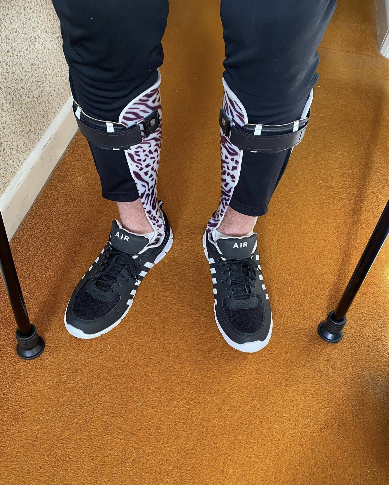
Orthotics
Orthotics (Greek: Ορθός, romanized: ortho, lit. 'to straighten, to align') also known as orthology is a medical specialty that focuses on the design and application of orthoses, sometimes known as braces, calipers, or splints. An orthosis is "an externally applied device used to influence the structural and functional characteristics of the neuromuscular and skeletal systems." Orthotists are medical professionals who specialize in designing orthotic devices such as braces or foot orthoses.
Orthotic devices are classified into four areas of the body according to the international classification system (ICS): orthotics of the lower extremities, orthotics of the upper extremities, orthotics for the trunk, and orthotics for the head. Orthoses are also classified by function: paralysis orthoses and relief orthoses.
Under the International Standard terminology, orthoses are classified by an acronym describing the anatomical joints they support. Some examples include KAFO, or knee-ankle-foot orthoses, which span the knee, ankle, and foot; TLSO, or thoracic-lumbar-sacral orthoses, supporting the thoracic, lumbar and sacral regions of the spine. The use of the International Standard is promoted to reduce the widespread variation in the description of orthoses, which is often a barrier to interpreting research studies.
The transition from an orthosis to a prosthesis can be fluid. An example is compensating for a leg length discrepancy, equivalent to replacing a missing part of a limb. Another example is the replacement of the forefoot after a forefoot amputation. This treatment is often made from a combination of a prosthesis to replace the forefoot and an orthosis to replace the lost muscular function (ortho prosthesis).[citation needed]
An orthotist is a specialist responsible for the customising, manufacture, and repair of orthotic devices (orthoses). The manufacture of modern orthoses requires both artistic skills in modeling body shapes and manual skills in processing traditional and innovative materials— CAD/CAM, CNC machines and 3D printing are involved in orthotic manufacture. Orthotics also combines knowledge of anatomy and physiology, pathophysiology, biomechanics and engineering.
In the United States, while orthotists require a prescription from a licensed healthcare provider, physical therapists are not legally authorized to prescribe orthoses. In the U.K., orthotists will often accept referrals from doctors or other healthcare professionals for orthotic assessment without requiring a prescription.
Orthoses are offered as:
Both custom-fabricated products and semi-finished products are used in long-term care and are manufactured or adapted by the orthotist or by trained orthopedic technicians according to the prescription. In many countries the physician or clinician defines the functional deviations in his prescription, e.g. paralysis (paresis) of the calf muscles (M. Triceps Surae) and derives the indication from this, e.g. orthotic to restore safety when standing and walking after a stroke. The orthotist creates another detailed physical examination and compares it with the prescription from the physician. The orthotist describes the configuration of the orthosis, which shows which orthotic functions are required to compensate for the functional deviation of the neuromuscular or skeletal system and which functional elements must be integrated into the orthosis for this. Ideally, the necessary orthotic functions and the functional elements to be integrated are discussed in an interdisciplinary team between physician, physical therapist, orthotist and patient.
Hub AI
Orthotics AI simulator
(@Orthotics_simulator)
Orthotics
Orthotics (Greek: Ορθός, romanized: ortho, lit. 'to straighten, to align') also known as orthology is a medical specialty that focuses on the design and application of orthoses, sometimes known as braces, calipers, or splints. An orthosis is "an externally applied device used to influence the structural and functional characteristics of the neuromuscular and skeletal systems." Orthotists are medical professionals who specialize in designing orthotic devices such as braces or foot orthoses.
Orthotic devices are classified into four areas of the body according to the international classification system (ICS): orthotics of the lower extremities, orthotics of the upper extremities, orthotics for the trunk, and orthotics for the head. Orthoses are also classified by function: paralysis orthoses and relief orthoses.
Under the International Standard terminology, orthoses are classified by an acronym describing the anatomical joints they support. Some examples include KAFO, or knee-ankle-foot orthoses, which span the knee, ankle, and foot; TLSO, or thoracic-lumbar-sacral orthoses, supporting the thoracic, lumbar and sacral regions of the spine. The use of the International Standard is promoted to reduce the widespread variation in the description of orthoses, which is often a barrier to interpreting research studies.
The transition from an orthosis to a prosthesis can be fluid. An example is compensating for a leg length discrepancy, equivalent to replacing a missing part of a limb. Another example is the replacement of the forefoot after a forefoot amputation. This treatment is often made from a combination of a prosthesis to replace the forefoot and an orthosis to replace the lost muscular function (ortho prosthesis).[citation needed]
An orthotist is a specialist responsible for the customising, manufacture, and repair of orthotic devices (orthoses). The manufacture of modern orthoses requires both artistic skills in modeling body shapes and manual skills in processing traditional and innovative materials— CAD/CAM, CNC machines and 3D printing are involved in orthotic manufacture. Orthotics also combines knowledge of anatomy and physiology, pathophysiology, biomechanics and engineering.
In the United States, while orthotists require a prescription from a licensed healthcare provider, physical therapists are not legally authorized to prescribe orthoses. In the U.K., orthotists will often accept referrals from doctors or other healthcare professionals for orthotic assessment without requiring a prescription.
Orthoses are offered as:
Both custom-fabricated products and semi-finished products are used in long-term care and are manufactured or adapted by the orthotist or by trained orthopedic technicians according to the prescription. In many countries the physician or clinician defines the functional deviations in his prescription, e.g. paralysis (paresis) of the calf muscles (M. Triceps Surae) and derives the indication from this, e.g. orthotic to restore safety when standing and walking after a stroke. The orthotist creates another detailed physical examination and compares it with the prescription from the physician. The orthotist describes the configuration of the orthosis, which shows which orthotic functions are required to compensate for the functional deviation of the neuromuscular or skeletal system and which functional elements must be integrated into the orthosis for this. Ideally, the necessary orthotic functions and the functional elements to be integrated are discussed in an interdisciplinary team between physician, physical therapist, orthotist and patient.