Hubbry Logo
Primer (molecular biology)Primer (molecular biology)Main
Open search
Primer (molecular biology)
Community hub
Primer (molecular biology)
logo
8 pages, 0 posts
0 subscribers
Be the first to start a discussion here.
Primer (molecular biology)
from Wikipedia
The DNA replication fork. RNA primer labeled at top.

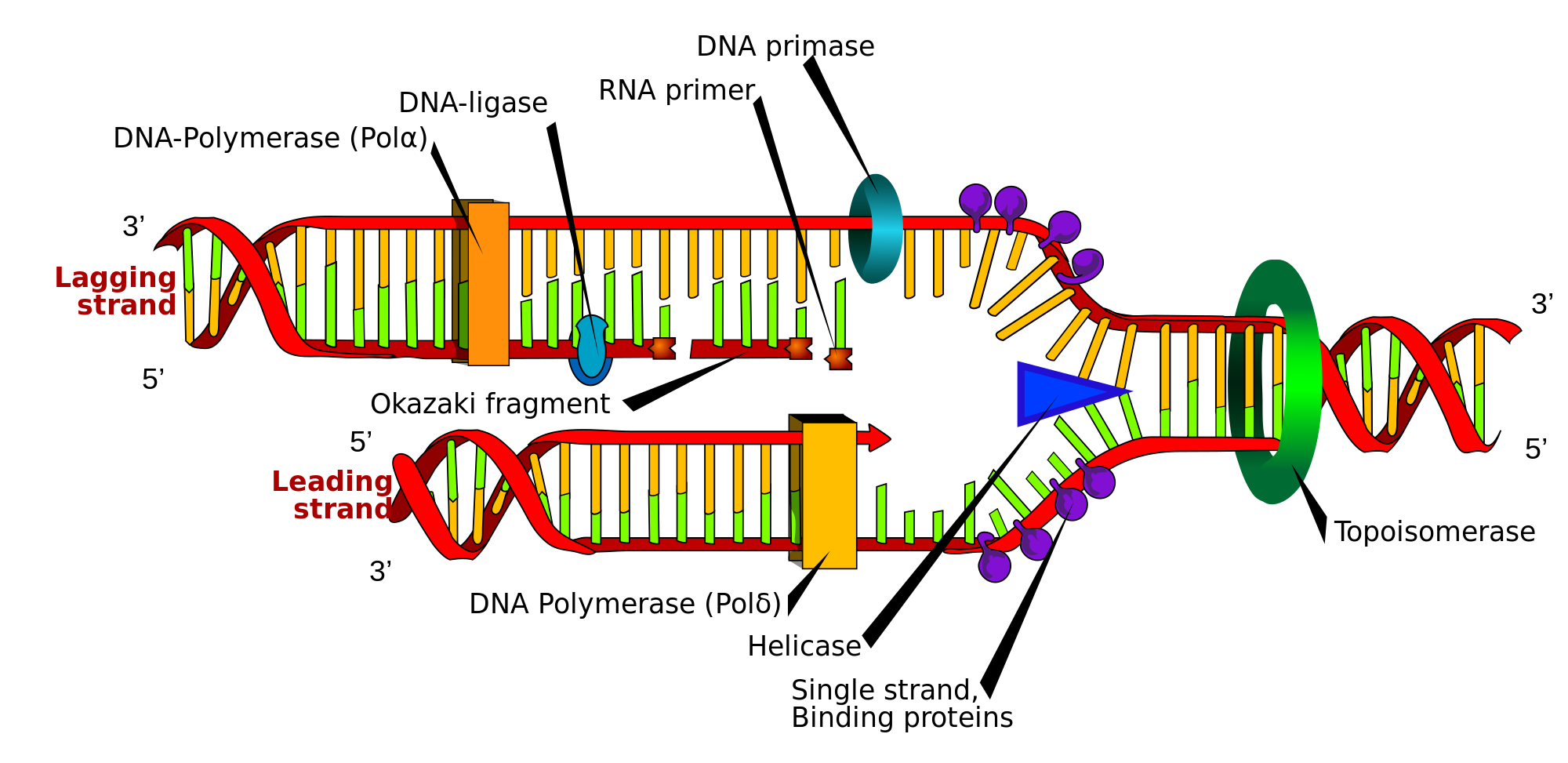
A primer is a short, single-stranded nucleic acid used by all living organisms in the initiation of DNA synthesis. A synthetic primer is a type of oligo, short for oligonucleotide. DNA polymerases (responsible for DNA replication) are only capable of adding nucleotides to the 3'-end of an existing nucleic acid, requiring a primer be bound to the template before DNA polymerase can begin a complementary strand.[1]

DNA polymerase adds nucleotides after binding to the RNA primer and synthesizes the whole strand. Later, the RNA strands must be removed accurately and replaced with DNA nucleotides. This forms a gap region known as a nick that is filled in using a ligase.[2] The removal process of the RNA primer requires several enzymes, such as Fen1, Lig1, and others that work in coordination with DNA polymerase, to ensure the removal of the RNA nucleotides and the addition of DNA nucleotides.

Living organisms use solely RNA primers, while laboratory techniques in biochemistry and molecular biology that require in vitro DNA synthesis (such as DNA sequencing and polymerase chain reaction) usually use DNA primers, since they are more temperature stable. Primers can be designed in laboratory for specific reactions such as polymerase chain reaction (PCR). When designing PCR primers, there are specific measures that must be taken into consideration, like the melting temperature of the primers and the annealing temperature of the reaction itself.

RNA primers in vivo

[edit]

RNA primers are used by living organisms in the initiation of synthesizing a strand of DNA. A class of enzymes called primases add a complementary RNA primer to the reading template de novo on both the leading and lagging strands. Starting from the free 3'-OH of the primer, known as the primer terminus, a DNA polymerase can extend a newly synthesized strand. The leading strand in DNA replication is synthesized in one continuous piece moving with the replication fork, requiring only an initial RNA primer to begin synthesis. In the lagging strand, the template DNA runs in the 5′→3′ direction. Since DNA polymerase cannot add bases in the 3′→5′ direction complementary to the template strand, DNA is synthesized 'backward' in short fragments moving away from the replication fork, known as Okazaki fragments. Unlike in the leading strand, this method results in the repeated starting and stopping of DNA synthesis, requiring multiple RNA primers. Along the DNA template, primase intersperses RNA primers that DNA polymerase uses to synthesize DNA from in the 5′→3′ direction.[1]

Another example of primers being used to enable DNA synthesis is reverse transcription. Reverse transcriptase is an enzyme that uses a template strand of RNA to synthesize a complementary strand of DNA. The DNA polymerase component of reverse transcriptase requires an existing 3' end to begin synthesis.[1]

Primer removal

[edit]

After the insertion of Okazaki fragments, the RNA primers are removed (the mechanism of removal differs between prokaryotes and eukaryotes) and replaced with new deoxyribonucleotides that fill the gaps where the RNA primer was present. DNA ligase then joins the fragmented strands together, completing the synthesis of the lagging strand.[1]

In prokaryotes, DNA polymerase I synthesizes the Okazaki fragment until it reaches the previous RNA primer. Then the enzyme simultaneously acts as a 5′→3′ exonuclease, removing primer ribonucleotides in front and adding deoxyribonucleotides behind. Both the activities of polymerization and excision of the RNA primer occur in the 5′→3′ direction,  and polymerase I can do these activities simultaneously; this is known as "Nick Translation".[1] Nick translation refers to the synchronized activity of polymerase I in removing the RNA primer and adding deoxyribonucleotides. Later, a gap between the strands is formed called a nick, which is sealed using a DNA ligase.

In eukaryotes the removal of RNA primers in the lagging strand is essential for the completion of replication. Thus, as the lagging strand being synthesized by DNA polymerase δ in 5′→3′ direction, Okazaki fragments are formed, which are discontinuous strands of DNA. Then, when the DNA polymerase reaches to the 5' end of the RNA primer from the previous Okazaki fragment, it displaces the 5′ end of the primer into a single-stranded RNA flap which is removed by nuclease cleavage. Cleavage of the RNA flaps involves three methods of primer removal.[3] The first possibility of primer removal is by creating a short flap that is directly removed by flap structure-specific endonuclease 1 (FEN-1), which cleaves the 5' overhanging flap. This method is known as the short flap pathway of RNA primer removal.[4] The second way to cleave a RNA primer is by degrading the RNA strand using a RNase, in eukaryotes it's known as the RNase H2. This enzyme degrades most of the annealed RNA primer, except the nucleotides close to the 5' end of the primer. Thus, the remaining nucleotides are displayed into a flap that is cleaved off using FEN-1. The last possible method of removing RNA primer is known as the long flap pathway.[4] In this pathway several enzymes are recruited to elongate the RNA primer and then cleave it off. The flaps are elongated by a 5' to 3' helicase, known as Pif1. After the addition of nucleotides to the flap by Pif1, the long flap is stabilized by the replication protein A (RPA). The RPA-bound DNA inhibits the activity or recruitment of FEN1, as a result another nuclease must be recruited to cleave the flap.[3] This second nuclease is DNA2 nuclease, which has a helicase-nuclease activity, that cleaves the long flap of RNA primer, which then leaves behind a couple of nucleotides that are cleaved by FEN1. At the end, when all the RNA primers have been removed, nicks form between the Okazaki fragments that are filled-in with deoxyribonucleotides using an enzyme known as ligase1, through a process called ligation.

Uses of synthetic primers

[edit]
Diagrammatic representation of the forward and reverse primers for a standard PCR

Synthetic primers are chemically synthesized oligonucleotides, usually of DNA, which can be customized to anneal to a specific site on the template DNA. In solution, the primer spontaneously hybridizes with the template through Watson-Crick base pairing before being extended by DNA polymerase. Both Sanger sequencing and next-generation sequencing require primers to initiate the reaction.[1]

PCR primer design

[edit]

The polymerase chain reaction (PCR) uses a pair of custom primers to direct DNA elongation toward each other at opposite ends of the sequence being amplified. These primers are typically between 18 and 24 bases in length and are complementary to the specific upstream and downstream sites of the sequence being amplified.[1]

Pairs of primers are designed to have similar melting temperatures since annealing during PCR occurs for both strands simultaneously. The melting temperature is not be either too much higher or lower than the reaction's annealing temperature. If annealing temperatures are too low, non-specific structures can form, reducing the efficiency of the reaction.[5]

Additionally, primer sequences need to be chosen to uniquely select for a region of DNA, avoiding the possibility of hybridization to a similar sequence nearby. A commonly used method for selecting a primer site is BLAST search, whereby all the possible regions to which a primer may bind can be seen. Both the nucleotide sequence as well as the primer itself can be BLAST searched. The free NCBI tool Primer-BLAST integrates primer design and BLAST search into one application,[6] as do commercial software products such as ePrime and Beacon Designer. In silico PCR may be performed to evaluate the specificity of designed primers.[7]

Selecting a specific region of DNA for primer binding requires some additional considerations. Regions high in mononucleotide and dinucleotide repeats should be avoided, as loop formation can occur and contribute to mishybridization. Primers that are complementary to each other can lead to the formation of primer-dimers, lowering the efficiency of the desired reaction.[8] Primers that are able to anneal to themselves can form internal hairpins and loops that hinder hybridization with the template DNA.[8]

When designing primers, additional nucleotide bases can be added to the back ends of each primer, resulting in a customized cap sequence on each end of the amplified region. One application for this practice is for use in TA cloning, a special subcloning technique similar to PCR, where efficiency can be increased by adding AG tails to the 5′ and the 3′ ends.[9]

Degenerate primers

[edit]

Some situations may call for the use of degenerate primers. These are mixtures of primers that are similar, but not identical. These may be convenient when amplifying the same gene from different organisms, as the sequences are probably similar but not identical. This technique is useful because the genetic code itself is degenerate, meaning several different codons can code for the same amino acid. This allows different organisms to have a significantly different genetic sequence that code for a highly similar protein. For this reason, degenerate primers are also used when primer design is based on protein sequence, as the specific sequence of codons are not known. Therefore, primer sequence corresponding to the amino acid isoleucine might be "ATH", where A stands for adenine, T for thymine, and H for adenine, thymine, or cytosine, according to the genetic code for each codon, using the IUPAC symbols for degenerate bases. Degenerate primers may not perfectly hybridize with a target sequence, which can greatly reduce the specificity of the PCR amplification.

Degenerate primers are widely used and extremely useful in the field of microbial ecology. They allow for the amplification of genes from thus far uncultivated microorganisms or allow the recovery of genes from organisms where genomic information is not available. Usually, degenerate primers are designed by aligning gene sequencing found in GenBank. Differences among sequences are accounted for by using IUPAC degeneracies for individual bases. PCR primers are then synthesized as a mixture of primers corresponding to all permutations of the codon sequence.

References

[edit]
[edit]
Revisions and contributorsEdit on WikipediaRead on Wikipedia
from Grokipedia
In , a primer is a short, single-stranded —typically 18 to 25 long—that provides a free 3'-hydroxyl group to initiate the synthesis of a strand by enzymes. Primers are essential in both natural and laboratory techniques, where they hybridize to a complementary template strand to define the starting point for nucleotide addition in the 5' to 3' direction. In cellular DNA replication, primers are naturally occurring RNA molecules synthesized by the enzyme primase, a specialized that does not require a preexisting primer itself. These RNA primers, about 5–10 nucleotides long in prokaryotes and up to 10 nucleotides in eukaryotes, are laid down on the lagging strand at intervals of 1000–2000 nucleotides in prokaryotes and 100–200 nucleotides in eukaryotes to initiate the formation of during discontinuous synthesis. Once extends the primer by adding deoxyribonucleotides, the RNA primer is removed by nucleases (such as RNase H and flap endonuclease in eukaryotes) and replaced with DNA, with the fragments joined by to form a continuous strand. Synthetic primers, usually designed as short DNA oligonucleotides, are widely used in vitro for applications such as the polymerase chain reaction (PCR), , and . In PCR, a pair of primers—one forward and one reverse—flanks the target DNA sequence, annealing to the denatured template during each cycle to enable exponential amplification of the region between them using a thermostable like . This technique, first demonstrated in 1985, revolutionized by allowing rapid production of millions of copies of specific DNA segments from minute starting material. Primer design is critical for specificity and efficiency, considering factors like melting temperature, GC content, and avoidance of secondary structures to ensure accurate hybridization and minimal off-target amplification.

Fundamentals of primers

Definition and role in DNA synthesis

In molecular biology, a primer is a short single-stranded nucleic acid sequence, either RNA or DNA, that hybridizes to a complementary region on a DNA template strand, providing a free 3' hydroxyl (OH) group essential for the initiation of DNA synthesis by DNA polymerase enzymes. Natural primers in cellular DNA replication are typically RNA, ranging from 5 to 12 nucleotides in length depending on the organism—for instance, approximately 10 nucleotides in eukaryotes and 10–12 in prokaryotes like Escherichia coli. In contrast, synthetic primers used in laboratory applications are DNA oligonucleotides, usually 18–25 nucleotides long, designed to anneal specifically to target sequences. The primary role of a primer is to enable processive DNA polymerization, as DNA polymerases lack the ability to initiate synthesis de novo on a bare single-stranded template; instead, they require the primer's 3' OH end to catalyze the addition of deoxyribonucleotide triphosphates in the 5' to 3' direction. During semi-conservative DNA replication, this function is critical for both strands at the replication fork: a single RNA primer initiates continuous synthesis on the leading strand, while multiple short RNA primers are laid down periodically (every 100–200 nucleotides in eukaryotes or 1,000–2,000 in prokaryotes) to start each discontinuous Okazaki fragment on the lagging strand. Without primers, replication would stall, as the antiparallel nature of DNA strands necessitates this discontinuous mechanism on the lagging template. The concept of primers arose from pioneering studies on in the 1960s and 1970s, building on the 1968 discovery of —short, discontinuous DNA segments on the lagging strand—by Reiji Okazaki and colleagues, who demonstrated replication proceeds in a 5' to 3' direction via pulse-labeling experiments in E. coli. Subsequent work by the Okazaki group in the early 1970s revealed these fragments are initiated by short primers attached to their 5' ends, resolving how DNA chain growth begins and confirming the involvement of in prokaryotic and viral replication systems. For example, each on the lagging strand is primed by an segment synthesized complementary to the template, allowing to extend it until the next primer site.

Biochemical requirements for priming

For a primer to function in DNA synthesis, it must hybridize to the single-stranded template DNA through specific Watson-Crick base pairing, in which (A) pairs with (T) or uracil (U) in RNA primers, and (G) pairs with (C), forming hydrogen bonds that stabilize a short double-stranded duplex. This complementary annealing creates a primer-template junction, positioning the primer's free 3' hydroxyl (OH) group at the end of the duplex, which is essential for subsequent nucleotide addition. The resulting structure mimics a naturally occurring double helix segment, ensuring accurate base selection and alignment for polymerase binding. DNA polymerases exhibit strict enzymatic specificity, requiring the primer-template junction to initiate synthesis; for instance, bacterial III and eukaryotic polymerases δ and ε cannot catalyze the formation of the initial without this pre-existing structure. These enzymes are incapable of de novo DNA synthesis—starting from free deoxynucleoside triphosphates (dNTPs)—primarily because the geometry prevents proper alignment of the first two , imposing a high barrier that cannot be overcome without the primer's 3' OH to act as the . This requirement evolved to enhance fidelity, as the junction provides a defined starting point that aligns the template for error-correcting mechanisms. In vivo, primers must possess an appropriate length to ensure functional stability while supporting efficient replication dynamics; typically, RNA primers range from 7 to 12 , with a minimum of 4-5 needed to form a stable initial duplex under physiological conditions. This optimal length balances hybridization specificity—reducing the risk of non-specific binding—and the error rate, as shorter primers allow more frequent initiation on the lagging strand to accommodate the replication fork's movement, while longer ones might increase mispairing potential. Thermodynamic stability of the primer-template duplex is further influenced by the melting (Tm), calculated based on composition where GC pairs contribute greater stability due to three hydrogen bonds compared to two for AT pairs, ensuring the complex remains intact at body (approximately 37°C) without excessive rigidity that could hinder extension. All DNA synthesis occurs unidirectionally in the 5' to 3' polarity, with the primer anchoring the process by exposing its 3' OH end as the site for nucleophilic attack on the α-phosphate of incoming dNTPs, releasing and extending the chain. This directionality is a universal feature of replicative polymerases, reflecting the enzyme's catalytic mechanism that favors forward progression and incorporates energy from dNTP to drive .

Natural RNA primers in vivo

Synthesis by primase enzyme

In , RNA primase is a specialized that synthesizes short RNA primers de novo during , without requiring a preexisting primer or template-directed like standard polymerases. In prokaryotes, such as , primase is encoded by the dnaG and functions as a monomeric that binds to single-stranded DNA (ssDNA) at replication origins or the start sites of on the lagging strand. The interacts closely with the DnaB , which unwinds the DNA duplex and stimulates primase activity by facilitating ssDNA access. The mechanism of primer synthesis by prokaryotic primase begins with recognition of specific ssDNA sequences, often those containing a 5'-CTG or 5'-CAG motif, where primase binds via conserved residues in its template-tracking site. Initiation occurs preferentially with nucleotides (ATP or GTP) at the 5' end, forming a dinucleotide that serves as the primer start; subsequent extension proceeds in the 5' to 3' direction, adding 5-12 ribonucleotides via a two-metal-ion mechanism involving magnesium ions to coordinate substrates. This process yields primers typically 10-12 nucleotides long, after which primase dissociates, allowing handover to for extension. In E. coli, primase operates rapidly but with relatively low fidelity, synthesizing one primer per Okazaki fragment approximately every 1,000-2,000 on the lagging strand, while the leading strand requires only a single primer at the origin. In eukaryotes, primase forms part of the heterotetrameric α-primase (Pol α-primase) complex, consisting of the catalytic primase subunits (PRIM1 and PRIM2) and polymerase subunits (POLA1 and POLA2), which synthesizes chimeric RNA-DNA primers. The subunit binds ssDNA, often in coordination with the CMG complex (comprising MCM2-7, CDC45, and GINS), and initiates synthesis de novo on single-stranded DNA templates, showing a preference for pyrimidine-rich sequences, starting with a nucleotide and extending an RNA segment of 7-12 in the 5' to 3' direction. Unlike prokaryotic primase, the eukaryotic version is slower and more processive, with the associated Pol α subunit immediately extending the RNA primer by 15-20 deoxynucleotides to form a hybrid primer of 20-30 nucleotides total, before dissociation triggered by a conformational shift from A-form to B-form . Primers are required once at each replication origin for the leading strand and repeatedly for every 100-200 nucleotides on the lagging strand, reflecting the shorter fragment lengths in eukaryotic genomes. Regulation of primase activity ensures coordination with the replication machinery; in prokaryotes, DnaG-primase association with DnaB and transient interactions with the β-clamp facilitate timely primer synthesis and polymerase handover at the . Eukaryotic Pol α-primase is regulated through cell cycle-dependent and binding to the CMG , promoting accurate priming while minimizing errors, in contrast to the faster, more error-prone prokaryotic system.

Removal and processing mechanisms

In the process of , particularly during the maturation of on the lagging strand, RNA primers synthesized by must be removed to ensure the continuity of the newly synthesized DNA strand. This removal and subsequent processing are critical steps that prevent the retention of segments in the , which could otherwise genomic stability. The mechanisms differ between prokaryotes and eukaryotes, involving specialized enzymes that coordinate cleavage, gap filling, and ligation. In prokaryotes, such as , (Pol I) plays a central role in primer removal through its 5'→3' activity coupled with activity, a process known as nick translation. Pol I initiates at the nick between the RNA primer and the downstream DNA, degrading the ribonucleotides while simultaneously synthesizing deoxyribonucleotides to replace them, thereby filling the gap without leaving unsealed breaks. Once the primer is excised and the gap filled, seals the remaining nick to form a , completing the fragment. This efficient, multifunctional action of Pol I ensures rapid maturation of . Eukaryotic primer removal is more complex and involves multiple enzymes to handle the longer and chromatin-associated replication. RNase H, specifically type 2 in eukaryotes, initiates the process by cleaving the RNA strand within the RNA-DNA hybrid, leaving typically a single ribonucleotide attached to the 5' end of the DNA. The remnant is then removed by flap endonuclease 1 (FEN1), which excises structured flaps generated during strand-displacement synthesis by DNA polymerase δ (Pol δ); FEN1 recognizes and cleaves these flaps at the junction of RNA and DNA. Pol δ fills the resulting gap with deoxyribonucleotides using dNTPs as substrates, while replication protein A (RPA) binds to the single-stranded DNA to stabilize the template and prevent secondary structures during this phase. Finally, I seals the nick, forming a continuous DNA strand. Incomplete or erroneous primer removal poses significant challenges to genomic integrity. Retained RNA primers can lead to RNA-DNA hybrids that trigger through error-prone repair or replication stalling. At chromosome ends, failure to fully process the terminal RNA primer on the lagging strand contributes to the end-replication problem, resulting in progressive shortening with each cell division, which limits cellular lifespan and promotes . These mechanisms were elucidated in the , building on Kornberg's foundational work on and its activities, which highlighted the enzyme's role in maintaining replication fidelity by removing RNA segments.

Synthetic DNA primers in laboratory techniques

Design principles for PCR primers

The design of synthetic DNA primers for (PCR) amplification requires careful consideration of several parameters to ensure efficient, specific, and robust amplification of the target sequence. Optimal primer length typically ranges from 18 to 25 , as this provides sufficient specificity while allowing stable hybridization under standard PCR conditions. A of 40-60% is recommended to achieve a melting (Tm) in the range of 50-60°C, promoting balanced stability without excessive secondary structure formation. To prevent primer-dimer formation, sequences should avoid self-complementarity, particularly at the 3' ends of forward and reverse primers. Specificity is paramount in PCR primer design, with the 3' end requiring perfect complementarity to the template DNA to enable efficient polymerase extension. Tools such as Primer3 facilitate the selection of unique primer sequences by evaluating potential off-target binding sites, while BLAST or Primer-BLAST is used to verify specificity against genomic databases, minimizing non-specific amplification. The annealing temperature is calculated based on the primer Tm using the Wallace rule: Tm = 4(G + C) + 2(A + T) in °C, with the annealing step typically set 3-5°C below the lower Tm to optimize hybridization. This empirical formula, derived from early oligonucleotide hybridization studies, provides a straightforward estimate for primers under 20 nucleotides. Several sequence features must be avoided to enhance primer performance and reduce artifacts. Primers should not contain runs of more than three identical bases, as these can lead to mispriming or slippage during amplification. Repetitive motifs, palindromic sequences, or potential secondary structures like hairpins should be minimized, as they promote non-specific binding or inhibit annealing; in silico prediction tools such as mfold can identify these risks. For added stability, a GC clamp—typically one or two G or C bases at the 3' end—enhances specific template binding without compromising the overall Tm. Optimization involves in silico validation followed by empirical testing to refine primer performance. Software like Primer3 integrates multiple criteria to rank candidate primers, allowing adjustments for specific applications such as quantitative PCR (qPCR), where primers are paired with probes containing fluorophores and quenchers for real-time detection. In multiplex PCR, primers must be designed to avoid overlap in binding sites and Tm values, ensuring simultaneous amplification of multiple targets without interference.

Degenerate and modified primers

Degenerate primers are synthetic designed with intentional sequence variations at specific positions to amplify target sequences that exhibit natural variability, such as conserved regions within families or homologous genes across . These variations are typically represented using IUPAC codes, where symbols like N denote any of the four bases (A, C, G, or T), R indicates A or G, and Y specifies C or T, resulting in a of multiple primer sequences in a single reaction. This approach reduces primer specificity to accommodate sequence uncertainty but enhances the yield of related amplicons, making it particularly useful for identifying and unknown or distantly related genes. The of degenerate primers in PCR was formalized in the early 1990s, building on hybridization techniques from the , with seminal guidelines emphasizing their design to minimize non-specific binding while maximizing coverage of target variants. In designing degenerate primers, bases like are often incorporated at ambiguous positions because can form stable base pairs with A, C, G, or T, effectively acting as a universal base that simplifies the primer mixture and improves annealing efficiency without excessive degeneracy. Alternatively, synthetic universal bases such as 5-nitroindole or 3-nitropyrrole can replace ambiguous sites, providing non-discriminatory pairing that maintains consistent temperatures (Tm) across variants and reduces the total number of primer sequences needed. These modifications were developed to address limitations in early degenerate designs, where high degeneracy (e.g., >256 variants) could lead to inefficient amplification. Tools like CODEHOP (consensus-degenerate hybrid primers) further refine this by aligning protein sequences to generate primers with a non-degenerate 5' consensus region for specificity and a degenerate 3' core for flexibility, enabling the cloning of novel genes in large families. Modified primers incorporate chemical alterations to the standard DNA backbone or termini to enhance stability, detection, or performance in challenging conditions. Phosphorothioate (PS) modifications replace oxygen atoms in the phosphodiester backbone with , conferring resistance to degradation, which is advantageous in applications involving cellular uptake or environmental samples where enzymes might degrade unmodified primers; early studies demonstrated that PS-modified primers improved PCR product yield by protecting against exonucleases during amplification. Labels such as or fluorescent dyes (e.g., FAM, HEX) are attached typically at the 5' end to facilitate downstream detection: enables streptavidin-based capture for assays like or solid-phase hybridization, while fluorescent tags allow real-time monitoring or fragment analysis via . Locked nucleic acids (LNAs) introduce a in the ring, locking it into a rigid C3'-endo conformation that boosts Tm by 3–8°C per substitution and improves mismatch discrimination, thus enhancing specificity in PCR for single-nucleotide polymorphisms or low-abundance targets. These specialized primers find key applications in evolutionary studies, where degenerate designs amplify orthologs across taxa to reconstruct phylogenies, and in , enabling broad-spectrum coverage of microbial communities without prior sequence knowledge—for example, degenerate primers targeting 16S rRNA have recovered diverse bacterial taxa from environmental samples. However, trade-offs include increased costs due to complex synthesis (e.g., degenerate mixtures can exceed $100 per nmol), potential for higher non-specific amplification leading to chimeric products or elevated error rates in downstream sequencing, and reduced efficiency in high-degeneracy scenarios that may require optimized annealing temperatures. Modified primers, while improving robustness, can sometimes inhibit activity if over-incorporated (e.g., multiple PS bonds reducing extension rates by 10–20%), necessitating empirical validation for each application.

Applications beyond PCR

Synthetic primers play a crucial role in DNA sequencing methods beyond amplification, providing the annealing sites necessary for chain extension and fragment analysis. In the chain-termination method developed by Sanger and colleagues in 1977, custom oligonucleotide primers anneal to single-stranded DNA templates adjacent to the region of interest, enabling DNA polymerase to incorporate chain-terminating dideoxynucleotides and generate a ladder of fragments for electrophoretic separation and sequence determination. Universal primers, such as those derived from the M13 bacteriophage vector (e.g., M13 forward: 5'-TGTAAAACGACGGCCAGT-3' and reverse: 5'-CAGGAAACAGCTATGAC-3'), have become standard for Sanger sequencing of inserts cloned into M13 or pUC-based plasmids, allowing consistent priming without redesign for each template. In next-generation sequencing (NGS) library preparation, adapter oligonucleotides—functioning as primers with partial double-stranded structures—are ligated to fragmented DNA ends, providing binding sites for subsequent amplification and sequencing primers during bridge amplification on flow cells. In and , primers are engineered for precise manipulation of DNA sequences. Site-specific primers oriented outward from a known insert enable inverse PCR, which amplifies flanking regions for cloning unknown sequences adjacent to characterized elements, such as transposon insertions. For , overlap extension techniques use primers with complementary overlapping regions at their 5' ends to assemble mutated fragments; the overlap (typically 15-25 bases) facilitates recombination during PCR, allowing insertion, deletion, or substitution of sequences. Additionally, primers with deliberate mismatches can direct the incorporation of desired changes during extension; in some methods, mismatches are placed at the 3' end, while in the QuikChange method, primers are fully complementary to the template except for the mutation site typically located in the middle of the primer, enabling amplification of the entire while embedding the edit. Beyond sequencing and , synthetic primers support probe-based detection and RNA synthesis in diagnostic and applications. In assays for real-time quantitative PCR diagnostics, primers flank the target amplicon while a fluorescently labeled probe hybridizes internally; during extension, the probe's 5' cleavage by releases the , enabling specific or detection with high sensitivity (e.g., limits of detection in the attomolar range for viral RNA). For transcription, PCR-generated templates incorporate T7 promoter sequences via forward primers (e.g., 5'-TAATACGACTCACTATAGGG-3' followed by a 5-10 base spacer and gene-specific sequence), allowing T7 to initiate capped or uncapped synthesis for applications like mRNA vaccines or production. Advancements as of 2025 have integrated synthetic primers with CRISPR-Cas systems for enhanced targeting precision and throughput. Primers designed to amplify (gRNA) templates with T7 promoters enable transcription of single guide RNAs (sgRNAs) that direct to specific genomic loci, facilitating knockouts or edits; optimized gRNA spacer sequences (20 nt) minimize off-target effects. Automation in leverages robotic pipetting and multiplex primer pools for parallel or variant library construction, accelerating studies by processing thousands of primer-template combinations daily.

References

Add your contribution
Related Hubs
User Avatar
No comments yet.