Hubbry Logo
Stasis dermatitisStasis dermatitisMain
Open search
Stasis dermatitis
Community hub
Stasis dermatitis
logo
7 pages, 0 posts
0 subscribers
Be the first to start a discussion here.
Stasis dermatitis
Stasis dermatitis
from Wikipedia
Stasis dermatitis
Other namescongestion eczema, gravitational dermatitis, gravitational eczema, stasis eczema, varicose eczema[1]
SpecialtyDermatology Edit this on Wikidata

Stasis dermatitis refers to the skin changes that occur in the leg as a result of "stasis" or blood pooling from insufficient venous return; the alternative name of varicose eczema comes from a common cause of this being varicose veins.[2]

Insufficient venous return results in increased pressure in the capillaries with the result that both fluid and cells may "leak" out of the capillaries. This results in red cells breaking down, with iron-containing hemosiderin possibly contributing to the pathology of this entity.[3]

Symptoms

[edit]

Stasis dermatitis may be characterized by:

  • Skin that appears thin, brown, and tissue-like, with possible skin lesions (macule or patches), red spots, superficial skin irritation, and/or darkening and/or thickening of the skin at the ankles or legs
  • Weak skin may ulcerate in some areas, and legs, ankles, or other areas may become swollen
  • Open sores, ulcers
  • Itching and/or leg pains
  • Sometimes pain may persist from swollen tissues and may feel like "stabbing" or "needle pricks"

If skin continues to deteriorate and breaks down, a venous ulcer (also known as a stasis ulcer) may form.[3] Without proper wound care, open cracks predispose patients for the entry of a bacterial infection, causing cellulitis in the leg.[4]

Diagnosis

[edit]

Stasis dermatitis is diagnosed clinically by assessing the appearance of red plaques on the lower legs and the inner side of the ankle. Stasis dermatitis can resemble several other conditions, such as cellulitis and contact dermatitis, and at times needs the use of a duplex ultrasound to confirm the diagnosis or if clinical diagnosis alone is not sufficient.[5]

Treatment

[edit]

Treatment may consist of topical applications of steroid-based creams and the use of compression stockings or intermittent pneumatic compression pumps, to help force the underlying buildup of fluids back out of the lower leg.[6]

Compression therapy should consist of moderate pressures and work best for ambulating patients.[5]

Ultimately, treating the underlying venous reflux is necessary to treat Stasis dermatitis. Invasive surgical procedures like saphenofemoral junction ligation with stripping were the norm for treatment in the past. However, less invasive methods are now more widely used. These newer methods include endovenous thermal ablation, ambulatory phlebectomy, and ultrasound foam sclerotherapy.[5]
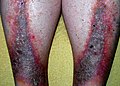
Complications

[edit]

If stasis dermatitis goes untreated, the patient is at risk of developing venous ulcers and Acroangiodermatitis.[6][5]

See also

[edit]

References

[edit]
[edit]
Revisions and contributorsEdit on WikipediaRead on Wikipedia
from Grokipedia
Stasis dermatitis, also known as venous stasis dermatitis or gravitational eczema, is a chronic inflammatory primarily affecting the lower legs due to impaired venous circulation and blood pooling from . It arises when faulty vein valves fail to pump blood effectively back to the heart, leading to increased pressure in the veins, fluid leakage into surrounding tissues, and subsequent skin changes. This condition is most common in older adults, with prevalence estimates of 6-7% in individuals over age 50 in the United States, rising to over 20% in those over 70, and a slight predominance in females. The primary causes of stasis dermatitis stem from venous hypertension, often resulting from deep vein , trauma, surgery, or prolonged immobility that damages vein walls or valves. Risk factors include aging, , , and occupations requiring extended standing or sitting, which exacerbate poor circulation and contribute to in the lower extremities. In advanced cases, the condition can progress due to secondary factors like or , further impairing lymphatic drainage and promoting inflammation. Symptoms typically begin with bilateral swelling around the ankles that worsens with dependency and improves with elevation, accompanied by pruritus (itching), erythema (redness), and scaling on the skin, often most pronounced on the medial malleoli (inner ankles). As the disease advances, patients may experience skin (reddish-brown discoloration from deposition), lichenification (thickening and hardening), and weeping or crusting erosions, which can lead to painful venous ulcers in severe instances. Complications such as secondary bacterial infections (e.g., ) or from topical treatments are common if unmanaged, significantly impacting through , sleep disruption, and mobility limitations. Diagnosis is primarily clinical, based on characteristic findings and a history of leg swelling, with Doppler often used to confirm underlying venous insufficiency or rule out . Treatment focuses on improving venous return through compression therapy (e.g., or bandages), leg elevation, and addressing with diuretics if needed, alongside topical corticosteroids for and emollients to restore . In cases of ulceration or , wound care, antibiotics, or surgical interventions like vein ablation may be required, though the condition is chronic and requires ongoing management to prevent recurrence. Prevention emphasizes measures such as regular exercise, weight control, and avoiding prolonged stasis, with early intervention for reducing progression risk.

Overview

Definition and Classification

Stasis dermatitis is a chronic inflammatory of the lower extremities resulting from (CVI) and venous due to venous . It represents the earliest cutaneous manifestation of underlying venous , manifesting as an eczematous eruption secondary to impaired venous return and blood pooling in the legs. Also known as venous eczema or gravitational dermatitis, it primarily affects the above the medial but can extend to other areas of the lower legs. As a subtype of eczematous dermatitis, stasis dermatitis is distinguished from primary forms such as atopic or by its secondary origin in vascular stasis rather than intrinsic immune dysregulation or external allergens. In the context of chronic venous disorders, it is classified under the Clinical-Etiologic-Anatomic-Pathophysiologic (CEAP) system as class C4, denoting pigmentation or eczema involving changes in the skin and secondary to venous hypertension. This classification highlights its position within the spectrum of venous insufficiency manifestations, preceding more advanced complications like or ulceration. The condition progresses through distinct stages reflecting the evolution of and tissue response. The acute features edematous, erythematous, and weepy lesions with possible vesiculation due to acute venous . The subacute involves drying, scaling, and excoriations as the resolves partially, while the chronic is marked by lichenification, , and from repeated episodes and ongoing stasis. Venous hypertension serves as the key driver, promoting leakage of fluid and inflammatory mediators into the .

Epidemiology

Stasis dermatitis, a cutaneous manifestation of , affects approximately 6-7% of adults over 50 years in the United States, equating to an estimated 15-20 million individuals. rises sharply with age, reaching over 20% in those over 70 years, reflecting the condition's strong association with advancing years and venous system deterioration. Global estimates mirror these patterns in aging populations, with —a key precursor—prevalent in up to 40% of women and 17% of men in Western countries. Demographically, stasis dermatitis exhibits a slight female predominance, attributed in part to factors like and multiparity that strain venous return. It is rare before age 50, with peak incidence in the elderly, and shows higher rates among individuals with , sedentary lifestyles, or a history of . Comorbidities such as and further elevate risk, contributing to disproportionate burden in these groups. Geographically, the condition is more prevalent in developed nations with aging demographics and lifestyles promoting , such as prolonged sitting or standing. Socioeconomic factors, including access to preventive care and or immobility, influence distribution, though data remain limited. The is expected to rise due to demographic shifts toward older age and increasing , driven by aging populations and sedentary lifestyles.

Pathophysiology

Etiology

Stasis dermatitis primarily arises from in the lower extremities, resulting from . This condition is driven by retrograde blood flow due to incompetent venous valves, valvular destruction, or venous obstruction caused by prior , surgery, or trauma. Several risk factors contribute to the development of and subsequent stasis dermatitis. A history of predisposes individuals to valvular damage and obstruction, while reflect underlying valvular incompetence. increases intra-abdominal pressure, impairing venous return, and prolonged standing or sitting promotes blood pooling in the legs. exacerbates these issues by elevating venous pressure through increased body mass and reduced mobility. The is multifactorial, involving such as familial venous insufficiency, which heightens susceptibility to valve incompetence. Iatrogenic causes, including saphenous vein harvesting during like coronary artery bypass grafting, can disrupt venous flow and lead to postoperative venous hypertension.

Mechanisms of Disease

Stasis dermatitis arises from chronic venous hypertension, often initiated by incompetent venous valves that permit retrograde blood flow and elevate hydrostatic pressure in the lower extremity veins. This increased pressure disrupts the integrity of walls, promoting leakage of plasma components, including fibrinogen, erythrocytes, and , into the surrounding interstitial tissues. While older hypotheses proposed that extravasated fibrinogen polymerizes into forming "fibrin cuffs" around capillaries that impede oxygen diffusion and cause hypoxia, this theory has been challenged as deposits are not unique to stasis dermatitis and do not fully explain the . The resultant hypoxic environment triggers an inflammatory cascade, beginning with the activation and trapping of leukocytes within the and perivascular spaces, facilitated by upregulation of molecules such as and VCAM-1. These leukocytes, along with infiltrating macrophages, release pro-inflammatory cytokines and matrix metalloproteinases (MMPs), such as MMP-1, MMP-2, and MMP-13, which degrade the and amplify tissue damage. Over time, this chronic inflammation leads to dermal through excessive deposition, accumulation from degraded erythrocytes, and sustained immune cell infiltration, fostering a cycle of ongoing skin remodeling. In advanced stages, these processes culminate in , a fibrosing marked by induration and scarring. At the molecular level, transforming growth factor-beta (TGF-β) plays a pivotal role in driving by stimulating differentiation into myofibroblasts, which produce excessive components. Concurrently, in the microvasculature exacerbates , contributing to persistent and further inflammatory perpetuation. These interconnected mechanisms underscore the progressive nature of stasis dermatitis as a manifestation of unresolved venous insufficiency.

Clinical Features

Symptoms

Stasis dermatitis primarily manifests through subjective symptoms that significantly affect patients' . The most common initial complaint is pruritus, or intense itching, which affects 55–66% of patients and can persist for years, often lasting up to 9 hours daily and worsening at night. This itching is linked to underlying in the skin. Patients also frequently report sensations of heaviness, aching, or cramping in the lower legs, particularly after prolonged standing or at the end of the day. Associated discomfort includes a burning or stinging sensation that arises from scratching the itchy areas, which can lead to further . Symptoms often exacerbate with factors such as or humid , dependency of the legs (e.g., prolonged standing or sitting), and high salt intake, which promotes fluid retention and . If the condition progresses to ulceration, patients experience escalating pain that intensifies the overall discomfort. These symptoms have notable patient-reported impacts, including sleep disturbances from nocturnal itching, burning, or pain, which contribute to daytime . Reduced mobility often results from leg fatigue and aching, limiting physical activities and daily functioning. The nature of the condition can lead to emotional effects such as , anxiety, and depression due to persistent discomfort and recurrent flares.

Signs

Stasis dermatitis commonly manifests with bilateral involvement of the lower extremities, predominantly affecting the medial malleolar region and potentially extending to the calf, foot, or encircling the ankle in more severe cases. Early physical findings include due to fluid accumulation, accompanied by erythematous, scaling, and eczematous patches on the skin. appears as reddish-brown discoloration from deposition, often speckled and localized over . In the acute phase, signs escalate to include weeping vesicles, bullae, and exudative plaques with crusting, reflecting intense inflammation and possible secondary infection. may worsen acutely, contributing to subcutaneous nodules or plaques. Visible varicosities are frequently observed, underscoring underlying venous insufficiency. Chronic progression reveals advanced features such as indurated, thickened with lichenification and from repeated , leading to a brawny, non-pitting . The lower often develops an inverted champagne bottle shape due to , characterized by calf and ankle , alongside a or bumpy texture. In longstanding cases, atrophie blanche presents as ivory-white atrophic patches surrounded by telangiectasias, indicating ischemic changes.

Diagnosis

History and Examination

The clinical assessment of stasis dermatitis begins with a detailed patient history to identify the onset, progression, and contributing factors to the condition. Clinicians inquire about the duration of symptoms, such as pruritus or skin changes, which often develop insidiously over months to years, primarily affecting the lower extremities. Patients are asked about patterns of leg swelling, including whether edema worsens with prolonged standing or sitting and improves with leg elevation, as this is characteristic of chronic venous insufficiency underlying the dermatitis. A history of deep vein thrombosis (DVT), varicose veins, or lower extremity trauma is elicited, as these increase the risk of venous hypertension leading to stasis dermatitis. Risk factors such as occupational demands involving extended periods of standing or sitting, obesity, older age, and comorbidities like congestive heart failure or hypertension are also assessed to contextualize the patient's venous disease burden. Red flags in the history include unilateral leg involvement, which may suggest venous obstruction or DVT rather than typical bilateral stasis, and rapid onset of symptoms, potentially indicating superimposed infection such as cellulitis. Physical examination focuses on the lower extremities to confirm features and rule out acute complications. begins with the distribution of changes, typically in the gaiter area around the medial and ankles, extending bilaterally up to the mid-calf in a stocking-like pattern, with early signs including erythematous, scaling patches and dependent . Chronic findings may include reddish-brown from deposition, as briefly noted in typical signs of advanced disease. assesses for tenderness over affected areas, which can indicate underlying or inflammation, and evaluates pitting by applying firm pressure to the medial or pretibial area for 5 seconds to check for persistent indentation. Limb circumference is measured at standardized points, such as the ankle and calf, to quantify severity and monitor progression, while is noted to differentiate warm, inflamed skin from cooler, chronic changes. Unilateral warmth or asymmetry on exam reinforces red flags from history, prompting further for obstruction or .

Diagnostic Investigations

The primary diagnostic investigation for stasis dermatitis involves duplex ultrasound, which serves as the gold standard for evaluating chronic venous insufficiency (CVI), the underlying cause of the condition. This non-invasive imaging modality assesses venous reflux by measuring the duration of retrograde flow after distal compression, with pathologic reflux defined as greater than 0.5 seconds in superficial veins or 1.0 second in deep veins such as the femoral or popliteal. It also evaluates valve competence through visualization of valve closure and detects obstructions by identifying flow abnormalities or compressibility of veins. Additionally, the ankle-brachial index (ABI) is routinely measured to exclude concomitant arterial disease, which could contraindicate compression therapy; an ABI value below 0.8 suggests arterial insufficiency and warrants further vascular evaluation. Further investigations are pursued when the diagnosis is atypical or to rule out mimics. Patch testing is recommended if allergic contact dermatitis is suspected, particularly given the disrupted skin barrier in stasis dermatitis that increases sensitization risk; it identifies specific allergens from topical agents commonly used in treatment. Skin biopsy is reserved for uncertain cases, revealing characteristic histologic features such as , epidermal acanthosis, dermal , perivascular lymphocytic infiltrates, and deposition from extravasated red blood cells. , an invasive contrast-based imaging technique, is rarely employed but may be considered in complex cases with suspected deep venous obstruction not fully characterized by . Diagnosis of stasis dermatitis requires clinical correlation using the CEAP classification system for chronic venous disorders, which standardizes assessment across clinical (C), etiologic (E), anatomic (A), and pathophysiologic (P) domains. Stasis dermatitis typically corresponds to class C4a, indicating pigmentation or eczema secondary to CVI, with etiology often primary (Ep) or secondary (Es) to venous reflux (Pr) or obstruction (Po) in superficial (As) or deep (Ad) veins. This framework ensures comprehensive evaluation beyond isolated tests, integrating findings to confirm venous etiology.

Management

Nonpharmacological Treatments

Compression therapy serves as the cornerstone of nonpharmacological management for stasis dermatitis, primarily by counteracting venous and reducing lower extremity . Graduated exerting 20-30 mmHg of pressure at the ankle are recommended for most patients to promote venous return and alleviate swelling, with application ideally performed in the morning before significant accumulates. For cases with more pronounced exudation or ulceration, multilayer inelastic bandages or an Unna boot—a zinc oxide-impregnated wrap providing sustained compression—can be employed to manage weeping lesions and support healing while minimizing irritation. Patient adherence to compression is crucial, as it directly correlates with reduced recurrence rates, though challenges such as discomfort necessitate education on proper fitting and use. Leg elevation and targeted exercises further enhance venous circulation and edema reduction, addressing the underlying pathophysiology of impaired venous return. Elevating the legs above heart level for at least 30 minutes, three to four times daily, facilitates gravitational drainage of fluid and can increase venous flow by up to 45%. Complementary exercises, such as ankle pumps (repeated dorsiflexion and plantarflexion) and supervised walking, activate the calf muscle pump to improve blood propulsion, with recommendations tailored to patient tolerance to avoid fatigue. Weight management counseling is also integral, as excess body weight exacerbates venous stasis; dietary guidance to reduce sodium intake supports overall fluid balance and long-term symptom control. Wound care in stasis dermatitis emphasizes gentle, non-traumatic interventions to prevent secondary and promote integrity, particularly when ulcers develop. Affected areas should undergo mild cleansing with lukewarm and a pH-neutral , followed by the application of non-adherent dressings to absorb without adhering to the bed. For ulcerated lesions, moist principles apply, using hydrocolloid or foam dressings under compression to foster formation. Patient education on daily —such as avoiding harsh , moisturizing with fragrance-free emollients, and promptly reporting signs of —empowers self-management and reduces complication risks.

Pharmacological Treatments

Topical therapies form the cornerstone of pharmacological management for stasis dermatitis, primarily targeting , pruritus, and skin barrier dysfunction. Mid- to high-potency topical corticosteroids, such as triamcinolone 0.1% ointment or , are recommended for acute flares to reduce , scaling, and itching, typically applied once or twice daily for 2 to 4 weeks to minimize risks of cutaneous , , or systemic absorption. Prolonged use should be avoided, with tapering to lower-potency agents like for maintenance in chronic cases. Emollients and barrier creams, such as white petrolatum or ceramide-based formulations, are essential adjuncts to hydrate the skin and prevent dryness, applied liberally after bathing and as needed throughout the day. For steroid-resistant or chronic inflammation, topical inhibitors like 0.1% ointment offer an alternative, applied twice daily, without the risk associated with corticosteroids. These agents are particularly useful in sensitive areas but may cause transient burning; they are off-label for stasis dermatitis but supported by their efficacy in other eczematous conditions. Systemic pharmacological options are reserved for complications or refractory cases. Antibiotics, such as oral cephalexin or (typically 500 mg four times daily for 7 to 10 days), are indicated for secondary bacterial infections like or impetiginization, guided by culture results when possible; topical may suffice for localized superinfections. , a rheologic agent (400 mg orally three times daily), improves and is used adjunctively to accelerate healing in stasis dermatitis, with meta-analyses demonstrating increased complete healing rates when combined with compression therapy. Diuretics, such as , may be employed cautiously in severe to reduce fluid overload, but only after addressing venous insufficiency and under monitoring for imbalances, as they do not treat the underlying . Compression therapy remains the primary nonpharmacological adjunct to enhance these treatments' efficacy.

Complications and Prognosis

Complications

One of the primary local complications of untreated or poorly managed stasis dermatitis is the development of venous leg ulcers, which arise due to chronic venous hypertension and persistent inflammation leading to breakdown. These ulcers typically occur on the medial and develop in approximately 1-2% of patients with venous skin changes annually. breakdown in stasis dermatitis also predisposes individuals to , a bacterial of the deeper skin layers that can cause acute worsening of symptoms and requires prompt intervention. Additionally, prolonged use of topical treatments for stasis dermatitis increases the risk of , often triggered by common allergens such as neomycin or in ointments. Chronic sequelae of stasis dermatitis include , a fibrosing that results in woody induration and hardening of the subcutaneous tissues, giving the lower leg an inverted champagne bottle appearance. This condition often coexists with atrophie blanche, characterized by white, atrophic, stellate scars surrounded by telangiectasias, particularly on the ankles. Secondary may develop as a consequence of longstanding venous insufficiency and lymphatic obstruction, exacerbating edema and tissue fibrosis. In cases of chronic ulcers associated with stasis dermatitis, there is a rare but serious risk of malignant transformation into , termed a Marjolin ulcer. Infection risks are heightened in stasis dermatitis due to impaired skin barrier function, leading to recurrent bacterial superinfections such as or , which can progress to systemic involvement. These superinfections often necessitate hospitalization for intravenous antibiotics, with diabetic patients facing particularly elevated risks owing to neuropathy and poor .

Prognosis

Stasis dermatitis is a chronic and relapsing condition characterized by persistent venous insufficiency, with effective management allowing for symptom control and prevention of progression in compliant patients. Without intervention, approximately 13% of patients with associated may progress to venous ulceration, while annual progression rates to ulcers in those with skin changes range from 1-2%. Mortality directly from stasis dermatitis is low, though it is associated with increased morbidity and mortality risks stemming from underlying comorbidities such as and . Several factors influence the long-term outcomes of stasis dermatitis. Early and treatment significantly improve by halting disease progression and reducing the of complications like ulceration. Poor adherence to , particularly compression, leads to worse outcomes, with compliance rates often below 50% due to discomfort and mobility challenges. Patients with factors including and a history of face a poorer , as these exacerbate venous . Remission of symptoms is achievable in some cases through vein procedures addressing the underlying insufficiency, such as endovenous ablation, which can improve skin changes and prevent recurrence. The condition imposes a substantial quality-of-life burden, with symptoms like pain, itching, swelling, and skin discoloration leading to impaired mobility, sleep disturbances, , and depression. Approximately 42% of patients report moderate-to-severe interference with daily activities and work. Adherence to compression therapy, as highlighted in studies up to 2023, can reduce the risk of recurrence by about 50%, underscoring its role in mitigating long-term impacts.

References

Add your contribution
Related Hubs
User Avatar
No comments yet.