Hubbry Logo
Digit (anatomy)Digit (anatomy)Main
Open search
Digit (anatomy)
Community hub
Digit (anatomy)
logo
8 pages, 0 posts
0 subscribers
Be the first to start a discussion here.
Digit (anatomy)
Digit (anatomy)
from Wikipedia
Hand

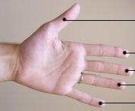
A digit is one of several most distal parts of a limb, such as fingers or toes, present in many vertebrates.

Names

[edit]

Some languages have different names for hand and foot digits (English: respectively "finger" and "toe", German: "Finger" and "Zeh", French: "doigt" and "orteil").

In other languages, e.g. Arabic, Russian, Polish, Spanish, Portuguese, Italian, Czech, Tagalog, Turkish, Bulgarian, and Persian, there are no specific one-word names for fingers and toes; these are called "digit of the hand" or "digit of the foot" instead. In Japanese, yubi (指) can mean either, depending on context.

Human digits

[edit]
Radiogram of a polydactyl left hand.
Mikhail Tal at the 1961 European chess championship.

Humans normally have five digits on each extremity. Each digit is formed by several bones called phalanges, surrounded by soft tissue. Human fingers normally have a nail at the distal phalanx. The phenomenon of polydactyly occurs when extra digits are present; fewer digits than normal are also possible, for instance in ectrodactyly. Whether such a mutation can be surgically corrected, and whether such correction is indicated, is case-dependent.[1] For instance the former chess world champion Mikhail Tal lived all his life with only three right-hand fingers.

Fingers Thumb Index Middle Ring Little
Toes Hallux Long Third Fourth Fifth

Brain representation

[edit]

Each finger has an orderly somatotopic representation on the cerebral cortex in the somatosensory cortex area 3b,[2] part of area 1[3] and a distributed, overlapping representation in the supplementary motor area and primary motor area.[4]

The somatosensory cortex representation of the hand is a dynamic reflection of the fingers on the external hand: in syndactyly people have a clubhand of webbed, shortened fingers. However, not only are the fingers of their hands fused, but the cortical maps of their individual fingers also form a club hand. The fingers can be surgically divided to make a more useful hand. Surgeons did this at the Institute of Reconstructive Plastic Surgery in New York to a 32-year-old man with the initials O. G.. They touched O. G.’s fingers before and after surgery while using MRI brain scans. Before the surgery, the fingers mapped onto his brain were fused close together; afterward, the maps of his individual fingers did indeed separate and take the layout corresponding to a normal hand.[5]

Evolution

[edit]
A reconstruction of Panderichthys.

Two ideas about the homology of arms, hands, and digits exist.

Until recently, few transitional forms were known to elaborate on this transition. One particular example is Panderichthys, a coastal fish from the Devonian period 385 million years ago. Prior to 2008, Panderichthys was interpreted as having a fin terminating at a single large plate surrounded by lepidotrichia (fin rays). However, a 2008 study by Boisvert et al. determined that this was mistaken. They discovered that the final bony portion of the fin in Panderichthys is split into at least four fin radials, bones similar to rudimentary fingers.[9]

Thus, in the evolution of tetrapods a shift occurred where the outermost rays of the fins were lost and replaced by the inner radials, which evolve into the earliest digits. This change is consistent with additional evidence from the embryology of actinopterygians, sharks and lungfish. Pre-existing distal radials in these modern fish develop in a very similar way to the digits of tetrapods.[9][10]

Several rows of digit-like distal fin radials are present in Tiktaalik, a much more complete Devonian vertebrate described in 2006. Though frequently described as the missing link between fishes and tetrapods, the exact relationship between Panderichthys, Tiktaalik, and tetrapods are yet to be fully resolved. Tiktaalik had some features of the forefin more similar to earlier fish, such as a large ulnare and a distinct axis of larger bones down the middle of the fin. According to Boisvert et al. (2008), "It is difficult to say whether this character distribution implies that Tiktaalik is autapomorphic, that Panderichthys and tetrapods are convergent, or that Panderichthys is closer to tetrapods than Tiktaalik. At any rate, it demonstrates that the fish–tetrapod transition was accompanied by significant character incongruence in functionally important structures."[9]p. 638.

Digit-like radials are also known in the rhizodont fish Sauripterus, though this is likely a case of convergent evolution. Elpistostege, a tetrapodomorph fish closely related to Tiktaalik, preserves one of the most tetrapod-like hands in any prehistoric fish. The hand of Elpisostege had 19 distal fin radials arranged into blocks up to four radials long. These sequential blocks of radials are very similar to digits.[11]

Bird and theropod dinosaur digits

[edit]

Birds and theropod dinosaurs (from which birds evolved) have three digits on their hands. The two digits that are missing are different: the bird hand (embedded in the wing) is thought to derive from the second, third and fourth digits of the ancestral five-digit hand. In contrast, the theropod dinosaurs seem to have the first, second and third digits. Recently a Jurassic theropod intermediate fossil Limusaurus has been found in the Junggar Basin in western China that has a complex mix: it has a first digit stub and full second, third and fourth digits but its wrist bones are like those that are associated with the second, third and fourth digits while its finger bones are those of the first, second and third digits.[12] This suggests the evolution of digits in birds resulted from a "shift in digit identity [that] characterized early stages of theropod evolution"[12]

See also

[edit]

Notes

[edit]
Revisions and contributorsEdit on WikipediaRead on Wikipedia
from Grokipedia
In , a digit is a finger-like projection at the distal end of a limb in vertebrates, serving as a terminal segment for grasping, manipulation, or locomotion. In humans, digits encompass the five s of the hand and the five s of the foot, collectively forming 10 digits per individual. The term "digit" applies generally to all such structures, whereas "finger" specifically denotes digits 2–5 of the hand and "toe" denotes digits 2–5 of the foot, with the thumb (pollex) and great toe (hallux) distinguished as digit 1. Human digits are structured around elongated bones called phalanges, articulated by synovial joints that enable flexion, extension, abduction, and adduction. and hallux each consist of two phalanges—a proximal phalanx and a distal phalanx—while digits 2–5 of both the hand and foot feature three phalanges: proximal, middle, and distal. These phalanges articulate proximally with the of the hand or of the foot at the metacarpophalangeal (MCP) or metatarsophalangeal (MTP) joints, respectively, and distally via interphalangeal joints. The total of 14 phalanges per hand and per foot accounts for 28 of the 56 phalangeal bones in the . Digits are richly innervated and vascularized to support sensory feedback and , with digital arteries and running along the lateral aspects to supply the skin, joints, and intrinsic muscles. at the distal tips provide protection and enhance tactile sensation, while the overall architecture allows for precise movements essential to daily activities. Variations such as (extra digits) or (fused digits) occur congenitally, highlighting the developmental plasticity of digit formation during embryogenesis.

Terminology

Names

In English anatomical terminology, the digits of the upper limb are commonly referred to as "fingers," while those of the lower limb are called "toes," with "finger" typically applying to digits 2 through 5 of the hand and "toe" to digits 2 through 5 of the foot; the general term "digit" encompasses all such structures on both limbs without distinction. The word "digit" derives from the Latin digitus, meaning "finger" or "toe," reflecting ancient associations with counting on the hands. Specific names exist for the first digits: the thumb is termed the "pollex" in Latin-derived medical nomenclature, possibly formed analogously to index from the verb pollere, "to be strong," emphasizing its robust function. Similarly, the big toe is the "hallux," from Late Latin (h)allux, a variant of hallus meaning "great toe," adapted by association with pollex. Linguistic variations appear across other languages, often unifying or specifying terms contextually. In Arabic, digits are generally "aṣābiʿ" (fingers), qualified as "aṣābiʿ al-yad" for hand digits and "aṣābiʿ al-qadam" for foot digits, literally "fingers of the hand/foot." Russian employs "palets" for both, distinguished as "palets ruki" (finger of the hand) or "palets nogi" (finger of the foot). Japanese uses "yubi" ambiguously for any digit, requiring clarification like "te no yubi" (hand finger) or "ashi no yubi" (foot finger) for precision. In French, "doigt" applies to hand digits, while foot digits are "doigts de pied" (fingers of the foot), maintaining a shared root without a fully distinct term for toes.

Numbering and Identification

In anatomical nomenclature, the digits of the human hand and foot are standardized using from I to V, beginning with the most lateral or preaxial digit and proceeding medially or postaxially. This system facilitates precise identification across medical disciplines, with digit I corresponding to (pollex) in the hand or the great toe (hallux) in the foot, digits II through IV representing the index, middle, and ring fingers/toes, and digit V denoting the (digitus minimus manus) or little toe (digitus minimus pedis). Manual digits (of the hand) are distinguished from pedal digits (of the foot) primarily by their positional orientation and associated terminology, though the Roman numeral scheme applies uniformly to both. The pollex serves as the preaxial digit I of the manus, enabling opposability, while the hallux functions analogously as digit I of the pes, contributing to bipedal ; the digiti minimi, as digits V, are the postaxial elements in both, often involved in fine motor tasks or balance. This distinction ensures clarity in descriptions of limb morphology, avoiding ambiguity between upper and lower extremity structures. In clinical and imaging contexts, such as and , digits are frequently identified via "ray" numbering, where each ray encompasses the corresponding metacarpal or metatarsal bone aligned with a digit, numbered I through V from the radial (thumb-side) or tibial (great toe-side) aspect. For instance, the first ray includes the first metacarpal and pollex, while the fifth ray comprises the fifth metacarpal and digitus minimus; this approach is essential for localizing fractures, deformities, or resections in radiographic evaluations like posteroanterior hand views. Ray numbering aligns with the Roman system but emphasizes the longitudinal axis of the limb segment for procedural precision.

Digits in Humans

Anatomy and Structure

Human digits, or fingers and toes, follow the typical pentadactyl arrangement, with five digits per limb in both the hand and foot. This structure consists of (or hallux in the foot) as the first digit and four lateral digits, enabling grasping in the hand and propulsion in the foot. The bony framework of the digits is primarily composed of phalanges and metacarpals in the hand or metatarsals in the foot. Each digit typically features three phalanges: the proximal phalanx closest to the palm or sole, the middle phalanx, and the distal phalanx at the tip. However, and hallux each have only two phalanges, lacking a middle one, which contributes to their enhanced mobility. The phalanges articulate with the metacarpals (in the hand) or metatarsals (in the foot) via joints, with the five metacarpals or metatarsals forming the palm or forefoot base, respectively. Foot digits generally have shorter phalanges compared to hand digits, supporting rather than fine manipulation. Joints within the digits include the metacarpophalangeal (MCP) or metatarsophalangeal (MTP) joints at the base, proximal interphalangeal (PIP) joints between proximal and middle phalanges, and distal interphalangeal (DIP) joints between middle and distal phalanges; the thumb and hallux feature an interphalangeal (IP) joint instead of PIP and DIP. These are synovial hinge joints stabilized by collateral ligaments on the sides and volar or plantar plates on the palmar/plantar surfaces, preventing excessive lateral deviation and hyperextension. Tendons from extrinsic muscles, such as the flexor digitorum profundus and superficialis for flexion, and extensor digitorum for extension in the hand, or flexor digitorum longus and extensor digitorum longus in the foot, cross these joints within synovial sheaths for smooth gliding. Intrinsic muscles like the lumbricals and interossei in the hand, or flexor digitorum brevis and interossei in the foot, provide fine control via short tendons inserting on phalangeal bases. The skin covering the digits is thick on the palmar and plantar surfaces, featuring friction ridges for grip, while dorsal skin is thinner. Nails, composed of keratinized plates produced by the nail matrix beneath proximal nail folds, overlie the distal phalanges, protecting the tips and aiding tactile sensation. Blood supply to the digits arises from digital arteries branching from the radial and ulnar arteries in the hand, or the dorsalis pedis and plantar arches in the foot, forming paired dorsal and palmar/plantar digital arteries that anastomose around each phalanx for redundancy. Innervation includes sensory supply from the median nerve to the thumb, index, middle, and radial ring fingers (palmar and distal dorsal), the ulnar nerve to the little and ulnar ring fingers, and the radial nerve to dorsal aspects of the thumb and index; in the foot, the medial plantar nerve supplies the hallux and medial digits, the lateral plantar nerve the lateral digits, and the deep fibular nerve the dorsal web spaces.

Variations and Anomalies

, a congenital condition involving supernumerary digits on the hands or feet, represents one of the most common limb anomalies. It is categorized into preaxial , which affects the radial or tibial side ( or great toe), and postaxial , which involves the ulnar or fibular side ( or ). Preaxial forms often result in or hallux duplication, while postaxial types typically feature an extra fifth digit that may be fully formed or rudimentary. The overall incidence of is approximately 1 in 500 to 1,000 live births, with variations by ethnicity—higher in African populations for postaxial hand and more prevalent in Caucasians for preaxial foot forms. Genetic factors play a significant role, particularly mutations in the GLI3 gene, which disrupt Sonic Hedgehog signaling and are linked to both isolated and syndromic , such as . Oligodactyly encompasses congenital reductions in digit number, leading to fewer than five digits per hand or foot, and is less common than . A prominent manifestation is , or split-hand/split-foot malformation (SHFM), characterized by absence of central digits (typically the second and third), resulting in a cleft appearance with fused or absent metacarpals and phalanges. This condition often affects both hands and feet asymmetrically and may involve nail dysplasia or reduced limb length. The incidence of SHFM is estimated at 1 in 90,000 live births, though broader rates are rarer and vary by subtype, such as ulnar ray deficiencies. exemplifies severe , sometimes presenting as a three-fingered hand configuration due to central ray aplasia. Syndactyly involves the partial or complete fusion of adjacent digits, often due to failure of tissue during embryogenesis, and can occur in isolation or with other anomalies like . It is classified as simple ( fusion only) or complex (bony union), and further as complete (extending to the digit tips) or incomplete (partial ). The third and fourth fingers are most commonly affected, with an incidence of about 1 in 2,000 to 3,000 live births, making it the second most frequent congenital hand malformation after . frequently co-occurs with in genetic syndromes, highlighting shared developmental pathways. Acquired digit variations arise postnatally from trauma, , or surgical intervention, contrasting with congenital forms. Amputations, often resulting from occupational injuries or vascular diseases, lead to partial or total digit loss and affect millions annually worldwide, with upper extremity cases comprising up to 90% of traumatic amputations in some populations. Reconstructive surgeries, such as pollicization, address acquired thumb loss by rotating and an adjacent finger (typically the index) to form a neothumb, restoring opposition and grip function with success rates exceeding 80% in functional outcomes. These procedures are also used for severe congenital but adapt well to traumatic defects, emphasizing the adaptability of hand .

Comparative Anatomy

In Non-Human Mammals

Non-human mammals display considerable variation in digit count and structure, often adapted to specific locomotor or ecological demands, contrasting with the consistent pentadactyl pattern in primates like humans. In perissodactyl ungulates, such as horses, only the third digit functions as the primary weight-bearing toe, with the second and fourth digits reduced to vestigial splint bones that provide lateral support but do not contact the ground. Artiodactyl ungulates, including cattle and deer, typically rely on two main digits—the third and fourth—for locomotion, while the first and fifth digits are absent or greatly diminished, and the second and fifth may persist as dewclaws. Primates outside humans generally maintain five digits per limb, with many species, such as Old World monkeys and apes, possessing an opposable first digit (thumb) that enables precise manipulation and grasping of objects or branches. Structural modifications further diversify digit function across mammalian orders. In carnivorans like felids, claws on digits II–V of both fore- and hindlimbs are retractile, sheathed within dermal pockets when idle to preserve sharpness for , prey capture, and traction; domestic cats, for example, have five digits on forelimbs and four on hindlimbs, with the first forelimb digit (dewclaw) non-retractile. Ungulates often feature hooves—keratinized sheaths encasing the distal phalanges of weight-bearing digits—for protection and efficient weight distribution on hard surfaces; in some like alpacas, the pedal digits show integrated development with partial soft-tissue fusion () between adjacent rays, enhancing stability. Elephants retain five digits per foot, each tipped with a broad, nail-like structure for gripping and digging, though phalangeal counts vary, with forefoot digits typically having three phalanges and hindfoot digits fewer. Specialized adaptations highlight functional specialization in other groups. Bats exhibit extreme elongation of manual digits II–V, which support the chiropatagium (wing membrane) essential for powered flight, while the first digit remains short and clawed for perching. In cetaceans, such as dolphins and whales, forelimb digits are severely reduced and embedded within a paddle-like flipper, with interdigital webbing and hyperphalangy (increased phalangeal number, often exceeding 14 per digit) providing hydrodynamic lift despite the loss of individual mobility. These variations underscore how digit morphology in non-human mammals optimizes for terrestrial cursoriality, arboreal dexterity, aerial locomotion, or aquatic propulsion.

In Birds and Theropods

In birds, the hindlimb digits are adapted for perching and locomotion, typically consisting of four toes: a backward-facing hallux (digit I) and three forward-facing toes (digits II, III, and IV). This anisodactyl configuration allows for secure gripping of branches, with the hallux providing opposition to the other digits. The phalangeal formula is usually 2-3-4-5 for digits I-IV, respectively, though variations exist across species; for example, in perching birds like passerines, the digits feature curved claws and flexible joints that enhance grasp stability. Multiarticular flexor muscles, such as the flexor digitorum longus and flexor hallucis longus, enable powerful closure around perches, with proximally inserted tendons optimizing passive locking during rest to prevent slippage without continuous muscle effort. In raptors, such as eagles and hawks, the digits are elongated and equipped with sharp talons for active grasping of prey, where distally inserted flexors contribute up to 64% of total flexor mass to facilitate forceful carrying. Theropod dinosaurs, the ancestral group to birds, exhibit progressive digit reduction in their forelimbs, evolving from the primitive five digits of early archosaurs to three functional digits (I, II, and III) with reduced or absent IV and V. This reduction occurred in stages: digits IV and V were lost near the base of , followed by further shortening of metacarpals III and IV in more derived lineages like . Fossil evidence from basal theropods, such as , shows retention of small digit V remnants, while advanced forms like display robust digits I-III specialized for grasping. In the hindlimbs, theropods maintained four functional toes (I-IV), similar to birds, with digit I often elevated off the ground for bipedal efficiency, though some early theropods like retained five toes. The homology of digits between birds and theropods remains controversial, particularly for forelimbs, where anatomical supports digits I, II, and III in theropods and birds, but embryological development in birds forms the wing digits from positions corresponding to II, III, and IV. This discrepancy, known as the frame-shift hypothesis, posits a homeotic transformation where digit identities shifted laterally during theropod to flight, reconciling fossil morphologies with developmental like HoxD patterns. The discovery of inextricabilis provides key , as it exhibits a reduced digit I alongside prominent digits II–IV, suggesting that tetanuran theropods, including avian ancestors, shared II-III-IV identities, with a gradual homeotic shift occurring in early theropod . Fossil records of digit loss, such as reduced IV and V in basal theropods like , further support this transitional reduction toward avian flight adaptations, where forelimb digits became integrated into structure while hindlimb digits specialized for perching.

In Other Tetrapods

In basal tetrapods, the earliest known forms exhibit , reflecting the evolutionary transition from fin-like structures to limbs with distinct digits. For instance, the fossil possessed eight digits on its forelimbs, while had eight digits on the forelimbs and seven on the hindlimbs, serving as precursors to the pentadactyl condition seen in later tetrapods. Among modern amphibians, digit configurations vary but generally follow a pattern of four digits on the forelimbs and five on the hindlimbs, as observed in most anurans (frogs) and salamanders. These digits often feature reduced phalangeal counts compared to amniotes, with anurans typically having two to three phalanges per digit to facilitate flexibility. between the digits is prevalent in many aquatic or semi-aquatic species, such as frogs, enhancing propulsion during swimming by increasing surface area for paddling. In non-avian reptiles, digit anatomy reflects diverse locomotor adaptations, with most retaining the ancestral five digits on both forelimbs and hindlimbs, often equipped with claws for traction. Crocodilians, by contrast, have five digits on the forelimbs (largely non-webbed) and four webbed digits on the hindlimbs, aiding in aquatic maneuvering and terrestrial stability. Snakes, as highly derived squamates, are typically limbless, having lost all digits through evolutionary reduction for burrowing or slithering. Specialized adaptations include the adhesive pads and subdigital setae in geckos, which, combined with retractable claws, enable climbing on vertical surfaces via van der Waals forces. In burrowing amphibians like , limbs and digits are severely reduced or absent, with vestigial structures in primitive genera supporting a lifestyle.

Neural Aspects

Brain Representation

In the primary somatosensory cortex (S1), located in the , the digits of the hand are represented in a somatotopic manner, with individual fingers mapped in an orderly sequence from (digit 1) to (digit 5), progressing from lateral to medial and inferior to superior along the posterior wall of the . This organization forms part of the "hand knob" region, a characteristic bulge in the cortex dedicated to . High-resolution functional MRI (fMRI) studies at 7T have confirmed this fine-grained mapping, resolving distinct representations for each digit within Brodmann areas 3b, 1, and 2 of S1. The cortical representation of the digits exhibits , where a disproportionately large area of S1 is devoted to the hands compared to other body parts, reflecting the high density of mechanoreceptors in the and the demands of fine tactile discrimination essential for manual dexterity. For instance, receives an enlarged mapping in areas 1 and 2, with magnification factors of approximately 2.3 and 2.4 relative to other digits, enabling enhanced sensory resolution for precision tasks. This scaling aligns with the somatosensory , where the hand occupies a significant portion of the cortical surface despite its small peripheral size. Evidence for this organization comes from neuroimaging techniques such as fMRI and , which have mapped digit-specific activations during tactile stimulation, demonstrating consistent contralateral representations across individuals. A notable case is that of patient O.G., the first successful bilateral pediatric hand transplant recipient, where pre-transplant amputation-induced reorganization in S1—such as invasion of the hand area by adjacent body part representations—was largely reversed post-transplant, restoring distinct digit maps as sensory input resumed. In non-human , such as macaques, a similar exists in S1 area 3b, with digits ordered from (lateral, anterior, inferior) to fifth digit (medial, posterior, superior), though the mapping is less fractionated than in humans due to differences in manual skill complexity.

Functional Implications

The digits of the human hand and foot play critical roles in , enabling high-resolution tactile discrimination and that inform neural feedback loops for environmental interaction. In the fingertips, the two-point discrimination threshold is notably finer, typically ranging from 2 to 3 mm, compared to 30-40 mm on the , allowing precise detection of spatial details such as texture or edges during . This acuity arises from dense innervation by mechanoreceptors, including Meissner corpuscles, which detect low-frequency vibrations (30-50 Hz) for flutter-like sensations, and Pacinian corpuscles, sensitive to higher frequencies (100-400 Hz) for rapid pressure changes, contributing to both tactile and subtle proprioceptive cues in digit positioning. These sensory inputs are processed via the to support adaptive behaviors, such as adjusting grip force based on object slipperiness. Motor control of digits integrates neural signals for both dexterous manipulation and locomotor stability. In the hand, the opposable facilitates fine motor tasks through coordinated neural commands from the , enabling precision grips that involve independent digit movements for activities like tool use or writing. This control relies on distributed cortical networks that modulate opposition to other fingers, enhancing overall hand dexterity. In the foot, toes contribute to locomotion by providing neural-driven arch support and propulsion; motor commands from the activate intrinsic foot muscles to maintain balance and absorb impact during , preventing excessive pronation or supination. Neural plasticity in digit-related circuits allows adaptation following injury or loss, reshaping sensory-motor representations to mitigate functional deficits. After digit amputation, cortical reorganization occurs in the somatosensory and motor cortices, where adjacent areas expand into the deafferented zone, often correlating with phantom limb sensations as perceptual echoes of the lost input. This plasticity can extend to prosthetic integration, where repeated use of myoelectric or robotic devices induces adaptive grasping patterns; for instance, training with multi-degree-of-freedom prosthetics promotes neural remapping in the motor cortex, improving independent finger control and reducing cognitive load for users. In clinical rehabilitation for digit injuries, such as tendon lacerations or nerve compressions, neural feedback mechanisms are harnessed to restore function through targeted therapies. Neurofeedback protocols, often using real-time EEG or fMRI to reinforce desired motor patterns, enhance recovery of fine digit movements by promoting synaptic strengthening in sensorimotor areas, as seen in post-stroke hand rehab where tactile cues amplify neural drive for grasping. These approaches leverage plasticity to reintegrate proprioceptive signals, reducing compensatory errors and improving long-term outcomes in activities of daily living.

Development and Evolution

Embryological Development

The development of digits begins with the formation of limb buds during early embryogenesis. In humans, upper limb buds emerge around the fifth week of from the , while lower limb buds appear shortly thereafter. These buds consist of a core of covered by , which proliferates to drive initial outgrowth. The apical ectodermal ridge (AER), a thickened ectodermal structure at the distal tip of the limb bud, plays a crucial role in directing proximal-distal outgrowth by secreting fibroblast growth factors (FGFs), such as FGF8 and , that maintain proliferation in the underlying progress zone—a region of undifferentiated mesenchymal cells. Digit patterning along the anterior-posterior axis is primarily regulated by Sonic hedgehog (SHH) signaling from the zone of polarizing activity (ZPA), a mesenchymal organizer in the posterior limb bud. SHH establishes digit identity in a concentration- and time-dependent manner, with higher posterior concentrations specifying digits like the pinky and lower anterior ones specifying the thumb; disruptions in SHH exposure duration can lead to sequential specification of digits from anterior to posterior. Along the proximal-distal axis, , particularly from the HoxA and HoxD clusters, confer positional identity to skeletal elements, with sequential expression domains ensuring proper segmentation from to phalanges; for instance, is critical for distal digit formation. Phalange differentiation involves the condensation of mesenchymal cells into cartilaginous precursors, followed by , but digit separation requires in interdigital regions. This interdigital necrosis, mediated by and lysosomal enzymes, sculpts free digits from an initial paddle-like structure; in humans, this process peaks between weeks 6 and 8 of , completing separation by around week 8. Developmental anomalies of digits often arise from disruptions in these signaling pathways. Removal or dysfunction of the AER, as seen in experimental models or genetic defects like those in DLX5/6, impairs outgrowth and can cause (split-hand/foot malformation) by failing to maintain the progenitor cell pool. Similarly, mutations in Gli3, a downstream effector of SHH signaling, lead to in mouse models by failing to repress anterior SHH targets, resulting in extra digits due to expanded digit-forming potential.

Evolutionary Origins

The evolutionary origins of digits trace back to the Late Devonian period, approximately 385 million years ago, when sarcopterygian fishes—lobe-finned vertebrates—began exhibiting fin structures with radials that foreshadowed the autopods of tetrapods. In these ancestral forms, such as Panderichthys, the pectoral fin contained four rows of radials embedded within the fleshy lobe, representing an intermediate stage between fin rays and true digits, rather than entirely novel structures. This configuration, dated to around 385 million years ago, suggests that digit precursors evolved as modifications of existing fin endoskeletons to support weight-bearing in shallow-water environments. Key fossil discoveries illuminate the phylogenetic transition from fins to limbs with digits. , from approximately 375 million years ago, possessed robust distal radials in its pectoral fin that functioned as digit-like elements, enabling the animal to prop itself up on substrates akin to early terrestrial propulsion. Similarly, Elpistostege watsoni, also Late Devonian in age, featured up to 19 radials organized into proximodistal rows, with distal elements branching in patterns homologous to early digits, bridging the gap between fish radials and tetrapod phalanges. The earliest undisputed tetrapods, such as Acanthostega gunnari from about 365 million years ago, displayed polydactylous limbs with eight digits on the forelimbs, indicating that digit diversification preceded refinement in the Devonian transition to land. Over subsequent evolutionary history, digit number reduced from more than eight in stem tetrapods to the pentadactyl condition characteristic of amniotes and crown-group tetrapods, a shift likely driven by optimizations for efficient and manipulation. This reduction stabilized around five digits by the period, enhancing stability and dexterity on land while minimizing energetic costs of excess phalanges. Homology debates persist, particularly regarding digits, where developmental and supports identities as digits II-III-IV rather than the ancestral I-II-III, reflecting frameshifts in patterning during avian evolution. Recent genetic studies have elucidated the molecular basis of this transition. A 2025 analysis revealed that the enhancers regulating Hoxd gene expression in digits were co-opted from an ancestral regulatory landscape originally associated with cloacal development in , enabling the of digit-specific patterning during the fin-to-limb transition. Adaptive pressures for digit emergence centered on facilitating weight support, paddling through vegetation, and eventual grasping in terrestrial habitats, marking the origin of digits around 375 million years ago as a pivotal innovation for invasion of land.

References

Add your contribution
Related Hubs
User Avatar
No comments yet.