Hubbry Logo
Retina horizontal cellRetina horizontal cellMain
Open search
Retina horizontal cell
Community hub
Retina horizontal cell
logo
8 pages, 0 posts
0 subscribers
Be the first to start a discussion here.
Retina horizontal cell
from Wikipedia
Horizontal cell
Plan of retinal neurons.
Details
SystemVisual system
LocationRetina
Identifiers
MeSHD051248
NeuroLex IDnifext_40
Anatomical terms of neuroanatomy

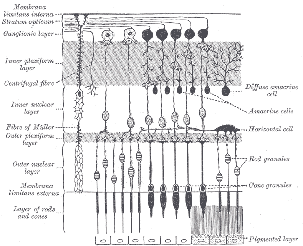
Horizontal cells are the laterally interconnecting neurons having cell bodies in the inner nuclear layer of the retina of vertebrate eyes. They help integrate and regulate the input from multiple photoreceptor cells. Among their functions, horizontal cells are believed to be responsible for increasing contrast via lateral inhibition and adapting both to bright and dim light conditions. Horizontal cells provide inhibitory feedback to rod and cone photoreceptors.[1][2] They are thought to be important for the antagonistic center-surround property of the receptive fields of many types of retinal ganglion cells.[3]

Other retinal neurons include photoreceptor cells, bipolar cells, amacrine cells, and retinal ganglion cells.

Structure

[edit]

Depending on the species, there are typically one or two classes of horizontal cells, with a third type sometimes proposed.[1][2]

Horizontal cells span across photoreceptors and summate inputs before synapsing onto photoreceptor cells.[1][2] Horizontal cells may also synapse onto bipolar cells, but this remains uncertain.[1][4]

There is a greater density of horizontal cells towards the central region of the retina. In the cat, it is observed that A-type horizontal cells have a density of 225 cells/mm2 near the center of the retina and a density of 120 cells/mm2 in more peripheral retina.[5]

Horizontal cells and other retinal interneuron cells are less likely to be near neighbours of the same subtype than would occur by chance, resulting in 'exclusion zones' that separate them. Mosaic arrangements provide a mechanism to distribute each cell type evenly across the retina, ensuring that all parts of the visual field have access to a full set of processing elements.[5] MEGF10 and MEGF11 transmembrane proteins have critical roles in the formation of the mosaics by horizontal cells and starburst amacrine cells in mice.[6]

Function

[edit]

Horizontal cells are depolarized by the release of glutamate from photoreceptors, which happens in the absence of light. Depolarization of a horizontal cell causes it to hyperpolarize nearby photoreceptors. Conversely, in the light, a photoreceptor releases less glutamate, which hyperpolarizes the horizontal cell, leading to depolarization of nearby photoreceptors. Thus, horizontal cells provide negative feedback to photoreceptors. The moderately wide lateral spread and coupling of horizontal cells by gap junctions, measures the average level of illumination falling upon a region of the retinal surface, which horizontal cells then subtract a proportionate value from the output of photoreceptors to hold the signal input to the inner retinal circuitry within its operating range.[1] Horizontal cells are also one of two groups of inhibitory interneurons that contribute to the surround of retinal ganglion cells:[2]

Illumination Center photoreceptor hyperpolarization Horizontal cell hyperpolarization Surround photoreceptor depolarization

The exact mechanism by which depolarization of horizontal cells hyperpolarizes photoreceptors is uncertain. Although horizontal cells contain GABA, the main mechanisms by which horizontal cells inhibit cones probably do not involve the release of GABA by horizontal cells onto cones.[4][7][8] Two mechanisms that are not mutually exclusive likely contribute to horizontal cell inhibition of glutamate release by cones. Both postulated mechanisms depend on the protected environment provided by the invaginating synapses that horizontal cells make onto cones.[4][9] The first postulated mechanism is a very fast ephaptic mechanism that has no synaptic delay, making it one of the fastest inhibitory synapses known.[4][10][11] The second postulated mechanism is relatively slow with a time constant of about 200 ms and depends on ATP release via Pannexin 1 channels located on horizontal cell dendrites invaginating the cone synaptic terminal. The ecto-ATPase NTPDase1 hydrolyses extracellular ATP to AMP, phosphate groups, and protons. The phosphate groups and protons form a pH buffer with a pKa of 7.2, which keeps the pH in the synaptic cleft relatively acidic. This inhibits the cone Ca2+ channels and consequently reduces the glutamate release by the cones.[4][11][12][13][14]

The center-surround antagonism of bipolar cells is thought to be inherited from cones. However, when recordings are made from parts of the cone that are distant from the cone terminals that synapse onto bipolar cells, center-surround antagonism seems to be less reliable in cones than in bipolar cells. As the invaginating synapses from horizontal cells are made onto cone terminals, the center-surround antagonism of cones is thought to be more reliably present in cone terminals.[15]

See also

[edit]

References

[edit]

Bibliography

[edit]
[edit]
Revisions and contributorsEdit on WikipediaRead on Wikipedia
from Grokipedia
Retinal horizontal cells are a class of inhibitory interneurons located in the outermost row of the inner nuclear layer of the vertebrate retina, where they receive direct glutamatergic input from rod and cone photoreceptors and form reciprocal synaptic connections with both photoreceptors and the dendrites of bipolar cells in the outer plexiform layer. These cells exhibit diverse morphologies across species, typically featuring extensive lateral dendritic processes that enable broad coverage of the retinal surface, with axon-bearing types connecting primarily to rods and axon-less types associating with cones; in mammals such as mice, a single axon-bearing type predominates, while primates and birds like chickens possess two to three distinct subtypes (e.g., H1 brush-shaped for cones, H2 stellate for rods and cones, and H3 candelabrum-shaped for cones). Horizontal cells are electrically coupled to one another via gap junctions, allowing synchronized activity over large areas, and they release the neurotransmitter GABA to provide both feedback inhibition to photoreceptors and feedforward inhibition to bipolar cells. The primary function of horizontal cells is to mediate , which sharpens visual contrast by implementing center-surround antagonism in the receptive fields of bipolar and ganglion cells, thereby enhancing and in the visual scene. This process involves measuring the average illumination across a surround and subtracting it from the center signal, a mechanism that also contributes to local gain control and adaptation to varying light levels. In species with , such as , specific horizontal cell subtypes facilitate color opponency by differentially connecting to cone types (e.g., L/M- or S-cones), modulating wavelength-specific signals before they reach bipolar cells. Additionally, horizontal cells participate in pathways that influence bipolar cell responses, further refining the transformation of photoreceptor signals into parallel ON and OFF pathways for . Horizontal cells originate from multipotent retinal progenitor cells during early retinogenesis, specified by transcription factors such as FoxN4 and Ptf1a, and undergo a unique bi-directional migration across the developing neuroepithelium before settling in the inner nuclear layer; molecular markers like Prox1, , Lhx1 (for axon-bearing types), and Isl1 (for axon-less types) distinguish their subtypes. Evolutionarily conserved across vertebrates, their diversity reflects adaptations to visual , with axon-less types often reduced or absent in nocturnal, rod-dominated retinas like those of . Notable properties include morphological plasticity, such as transient neurite extension during formation, and the rare ability of differentiated horizontal cells to re-enter the , which has been linked to the pathogenesis of in experimental models. Disruptions in horizontal cell function, as seen in certain retinal degenerations or genetic mutations, can impair contrast sensitivity and color perception, underscoring their essential role in .

Anatomy

Location in the Retina

Horizontal cells are laterally interconnecting neurons situated in the inner nuclear layer (INL) of the vertebrate , where they contribute to lateral processing of visual signals from photoreceptors. Their cell bodies are positioned primarily in the middle to outer portions of the INL, adjacent to the outer plexiform layer (OPL). From these somata, horizontal cells extend broad dendritic processes into the OPL, enabling interactions with rod and terminals. The spatial distribution of horizontal cells varies by and retinal eccentricity, reflecting adaptations to visual . In the cat retina, for instance, A-type horizontal cells exhibit a density gradient, reaching approximately 225 cells/mm² in central regions and declining to about 120 cells/mm² peripherally, which is compensated by expanded dendritic fields to maintain consistent coverage. By contrast, in the mouse retina, horizontal cells form a relatively uniform distribution across the retinal expanse, with densities averaging around 900 cells/mm². This even spacing is achieved through a tiled arrangement, where cells tile the retina without significant gaps or overlaps. The mosaic patterning of horizontal cells relies on mechanisms that enforce non-overlapping territories, including the formation of exclusion zones around each cell body. These zones prevent homotypic neighbors from encroaching, ensuring efficient sampling of photoreceptor inputs. In mice, this organization is critically dependent on the transmembrane proteins MEGF10 and MEGF11, which mediate repulsive homotypic interactions during postnatal development; double of these genes disrupts the mosaic, leading to clustered distributions and reduced uniformity.

Morphological Features

Retinal horizontal cells exhibit a distinctive wide-field morphology characterized by broad, bushy dendritic fields that typically span 100–300 micrometers in mammals, enabling extensive coverage across arrays of photoreceptors. These dendrites emerge directly from the soma and branch extensively in a planar fashion within the outer plexiform layer, forming dense arborizations that overlap with those of neighboring cells to facilitate lateral integration. The cell bodies are relatively large, oval or polygonal, with diameters of 10–20 micrometers, containing pale nuclei and abundant rich in mitochondria. In certain horizontal cells, particularly axon-bearing types found across vertebrates including mammals, long and thin processes extend from the soma, often reaching lengths of 200–300 micrometers or more, and terminate in separate synaptic fields distinct from the dendritic arbor. These axons maintain a narrow and do not branch extensively, contrasting with the bushy dendritic structure. Horizontal cell processes form specialized invaginating synapses at the base of photoreceptor terminals, inserting into the surrounding the ribbon synapse to establish close associations with photoreceptor and bipolar cell processes in synaptic triads. Ultrastructurally, these contacts feature lateral elements positioned adjacent to the synaptic ribbon, with horizontal cell dendrites penetrating the photoreceptor pedicle or spherule without forming conventional active zones. Horizontal cells are interconnected via extensive gap junctions, which appear as large plaques under electron microscopy and allow for the formation of electrical syncytia among coupled cells; notably, direct chemical synapses between horizontal cells themselves are absent. These gap junctions, mediated by connexins such as Cx57 in mammalian B-type cells, exhibit a narrow 2–4 nm extracellular gap and selectively couple cells of the same morphological class. The overall supports a wide-field design optimized for lateral spread, with cell densities varying by species and retinal region but typically ranging from 200–300 cells per mm² in mammals such as cats, and up to 1,000–2,000 cells per mm² in mice. This density, combined with dendritic overlap, results in comprehensive tiling of the photoreceptor layer.

Subtypes and Diversity

Horizontal cells in the vertebrate retina display notable diversity in subtypes, which varies by species and correlates with visual , such as cone abundance and demands. In mammals, there are typically two main subtypes: A-type horizontal cells, which are axonless and primarily connect to cones in a stratified manner within the outer , and B-type horizontal cells, which are axon-bearing and contact both and cones. A third subtype, C-type, has been proposed in some species like rabbits but remains debated due to inconsistent morphological and connectivity evidence. In , including macaques and humans, the subtypes are designated H1 and H2; H1 cells receive input predominantly from long- and medium-wavelength-sensitive () cones, while H2 cells contact short-wavelength-sensitive (S) cones, reflecting adaptations for trichromatic vision. Non-mammalian vertebrates, particularly fish, exhibit greater subtype diversity, with up to six morphological types identified in species like and , classified as monophasic (L-type, responding to all s), biphasic (C1-type, opponent to /), and triphasic (C2-type, opponent involving ). These classifications arise from differential connectivity, enabling color opponency; for instance, monophasic H1 cells contact all types via broad dendritic fields, while biphasic and triphasic cells show selective innervation for . In contrast to the simpler mammalian repertoire, fish horizontal cells often include dedicated rod-connected subtypes alongside -specific ones, supporting tetrachromatic vision. Morphological features underpin functional specialization across subtypes; in mammals, axon-bearing B-type (or H1) cells feature distinct dendritic arbors for cone synapses and axonal terminals for rod contacts, allowing segregated signaling pathways for scotopic and photopic conditions. This separation is less pronounced in axonless A-type cells, which focus on cone-driven surround inhibition. In fish, similar arbor distinctions in biphasic and triphasic cells facilitate chromatic antagonism. Molecular markers further highlight subtype heterogeneity. Connexin 57 (Cx57) is expressed in horizontal cell gap junctions across mammals, enabling electrical coupling essential for lateral spread of signals, with subtype-specific localization in axon terminals of B-type cells. The PROX1 serves as a pan-horizontal cell marker for identity and differentiation in mice and chickens, regulating progenitor exit from the to generate these . Neurotransmitter expression shows variation, with horizontal cells primarily utilizing GABA for inhibitory feedback, though some subsets co-express glycine transporters, suggesting potential glycinergic modulation in specific contexts. Recent genetic studies in mice reveal that horizontal cell numbers vary significantly across strains, independent of overall retinal size, through modulation of regulators like PROX1, which controls proliferation and genesis without altering progenitor pools. This heritable diversity, spanning over twofold differences, underscores evolutionary flexibility in populations.

Physiology

Response to Photoreceptor Input

In the dark, photoreceptors continuously release glutamate, which binds to ionotropic glutamate receptors, primarily and kainate subtypes, on the dendrites of horizontal cells, leading to an influx of cations and of these cells. This depolarized state in darkness reflects the tonic input from multiple connected photoreceptors, with horizontal cells exhibiting a resting around -30 to -40 mV. Upon light exposure, photoreceptors hyperpolarize and reduce glutamate release, causing the ionotropic receptors on horizontal cells to close and resulting in hyperpolarization of these cells. This generates graded changes known as S-potentials, which are sustained and propagate laterally through gap junctions connecting horizontal cells, allowing for spatial integration of signals across the retinal layer. Horizontal cell S-potentials are characteristically slow and sustained, lasting as long as the light stimulus, in contrast to the faster, transient responses of bipolar cells. These potentials typically reach amplitudes of 20-40 mV, with a reversal potential near -20 mV, driven by changes in cation conductance. Each horizontal cell sums inputs from numerous photoreceptors—up to 100 cones in some species—via extensive dendritic contacts, enabling averaging of local light levels and contributing to receptive field expansion. Response profiles vary across species: mammalian horizontal cells often show monophasic hyperpolarizing responses similar to rod-driven signals, while non-mammalian species exhibit biphasic or triphasic patterns that reflect color-specific opponency, such as hyperpolarization to one and depolarization to another.

Inhibitory Mechanisms

Horizontal cells in the exert inhibitory feedback to photoreceptors primarily through mechanisms that modulate synaptic transmission at the outer plexiform layer. Upon hyperpolarization in response to , horizontal cells reduce calcium influx into photoreceptor terminals, thereby decreasing glutamate release and enhancing contrast sensitivity. This process involves reciprocal synapses where horizontal cell in the dark promotes photoreceptor calcium entry, while hyperpolarization reverses this effect to suppress it. A key component of this inhibition is fast ephaptic feedback, which operates without chemical synaptic transmission and relies on electrical field effects at invaginating synapses. Horizontal cells modulate the extracellular potential in the synaptic cleft through hemichannels, such as connexin 57, altering photoreceptor membrane voltage with minimal delay—on the order of milliseconds. This mechanism contributes to rapid surround antagonism in receptive fields, independent of release. In contrast, slow feedback inhibition involves proton (H⁺) buffering mediated by ATP release from horizontal cells, which acidifies the and shifts the activation curve of photoreceptor calcium channels. Horizontal cells release ATP via hemichannels during , leading to that generates protons and a pH drop in the synaptic cleft; this effect exhibits a delay of approximately 200 ms and persists for seconds. Such pH-mediated modulation fine-tunes photoreceptor output over longer timescales compared to ephaptic effects. Although horizontal cells express GABA and possess vesicular release machinery, GABAergic inhibition remains controversial and appears minor relative to ephaptic and ATP/proton pathways. In mammalian retinas, GABA may act unconventionally through autaptic mechanisms on horizontal cells themselves, potentially influencing regulation via sodium-hydrogen exchangers rather than direct photoreceptor inhibition. Evidence for significant direct GABA release to photoreceptors is limited, with studies suggesting it does not substantially contribute to feedback strength. Recent research has identified distinct synaptic sites on horizontal cells for global (wide-field) and local (fine-scale) inhibition in the mammalian , enabling parallel processing of surround signals. Global feedforward occurs at surface synapses with bipolar cells, while local feedback to photoreceptors happens at invaginating contacts, allowing horizontal cells to mediate both broad antagonism and precise spatial tuning without interference.

Role in Visual Processing

Horizontal cells play a crucial role in shaping through , which enhances spatial contrast and in retinal ganglion cells via center-surround antagonism. This process involves horizontal cells receiving input from photoreceptors and providing inhibitory feedback, thereby suppressing activity in surrounding regions relative to the center of a stimulus and improving the detection of boundaries and fine details in images. In the absence of horizontal cells, as observed in depletion models, ganglion cell receptive fields expand and surround inhibition weakens, leading to reduced contrast sensitivity and altered tuning. Horizontal cells also contribute to global light adaptation by averaging light levels across wide retinal areas, which adjusts photoreceptor sensitivity to the mean and prevents response saturation over varying background intensities. This mechanism modulates synaptic gain at the photoreceptor-to-bipolar cell through feedback pathways, such as ephaptic and pH-mediated signals, ensuring stable visual output across ambient light changes. For instance, in models lacking horizontal cells, the shift in half-maximum response intensity to background light fails to occur, impairing overall retinal . In species with multiple horizontal cell subtypes, such as biphasic and triphasic cells in fish and turtle retinas, these interneurons facilitate color opponency by providing selective feedback to cones, contributing to red-green and blue-yellow processing pathways. Biphasic cells, which reverse polarity around 600 nm, oppose red and green cone signals to generate red-green antagonism, while triphasic cells, with reversals near 500-530 nm and 650-670 nm, integrate blue, green, and red inputs for broader spectral discrimination. This feedback propagates opponency to bipolar and ganglion cells, enhancing chromatic discrimination in visual perception. Additionally, the bushy dendrites of horizontal cells synchronize photoreceptor activity in the outer plexiform layer, coordinating burst firing in bipolar cells to facilitate efficient signal transmission. By hyperpolarizing connected photoreceptors, these cells align their outputs to produce coherent responses, such as the negative a-wave in electroretinograms, which would otherwise remain desynchronized and ineffective. Dysfunction of horizontal cells is linked to certain retinal pathologies, including rod dystrophies that manifest as incomplete night blindness and defects from drug exposures like , though such impairments are rare as a primary cause and often secondary to broader outer retinal disruptions. In these conditions, abnormal rod-cone interactions elevate electroretinogram ratios, indicating reduced horizontal cell-mediated modulation.

Development and Genetics

Embryonic Origin

Horizontal cells in the originate from multipotent retinal progenitor cells (RPCs) located in the pseudostratified neuroepithelium of the optic cup. In mice, these progenitors generate horizontal cells primarily between embryonic days 11 and 15 (E11–E15), with peak production around E13–E14, marking them as early-born neurons in the retinal . This timeline is conserved across s, including chicks and , where horizontal cell genesis similarly occurs during mid-embryogenesis as part of the initial wave of production following retinal cells and photoreceptors. Among inner nuclear layer (INL) neurons, horizontal cells differentiate early and migrate to their mature position immediately after their final mitotic division. Newborn horizontal cells exhibit bi-directional migration, traversing the thickness of the neuroepithelium from their peripheral birth sites toward the inner , temporarily approaching the nascent cell layer before reversing direction to settle in the outermost row of the INL. This migratory behavior, completed by postnatal days 5–7 (P5–P7) in , precedes the generation of most bipolar cells and aligns with the overall temporal sequence where horizontal cells emerge after photoreceptors but before rod photoreceptors, which are predominantly postnatal. By embryonic day 18 (E18) in mice, horizontal cell somata are positioned within the outer neuroblastic layer, and their dendritic processes begin extending toward the nascent outer plexiform layer (OPL) to establish initial contacts with photoreceptor terminals. Full morphological maturation, including the elaboration of extensive horizontal processes and the formation of synaptic triads in the OPL, is achieved by postnatal day 10 (P10) in rodents. During this postnatal phase, gap junctions—primarily composed of connexin 57 (Cx57)—assemble between horizontal cell processes, forming electrically coupled syncytia that facilitate lateral integration across the network. Subtype heterogeneity among horizontal cells begins to emerge in late embryogenesis, as post-mitotic cells respond to local microenvironmental cues that guide the diversification into axon-bearing and axonless morphologies. This early divergence sets the stage for the distinct functional specializations observed in maturity, contributing to the precise tiled mosaic arrangement in the outer INL.

Genetic Regulation and Molecular Markers

The PROX1 plays a central role in specifying horizontal cell fate during retinal development in mammals. Expressed in postmitotic precursors, PROX1 promotes the differentiation of retinal cells into horizontal cells by regulating exit and inhibiting alternative fates such as photoreceptor or bipolar cell lineages. In Prox1-null mice, horizontal cells are completely absent, demonstrating its essential function, while forced expression of Prox1 is sufficient to induce horizontal cell genesis from progenitors. Genetic factors also modulate horizontal cell density across mouse strains, with numbers varying nearly twofold (from approximately 9,900 to 18,500 cells per ) independent of overall retinal size, indicating regulation at the level of competence rather than expansion. This variation exhibits high (h² = 0.89) and is influenced by quantitative trait loci, such as one on linked to the , where a single nucleotide polymorphism in the 5' untranslated region alters expression and thereby horizontal cell production. Although Prox1 itself is critical for initial specification, enhancers regulating its dosage may contribute to fine-tuning density, though specific loci remain under investigation. Molecular markers distinguish horizontal cell identity and subtypes, including LIM homeodomain proteins like LHX1 (also known as Lim1), which is specifically expressed in horizontal cells from early differentiation onward and is required for their proper laminar positioning within the inner nuclear layer. LHX1 helps establish subtype-specific features, such as dendritic arborization patterns that underlie connections to or rod photoreceptors. coupling, essential for , is mediated by connexin 57 (Cx57, encoded by Gjc1), which forms homotypic channels exclusively in horizontal cell networks; Cx57 knockout disrupts coupling and sizes without altering cell number. Additionally, serves as a calcium-binding marker in horizontal cells across species, including mice, labeling populations involved in photoreceptor feedback and aiding identification in immunohistochemical studies. Recent genetic studies since 2011 have revealed additional regulators of horizontal cell organization. Transmembrane proteins MEGF10 and MEGF11 mediate homotypic repulsion between horizontal cells, ensuring non-overlapping mosaic spacing critical for uniform coverage of the ; double in mice leads to clustered distributions and reduced tiling efficiency, modeling aspects of retinal degeneration where disrupted spacing impairs visual processing.

References

Add your contribution
Related Hubs
User Avatar
No comments yet.