Hubbry Logo
PannexinPannexinMain
Open search
Pannexin
Community hub
Pannexin
logo
8 pages, 0 posts
0 subscribers
Be the first to start a discussion here.
Pannexin
Pannexin
from Wikipedia

Pannexin
Identifiers
SymbolPannexin
InterProIPR039099
TCDB1.A.25
Available protein structures:
PDB  IPR039099  
AlphaFold
pannexin 1
Identifiers
SymbolPANX1
NCBI gene24145
HGNC8599
OMIM608420
RefSeqNM_015368
UniProtQ96RD7
Other data
LocusChr. 11 q14-q21
Search for
StructuresSwiss-model
DomainsInterPro
pannexin 2
Identifiers
SymbolPANX2
NCBI gene56666
HGNC8600
OMIM608421
RefSeqNM_052839
UniProtQ96RD6
Other data
LocusChr. 22 q13
Search for
StructuresSwiss-model
DomainsInterPro
pannexin 3
Identifiers
SymbolPANX3
NCBI gene116337
HGNC20573
OMIM608422
RefSeqNM_052959
UniProtQ96QZ0
Other data
LocusChr. 11 q24.2
Search for
StructuresSwiss-model
DomainsInterPro

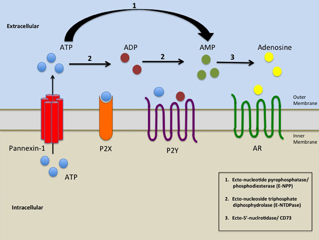
Pannexins (from Greek 'παν' — all, and from Latin 'nexus' — connection) are a family of vertebrate proteins identified by their homology to the invertebrate innexins.[1] While innexins are responsible for forming gap junctions in invertebrates, the pannexins have been shown to predominantly exist as large transmembrane channels connecting the intracellular and extracellular space, allowing the passage of ions and small molecules between these compartments (such as ATP and sulforhodamine B).

Three pannexins have been described in Chordates: Panx1, Panx2 and Panx3.[2]

Function

[edit]

Pannexins can form nonjunctional transmembrane channels for transport of molecules of less than 1000 Da. These hemichannels can be present in plasma, endoplasmic reticulum (ER) and Golgi membranes. They transport Ca2+, ATP, inositol triphosphate and other small molecules and can form hemichannels with greater ease than connexin subunits.[3] Pannexin 1 and pannexin 2 underlie channel function in neurons and contribute to ischemic brain damage.[4]

Pannexin 1 has been shown to be involved in early stages of innate immunity through an interaction with the P2X7 purinergic receptor. Activation of the pannexin channel through binding of ATP to P2X7 receptor leads to the release of interleukin-1β.[5]

Hypothetical roles of pannexins in the nervous system include participating in sensory processing, synchronization between hippocampus and cortex, hippocampal plasticity, and propagation of calcium waves. Calcium waves are supported by glial cells, which help maintain and modulate neuronal metabolism. According to one of the hypotheses, pannexins also may participate in pathological reactions, including the neural damage after ischemia and subsequent cell death.[6]

Pannexin 1 channels are pathways for release of ATP from cells.[7]

Relationship to connexins

[edit]

Intercellular gap junctions in vertebrates, including humans, are formed by the connexin family of proteins.[8] Structurally, pannexins and connexins are very similar, consisting of 4 transmembrane domains, 2 extracellular and 1 intracellular loop, along with intracellular N- and C-terminal tails. Despite this shared topology, the protein families do not share enough sequence similarity to confidently infer common ancestry.

The N-terminal portion (Pfam PF12534) of VRAC-forming LRRC8 proteins like LRRC8A may also be related to pannexins.[9]

The structure of a Xenopus tropicalis (western clawed frog) pannexin (PDB: 6VD7​) has been solved. It forms a heptameric disc. The human version (PDB: 6M02​) is similar.[10][11]

Clinical significance

[edit]

Truncating mutations in pannexin 1 have been shown to promote breast and colon cancer metastasis to the lungs by allowing cancer cells to survive mechanical stretch in the microcirculation through the release of ATP.[12]

Pannexins may be involved in the process of tumor development. Particularly, PANX2 expression levels predict post diagnosis survival for patients with glial tumors.

Probenecid, a well-established drug for the treatment of gout, allows for discrimination between channels formed by connexins and pannexins. Probenecid does not affect channels formed by connexins, but it inhibits pannexin-1 channels.[13]

References

[edit]

Further reading

[edit]
Revisions and contributorsEdit on WikipediaRead on Wikipedia
from Grokipedia
Pannexins are a family of three vertebrate-specific channel proteins—Pannexin-1 (Panx1), Pannexin-2 (Panx2), and Pannexin-3 (Panx3)—that oligomerize into heptameric large-pore channels in the plasma , enabling the efflux of small molecules such as ATP, nucleotides, , and ions up to approximately 1.5 kDa to mediate autocrine and . These channels, distinct from connexins that form intercellular gap junctions, primarily function as single- conduits for release under physiological and pathological conditions. Encoded by genes on chromosomes 11q21 (Panx1), 11q24.2 (Panx3), and 22q13.33 (Panx2), pannexins exhibit tissue-specific expression: Panx1 is ubiquitous, Panx2 predominates in the , and Panx3 is enriched in , , and osteoblasts. Structurally, each pannexin subunit features a tetraspan with four transmembrane α-, two extracellular loops (ECL1 and ECL2), an intracellular , and a , with sites on ECL2 influencing channel trafficking and activity. Cryo-electron microscopy (cryo-EM) studies have resolved the heptameric architecture of Panx1 and Panx3 at near-atomic resolution (2.9–3.8 ), revealing a central hourglass-shaped pore with unique ion selectivity motifs, such as a residue in the pore-lining that confers permeability to ATP and . Channel activation is tightly regulated by post-translational modifications, including caspase-3/7-mediated cleavage of the Panx1 (at residues 376–379), , elevated intracellular calcium, mechanical stress, and interactions with proteins like P2X7 receptors. Inhibitors such as and probenecid, along with the mimetic 10Panx, block these channels, highlighting their pharmacological targetability. Pannexins play pivotal roles in intercellular communication, contributing to processes like synaptic plasticity, immune cell activation, insulin secretion in pancreatic β-cells, and tissue repair. For instance, Panx1 facilitates ATP release to propagate calcium waves in astrocytes and T-lymphocytes, while Panx3 supports ATP-dependent signaling in skeletal muscle contraction and bone development. Dysregulated pannexin activity is implicated in numerous diseases, including inflammation (e.g., ischemia-reperfusion injury and airway hyperreactivity), tumorigenesis (e.g., promoting proliferation in pancreatic and hepatocellular carcinomas), and neurodegeneration (e.g., stroke and seizures), where excessive ATP release amplifies purinergic signaling cascades. Recent advances in structural biology and targeted inhibition underscore their therapeutic potential in modulating inflammatory and oncogenic pathways.

Discovery and Classification

Historical Background

The pannexin family of proteins was first identified in through a bioinformatics approach that searched genomes for homologs of innexins, the proteins responsible for forming s in non-chordates. Yuri Panchin and colleagues discovered three genes in the —initially termed pannexin1 (Panx1), pannexin2 (Panx2), and pannexin3 (Panx3)—which encoded proteins sharing sequence similarity with innexins, particularly in predicted transmembrane domains. This finding suggested pannexins as a potential second family of gap junction proteins in vertebrates, distinct from the well-known connexins. Subsequent efforts focused on and sequencing these genes across vertebrates to confirm their conservation and expression. In 2003, Roberto Bruzzone and team Panx1 and Panx2 from rat cDNA, revealing their predominant expression in neural tissues and structural features indicative of channel-forming capabilities. Shortly after, in 2004, Anna Baranova et al. the human PANX1, PANX2, and PANX3 genes, demonstrating their homology to innexins and presence in various tissues, including , , and intestine, thus establishing pannexins as a conserved family in mammals. These studies provided the full-length sequences necessary for functional investigations. Early functional characterization between 2002 and 2005 utilized systems to test pannexins' channel properties. When expressed in oocytes, Panx1 induced large membrane currents sensitive to blockers like , indicating its ability to form functional hemichannels. Heteromeric combinations of Panx1 and Panx2 in oocytes produced channels with distinct pharmacological profiles, further supporting their role in intercellular communication, though Panx2 alone showed limited activity. These studies positioned pannexins as viable candidates for forming non-selective pores permeable to ions and small molecules. By 2010, research had shifted the understanding of pannexins from hypothetical formers to primarily non-junctional membrane channels. Electrophysiological and dye-uptake experiments demonstrated that Panx1, in particular, functions as an ATP-release pathway in single cells, independent of cell-cell contact, as evidenced by its involvement in the P2X7 receptor pore complex. This paradigm change was solidified through reviews and studies highlighting pannexins' roles in single-membrane channels rather than intercellular junctions, distinguishing them from connexins.

Nomenclature and Isoforms

The pannexin (PANX) family comprises three isoforms in humans—PANX1, PANX2, and PANX3—naming conventions that reflect their sequential discovery and structural homology to innexins, with the "Panx" prefix denoting their broad phylogenetic distribution across chordates. These s are mapped to distinct chromosomal loci: PANX1 at 11q21, PANX2 at 22q13.33, and PANX3 at 11q24.2. The corresponding proteins exhibit varying molecular weights, with Panx1 at approximately 48 (426 ), Panx2 at approximately 68 (676 ), and Panx3 at approximately 38 (392 ). All isoforms share a conserved topology featuring four transmembrane domains, two extracellular loops, and intracellular N- and C-termini, which underpin their channel-forming potential. Sequence homology among the isoforms is moderate, with Panx1 and Panx2 sharing about 50% identity in their transmembrane regions, while Panx3 displays roughly 30% overall identity with the other two, and lower similarity (around 27%) with Panx2 specifically. This divergence is particularly evident in the intracellular tails, where Panx2 has a notably longer , contributing to isoform-specific functional nuances. Panx1 remains the most extensively studied and ubiquitously expressed isoform, often serving as the prototypical model, whereas Panx2 is predominantly associated with neuronal contexts and Panx3 with specialized tissues such as and . At the genetic level, each PANX gene exhibits a distinct exon-intron organization. PANX1 spans 7 s and 6 introns, PANX2 comprises 4 s and 3 introns, and PANX3 consists of 4 s and 3 introns. variants have been identified primarily for PANX1, including two major isoforms (Panx1a and Panx1b) arising from differential processing of exon 5, which may influence channel trafficking or activity; fewer variants are reported for PANX2 and PANX3, though tissue-specific splicing events suggest potential regulatory diversity across isoforms.

Molecular Structure

Protein Architecture

Pannexin proteins share a conserved core topology consisting of four α-helical transmembrane domains (TM1–TM4), two extracellular loops (E1 and E2) with conserved residues forming intramolecular bonds (e.g., Cys66–Cys267 and Cys84–Cys248 in Panx1), and intracellular N- and C-termini. The transmembrane helices form a bundle that anchors the protein in the , while the extracellular loops connect TM1–TM2 (E1) and TM3–TM4 (E2), contributing to the protein's structural integrity and potential interactions with the extracellular environment. The N-terminus is typically short and intracellular, often forming a helical segment near the pore entry, whereas the C-terminus is longer and cytoplasmic, playing roles in regulatory interactions. Key structural motifs include the N-glycosylation site on the E2 loop of Panx1 at asparagine 254 (Asn254), which is essential for proper folding, trafficking to the plasma membrane, and preventing retention in the . In Panx1, the contains phosphorylation sites that modulate channel activity through kinase-dependent mechanisms during cellular signaling events. These motifs underscore the biophysical properties of pannexins, influencing stability and responsiveness to cellular cues. Isoform variations, such as differences in C-terminal length among Panx1, Panx2, and Panx3, can alter domain flexibility without disrupting the overall transmembrane architecture. Cryo-EM structural models have provided atomic-level insights into the architecture, with the Panx1 (PDB ID: 6M02) revealing a compact where the adopts a tilted bundle configuration, and the extracellular domain extends outward in a manner that supports the bell-shaped overall protomer form. Post-translational modifications, particularly N-glycosylation on E2, impact trafficking by promoting Golgi processing and surface expression, while on intracellular sites fine-tunes activity in response to stimuli like or . These features highlight how the protein's architecture balances structural rigidity with regulatory plasticity.

Channel Assembly

Pannexin channels assemble as heptameric oligomers, consisting of seven identical or mixed subunits that form a transmembrane disc-like structure. This stoichiometry distinguishes pannexins from related channels, which typically form hexameric assemblies. Cryo-electron microscopy (cryo-EM) structures of Pannexin 1 (Panx1), Pannexin 2 (Panx2), and Pannexin 3 (Panx3) confirm this heptameric organization, with each subunit featuring four transmembrane helices (TM1–TM4) arranged symmetrically around a central pore axis. The assembled channel forms a large pore with an estimated diameter of approximately 1–2 nm, permitting the passage of ions and metabolites up to about 1 in size. Structural analyses reveal a bell-shaped extracellular vestibule, widest in the (up to ~10 hydration ), narrowing at the TM1 constriction site formed by residues such as 74 in Panx1 or 74 in Panx3. This architecture supports non-selective permeation while maintaining structural integrity across the membrane bilayer. Biogenesis of pannexin channels occurs primarily in the (ER) and Golgi apparatus, where monomers oligomerize into heptamers prior to trafficking to the plasma membrane. N-linked glycosylation at specific asparagine residues, such as N86 in Panx2, facilitates proper folding and vesicular transport along the secretory pathway. Once at the plasma membrane, the channels integrate into lipid bilayers to enable surface expression and function. Panx1 predominantly forms homomeric heptamers, as evidenced by homogeneous cryo-EM reconstructions. However, potential heteromeric assemblies with Panx2 have been observed in co-expression systems, yielding channels with altered properties such as reduced conductance compared to Panx1 homomers. Channel stability is reinforced by conserved disulfide bonds in the extracellular loops, which cross-link the first (EL1) and second (EL2) loops to maintain the pore's conformational rigidity. For instance, in Panx2, bonds between cysteines C81–C279 and C99–C259 stabilize the extracellular domain, preventing unfolding and ensuring heptameric integrity. Similar pairings, such as C66–C261 and C84–C242 in Panx3, underscore this mechanism across isoforms.

Expression and Localization

Tissue Distribution

Pannexins exhibit distinct tissue distribution patterns across vertebrate species, with isoform-specific expression profiles that reflect their specialized roles. Panx1 displays the broadest and most ubiquitous expression among the three isoforms, detectable in a wide array of mammalian tissues including the brain, heart, skeletal muscle, skin, testis, ovary, placenta, thymus, prostate, lung, liver, small intestine, pancreas, spleen, colon, kidney, cochlea, and vascular components such as blood endothelium, erythrocytes, and smooth muscle cells. High levels of Panx1 mRNA and protein are particularly noted in the brain, heart, and immune-related tissues like the spleen and thymus, as well as in renal structures where it is broadly present in vascular endothelial and smooth muscle cells, proximal tubules, podocytes, cortical collecting ducts, and the juxtaglomerular apparatus. According to GTEx data, Panx1 shows moderate to high median expression (nTPM >10) across most tissues, with elevated levels in the brain cortex and heart ventricle establishing its foundational presence in excitable and immune-responsive systems. In contrast, Panx2 expression is predominantly restricted to the (CNS), with robust mRNA and protein levels in brain regions such as the , , medulla, occipital pole, , , , hippocampus, , and , as well as in ocular tissues. Within the CNS, Panx2 is notably expressed in , , neurons, and neural progenitor cells, supporting its involvement in glial-neuronal interactions. Low-level expression occurs outside the CNS in tissues like the , , liver, , and , indicating a more ubiquitous protein distribution than initially anticipated, though CNS predominance persists. GTEx analysis confirms this pattern, with Panx2 overexpressed in the brain cortex (fold-change x5.0) and (x4.5) relative to other tissues, and median RPKM up to 35.4 in cerebellar samples. Panx3 shows the most restricted distribution, primarily in skeletal and epithelial tissues including osteoblasts, chondrocytes, synovial fibroblasts, , , , Leydig cells, , and , with additional presence in the heart ventricle and low levels in , liver, , , and . In the kidney, Panx3 is confined to the and cortical . GTEx data highlight overexpression in testis (x7.0), minor (x6.8), and (x4.9), underscoring its specialization in reproductive and glandular tissues alongside musculoskeletal structures. Developmentally, pannexin expression is upregulated during embryogenesis, particularly in neural and skeletal tissues. Panx1 mRNA is prominent in embryonic heads and bodies from E9.5 to E12.5, with high levels in the developing that decline postnatally in the brain. Panx2 expression is low prenatally but increases postnatally in the CNS. Panx3 reaches peak levels during embryogenesis, exceeding Panx1 at E13 in chickens, and plays a key role in late-stage bone growth in avian species, where its knockdown reduces endochondral volume by approximately 20%. These patterns vary across , with Panx3 showing enhanced expression in avian developing compared to mammals.

Subcellular Localization

Pannexin channels display isoform-specific subcellular localizations that influence their roles in cellular signaling. Pannexin 1 (Panx1) is predominantly localized to the plasma membrane, forming non-junctional hemichannels that facilitate ATP release and permeation. Panx1 can also reside in the (ER) and Golgi apparatus, where it participates in intracellular and metabolite exchange. Pannexin 2 (Panx2) exhibits primarily intracellular localization, including the ER, endosomal vesicles, and ER-mitochondria contact sites (MAMs), with limited presence at the plasma membrane in specific cell types such as neurons. In the , Panx2 is enriched in and associates with sheaths, supporting roles in neuroglial communication. Pannexin 3 (Panx3) localizes mainly to the plasma membrane in cells like osteoblasts and chondrocytes, but also functions in the ER as a calcium-permeable channel. Trafficking of pannexins begins in the ER, where all isoforms undergo initial high-mannose (Gly1 form) and are packaged into COPII vesicles for export. For Panx1 and Panx3, Golgi apparatus processing converts the Gly1 form to complex-glycosylated Gly2, enabling vesicular transport and insertion into the plasma membrane; immature or glycosylation-deficient forms are retained in the ER and targeted for degradation. Panx2 typically remains in the Gly1 form, restricting it to intracellular compartments without Golgi maturation. Dynamic relocalization occurs in response to cellular stimuli. In immune cells, such as T lymphocytes, Panx1 translocates to the immune synapse upon T-cell receptor activation, often coupled with ATP signaling, to support calcium influx and intercellular communication. Post-activation, Panx1 can internalize via endocytosis to endolysosomal compartments, modulating channel availability.

Physiological Functions

Ion and Molecule Permeation

Pannexin channels, particularly Pannexin 1 (Panx1), function as large-pore membrane channels that facilitate the permeation of a diverse array of ions and small molecules across the plasma membrane. These channels exhibit non-selective permeability, allowing the passage of anions such as (Cl⁻) and (I⁻), as well as cations including sodium (Na⁺), (K⁺), and calcium (Ca²⁺). Additionally, Panx1 channels are notably permeable to like (ATP) and uridine triphosphate (UTP), and certain metabolites such as glutamate, with a up to approximately 1 kDa. This broad permeability profile is supported by structural studies revealing multiple ion pathways, including a main central pore and peripheral side tunnels that accommodate these permeants under specific activation conditions. The biophysical properties of Panx1 channels include a single-channel conductance typically around 50 pS, with reports ranging from ~15 pS at negative potentials to ~90 pS at positive potentials, reflecting voltage-dependent outward rectification and asymmetric ion flow; larger conductances up to 500 pS reported in earlier studies are controversial and not consistently observed in recent electrophysiological analyses. Selectivity is generally non-selective for monovalent ions but shows a for anions in some configurations, modulated by key residues like E414 and R75; for instance, mutations such as W74A can equalize permeability to and . The pore architecture features a cutoff of about 1.2 nm, enabling diffusion of ATP-sized molecules while restricting larger entities, as determined by cryo-EM structures of the heptameric assembly. In physiological contexts, Panx1-mediated permeation drives significant flux rates, such as ATP release from neurons during or stress, contributing to intercellular signaling. Similarly, calcium influx through activated Panx1 channels occurs prominently in inflammatory responses, where elevated Ca²⁺ entry amplifies immune in various cell types. These flux dynamics underscore the channel's role in rapid, paracrine communication without delving into downstream effects.

Cellular Processes

Pannexins facilitate intercellular communication primarily through the release of ATP, enabling autocrine and in various cell types. In neurons, Panx1 channels mediate ATP efflux that activates purinergic receptors, supporting synaptic transmission and network activity. In immune cells, such as macrophages and T cells, Panx1-driven ATP release promotes , activation, and immune cell recruitment, thereby coordinating inflammatory responses and tissue . Intracellularly, pannexins contribute to calcium signaling and programmed cell death. Panx1 channels localized to the endoplasmic reticulum (ER) permit calcium release, which regulates apoptosis by modulating caspase activation and mitochondrial pathways in response to cellular stress. In the hippocampus, Panx1 influences synaptic plasticity by controlling ATP-dependent purinergic signaling; blockade or knockout of Panx1 enhances long-term potentiation (LTP) and alters the threshold for long-term depression (LTD), thereby stabilizing neuronal excitability and learning mechanisms. Tissue-specific functions highlight pannexin isoforms in specialized . Panx2 is expressed in and neural progenitors, where it may contribute to development through ATP-mediated cellular communication. Panx3, prominent in skeletal tissues, inhibits osteoblast proliferation by downregulating Wnt/β-catenin and PKA/CREB pathways, promoting exit and differentiation into mature bone-forming cells. In sensory and vascular , pannexins maintain physiological signaling. In , Panx1 hemichannels in type II receptor cells release ATP upon gustatory stimulation, transmitting signals to afferent via purinergic activation. In endothelial cells, recent studies show Panx1-mediated ATP release regulates vascular tone by activating P2Y receptors and downstream calcium entry, influencing arterial reactivity and preventing excessive in systemic circulation.

Regulation of Activity

Activation Mechanisms

Pannexin channels, particularly Pannexin 1 (Panx1), are activated through multiple endogenous mechanisms that respond to cellular stress, signaling cues, and physiological changes, enabling the release of ATP and other metabolites. These pathways are tightly regulated by the channel's C-terminal domain, which can act as an autoinhibitory plug in its resting state. Mechanosensitivity represents a key trigger for Panx1, especially in vascular environments where mechanical forces like stretch influence channel opening. In vascular cells, mechanical stretch induces Panx1 activity, leading to increased permeability and ATP release, as demonstrated by enhanced dye uptake (e.g., ) under hypotonic conditions simulating stretch. This process can be modulated by events, such as PKA-dependent modification at residues T302 and S328, which reduce stretch-induced . Studies in cell models expressing Panx1 confirm that negative pressure (∼40 mbar) elicits single-channel currents of ∼475 pS, underscoring the channel's intrinsic mechanosensitive properties. Ligand gating of Panx1 occurs prominently through interactions with purinergic receptors, notably the P2X7 receptor. Extracellular ATP binding to P2X7 activates Panx1 by promoting channel association and downstream signaling, without direct cleavage of the Panx1 . This pathway facilitates ATP-dependent dye uptake and IL-1β release in macrophages, where Panx1 co-immunoprecipitates with P2X7 and inhibition of Panx1 blocks these responses. Additionally, ATP via P2Y receptors elevates intracellular Ca²⁺, further enhancing Panx1 opening in various cell types. Recent evidence indicates that human Panx1 is not directly phosphorylated by Src family kinases at previously proposed tyrosine residues, challenging earlier models of this signaling pathway. Voltage dependence modulates Panx1 activity, with promoting outward currents and hyperpolarization restricting permeation. Panx1 exhibits rectification, showing larger unitary conductances (∼96 pS) at positive potentials (+50 to +80 mV) compared to smaller inward conductances (∼15 pS) at negative potentials (-50 to -80 mV), though open probability remains voltage-independent. This asymmetry arises from the channel's pore architecture rather than true gating, allowing enhanced and during . Other activation mechanisms include proteolytic cleavage and . During , 3 and 7 cleave the Panx1 at the DVVD motif (residues 376–379), removing autoinhibition and irreversibly activating the channel to release ATP. Isoform differences influence sensitivity; for instance, Panx3 is less responsive to extracellular ATP compared to Panx1, with its more tied to and intracellular ATP regulation in chondrocytes, reflecting distinct regulatory motifs.

Inhibitors and Modulators

Pannexin channels, particularly Panx1, are subject to inhibition by various pharmacological agents that target specific structural domains, such as the extracellular loops (ECLs). , a derivative of glycyrrhetinic acid originally used for its properties, inhibits Panx1 and Panx2 channels with an of approximately 5 μM by binding to ECL1 and ECL2, thereby closing the channel pore and reducing ATP release. Similarly, probenecid, a uricosuric drug employed in treatment to enhance excretion, acts as a Panx1 blocker with an ranging from 150 to 360 μM, primarily through interactions with ECL1 that modulate gating without significantly affecting channels. The 10Panx1 , a synthetic decapeptide (WRQAAFVDSY) mimicking the first extracellular helical region (residues 74–83) of Panx1, inhibits channel activity at concentrations around 10 μM by sterically hindering pore formation or disrupting channel assembly, thereby blocking ATP-mediated processes like IL-1β release. Endogenous factors also contribute to pannexin modulation, with extracellular ATP itself providing by inhibiting Panx1 at millimolar concentrations, a mechanism that prevents excessive ATP release during channel activation. High extracellular concentrations, typically above 60 mM, have been observed to influence Panx1 activity indirectly by altering the balance of inhibitory ATP effects, though direct inhibition remains context-dependent. Isoform-specific modulation includes sensitivity in Panx2, where divalent cations like Mg²⁺ can exert inhibitory effects on channel currents, distinguishing it from Panx1 behavior in neuronal contexts. Allosteric modulators fine-tune pannexin function through environmental cues. Acidic extracellular (below 7.0) closes Panx1 channels by altering gating mechanisms, potentially protecting cells from excessive release during inflammatory or ischemic conditions. lipids, particularly , reduce Panx1 activity by stabilizing the channel in a closed conformation; depletion of enhances dye uptake, ATP release, and ionic currents in and neurons, underscoring cholesterol's role in suppressing pathological overactivation. Recent advancements as of 2025 have identified novel Panx1 inhibitors for ischemia-reperfusion , including nanobody-based agents like Nb1 and Nb9 that target extracellular domains to mitigate cardiac damage by blocking ATP release and . Small-molecule naphthyridones and optimized stapled 10Panx1 analogues, such as SBL-PX1-42, demonstrate improved potency and stability, inhibiting Panx1-mediated signaling in multi-organ ischemia models by engaging transmembrane (TM) and ECL regions to alleviate and . These developments highlight the potential for isoform-selective modulators in therapeutic contexts, though challenges in specificity and off-target effects persist.

Comparison with Other Channel Proteins

Relation to Connexins

Pannexins and connexins share structural similarities in their membrane topology, both featuring four transmembrane domains with intracellular N- and C-termini, which enable the formation of oligomeric channels permeable to ions and small molecules. However, pannexins, particularly Panx1, can assemble into both hexameric and heptameric structures, with the latter being predominant as revealed by recent cryo-electron microscopy and functional studies, contrasting with the exclusively hexameric assembly of connexins. Despite these architectural parallels, pannexins and connexins exhibit no , underscoring their independent evolutionary origins. Functionally, pannexins primarily form single-membrane channels that mediate the release of intracellular molecules such as ATP into the , without docking to form intercellular . In contrast, connexins assemble into both hemichannels and paired that directly connect the cytoplasms of adjacent cells, facilitating direct intercellular exchange. This distinction has led to early misconceptions, where pannexins were initially regarded as "vertebrate innexins" due to their sequence similarity to proteins, though they do not participate in formation in . Evolutionarily, pannexins and connexins represent , developing similar channel functions through unrelated genetic lineages; pannexins are more closely related to the innexin family found in . This convergence is evident in their shared roles in intercellular signaling, yet pannexins have diverged to function predominantly as plasma membrane channels in chordates. Pharmacological differences further highlight their distinct identities: probenecid selectively inhibits pannexin channels without affecting connexin-based structures, aiding in experimental discrimination between the two.

Relation to Innexins

Pannexins were identified in s through their sequence similarity to innexins, the proteins that form gap junctions. Sequence alignments reveal approximately 25-33% identity between pannexins and innexins, establishing pannexins as the orthologs of these channel proteins. This homology underscores a shared evolutionary lineage within the innexin/pannexin superfamily, distinct from other families. Structurally, pannexins and innexins exhibit conserved topological features, including four transmembrane domains, two extracellular loops, and intracellular amino- and carboxyl-terminal tails. Both families also possess conserved residues in their extracellular loops, which may contribute to channel assembly and stability. Functionally, these proteins form channels permeable to ions, ATP, and other small molecules up to approximately 1 kDa, enabling intercellular or . Evolutionarily, innexins serve as the primary gap junction proteins in invertebrates, such as , where they facilitate direct electrical and metabolic coupling between cells. In vertebrates, pannexins have diverged, retaining channel-forming capabilities but losing the ability to dock and form intercellular s, likely due to N-glycosylation sites that prevent intermembrane interactions. This adaptation reflects a broader evolutionary shift in chordates, where pannexins primarily function as non-junctional hemichannels. Despite these differences, pannexins and innexins share functional analogies in neuronal signaling, where both contribute to rapid communication via and ATP release in neural tissues. However, pannexins have specialized for non-junctional roles in vertebrates, such as , contrasting with the junctional emphasis of innexins in .

Pathophysiological Roles

Involvement in Diseases

Dysregulated pannexin activity, particularly of Panx1, contributes to neuronal damage in ischemic conditions such as , where channel opening leads to ATP release that exacerbates and . In models of cerebral ischemia, Panx1-mediated ATP efflux activates purinergic receptors on and neurons, promoting and secondary injury. Similarly, in , Panx1 channels sustain hyperexcitability by facilitating ATP release that modulates excitatory-inhibitory balance, with genetic deficiency reducing activity in mouse models. Panx1 inhibition has shown effects by limiting ATP-dependent signaling in neuronal networks. For Panx2, reduced expression or potential loss-of-function alterations in s correlate with enhanced tumor growth, as overexpression suppresses in vitro. In cancer, Panx1 promotes in and colon cancers through ATP signaling that enhances tumor and immune evasion. A truncated Panx1 variant (Panx1^{1-89}) enriched in cells forms constitutively active channels, driving ATP release and even without external stimuli. In colon cancer, elevated Panx1 expression in tumor cells correlates with advanced disease stage and poorer survival, facilitating ATP-mediated proliferation and . For , Panx1 upregulation supports cell growth and migration across tumor regions, while Panx3 overexpression inhibits proliferation in oral variants by promoting via AKT/ pathway suppression. Panx1 plays a key role in inflammatory and immune pathologies by enabling activation, where channel-mediated ATP release during or triggers caspase-1-dependent maturation. In , Panx1 contributes to disease progression by amplifying lung injury through ATP efflux and signaling, with dual roles in and recovery observed in recent models. Inhibition of Panx1 reduces -driven inflammation and in -associated . In renal diseases, Panx1 promotes kidney fibrosis following acute injury via noncanonical functions that induce and deposition, independent of its channel activity. Vascular complications in involve elevated Panx1 in arterial myocytes, which modulates myogenic tone and impairs blood flow regulation through ATP-sensitive complexes affecting cAMP and activity. In musculoskeletal disorders, Panx3 deficiency accelerates development, leading to erosion, , and degeneration in models. Global Panx3 exacerbates age- and injury-induced by disrupting ATP-mediated signaling.

Therapeutic Implications

Pannexin channels, particularly Pannexin 1 (Panx1), have emerged as promising therapeutic targets due to their role in propagating inflammatory signaling in various pathologies. Probenecid, a repurposed uricosuric drug acting as a Panx1 inhibitor, has shown neuroprotective effects in preclinical models of ischemic stroke by reducing inflammation and brain edema. In rat models of transient global cerebral ischemia/reperfusion injury, probenecid administration before or up to 6 hours after ischemia mitigated neuronal damage and improved outcomes, highlighting its potential for acute neuroinflammatory conditions. Recent 2025 studies on novel Panx1 blockers, such as nanobody-based inhibitors, demonstrate increased survival in cardiac ischemia/reperfusion models by enhancing cardioprotection, addressing limitations of earlier nonspecific agents. These findings extend to multi-organ ischemic diseases, where Panx1 inhibition reduces inflammation and improves organ function across affected tissues. In cancer, targeting Panx1 offers opportunities to curb tumor progression. Knockdown of Panx1 in cell lines, such as B16-BL6, significantly reduces , invasion, and tumorigenic properties by limiting ATP release and altering cellular phenotypes toward less malignant states. Similarly, in cells, Panx1 inhibition suppresses ERK1/2-mediated migration and invasion, suggesting broad applicability in ATP-dependent tumor motility. For Pannexin 2 (Panx2), bioinformatic analyses identify it as a prognostic in lower-grade gliomas, where its expression correlates with altered molecular pathways, immune processes, and poorer patient outcomes, potentially guiding targeted therapies. Emerging research underscores Panx1 antagonists in sepsis management and Panx3 modulation for skeletal disorders. In 2025 reviews of -induced acute , Panx1 exhibits dual roles—exacerbating early but aiding recovery through epithelial repair—positioning selective antagonists like probenecid to optimize resolution while minimizing initial , as evidenced by reduced IL-1β release and improved function in murine models. For diseases, Panx3 regulates and differentiation via ATP-mediated Ca²⁺ signaling, and its modulation in arthritic conditions activates P2 receptors to influence ERK1/2 pathways, offering potential for therapies in and related musculoskeletal pathologies. Therapeutic development faces challenges, including achieving specificity over connexins—despite structural similarities in hemichannel formation, pannexins lack , yet current inhibitors like often cross-react. Delivery issues, such as stability of novel agents like nanobodies, limit clinical translation, necessitating advanced formulations for targeted organ delivery. Preclinical efficacy is promising in (AKI) models, where Panx1 inhibition with or genetic ablation protects renal function by curbing ATP efflux, mitochondrial dysfunction, and , reducing tissue damage in sepsis-associated AKI.

References

Add your contribution
Related Hubs
User Avatar
No comments yet.