Hubbry Logo
JointJointMain
Open search
Joint
Community hub
Joint
logo
8 pages, 0 posts
0 subscribers
Be the first to start a discussion here.
Joint
Joint
from Wikipedia
Not found
Revisions and contributorsEdit on WikipediaRead on Wikipedia
from Grokipedia
A joint, also known as an articulation, is the region where two or more bones meet and connect in the , enabling support, stability, and movement. These structures are essential for locomotion and daily activities, with most joints being mobile to allow varying degrees of motion between the connected bones. Joints are classified both histologically, based on the type of binding the bones, and functionally, based on the degree of movement they permit. Histologically, there are three main types: fibrous joints, connected by dense fibrous with little to no movement (e.g., sutures in the ); cartilaginous joints, linked by for limited mobility (e.g., the ); and synovial joints, the most common and freely movable type, characterized by a fluid-filled cavity. Functionally, joints are categorized as synarthroses (immovable), amphiarthroses (slightly movable), or diarthroses (freely movable), with synovial joints falling into the latter group. 
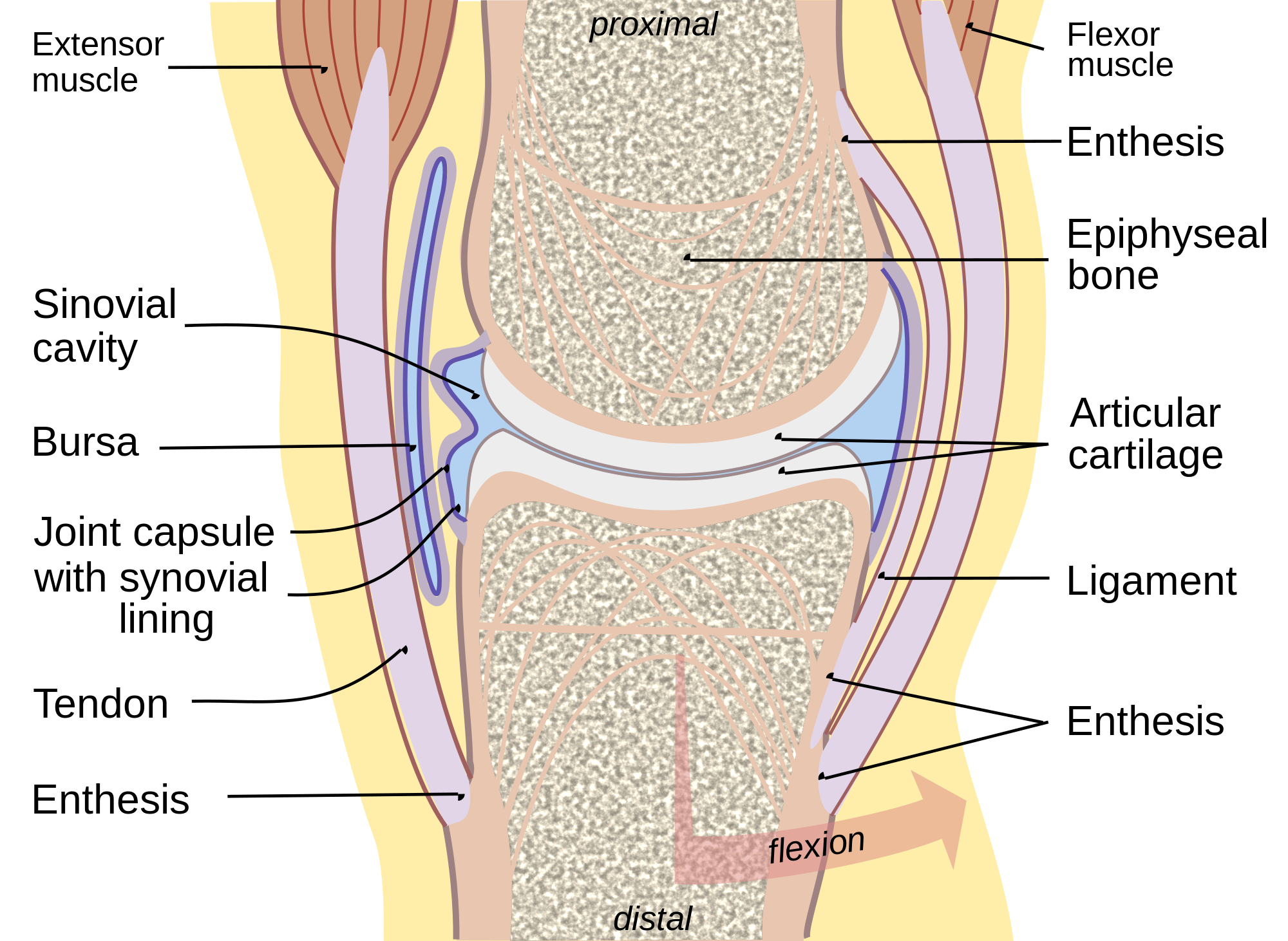
Synovial joints, which include subtypes such as (e.g., ), ball-and-socket (e.g., ), pivot (e.g., ), condyloid (e.g., ), saddle (e.g., ), and planar (e.g., intercarpal), feature a complex structure for optimal function. Key components include the articular capsule (a fibrous outer layer reinforced by ligaments), the (which secretes lubricating ), covering the ends to reduce friction, and the joint cavity containing the fluid. This design allows for smooth, low-friction movement while providing stability against dislocation.

Overview

Definition and General Characteristics

A joint is defined as the point of connection between two or more bones in the body, serving as a critical interface that facilitates both movement and structural stability. These connections are essential components of the skeletal system, where they enable coordinated actions such as walking and grasping while maintaining the body's overall form. Joints are broadly categorized into three structural types based on the linking the bones: fibrous joints, which are united by dense fibrous ; cartilaginous joints, connected by ; and synovial joints, featuring a fluid-filled cavity that allows greater . In terms of general characteristics, joints are integral to the skeletal system's roles in locomotion—by permitting limb and trunk movements—and in providing support, as they distribute mechanical loads across the body to prevent collapse under weight and stress. The human body contains approximately 360 joints, varying widely in their degree of mobility to balance flexibility with protection of vital structures. Functionally, joints range from immovable synarthroses, which offer rigid stability like those in the skull, to freely movable diarthroses, which support dynamic activities through synovial mechanisms. This classification by tissue type and movement degree underscores joints' adaptability, with synovial types comprising the majority for everyday mobility. Joints' design ensures efficient force transmission during activities, often reinforced by surrounding ligaments for added stability without compromising essential motion.

Importance in the Body

Joints play a crucial role in enabling bodily movement by allowing bones to articulate smoothly, thereby facilitating locomotion, manipulation of objects, and other essential activities. They absorb and distribute mechanical forces during physical exertion, preventing excessive stress on bones and tissues, while also contributing to the maintenance of posture through stable alignment of the skeletal framework. This integration with skeletal muscles and the allows for coordinated actions, where joint feedback informs muscle contractions to achieve precise control over body positioning and motion. Systemically, joints are vital for overall mobility and balance, enabling individuals to perform daily activities such as walking, standing, and reaching, which are foundational to . Optimal joint function supports dynamic equilibrium during movement, reducing the risk of falls and enhancing physical stability in varied environments. Impairments in joint health can significantly diminish by limiting participation in social, occupational, and recreational pursuits, underscoring their broad impact on physical and psychological well-being. Joints serve as primary sites for , where sensory receptors detect position, orientation, and motion to provide the with real-time data for reflexive adjustments in posture and . Additionally, they house nociceptors that signal in response to or overload, alerting the body to potential and prompting protective behaviors. In , joints underwent adaptations to support and upright posture, such as modifications in the hip and to efficiently transfer weight and enable energy-efficient walking over long distances. within certain joints aids lubrication to minimize wear during these functions.

Anatomy

Basic Components

Joints are formed by the articulation of two or more , which serve as the primary structural elements providing the framework for movement and support throughout the body. covers the opposing surfaces at the joint, creating a smooth, low-friction interface that facilitates gliding and absorbs mechanical shock during loading. The , a fibrous envelope surrounding the articulation, encloses the joint space and contributes to its overall stability by containing the synovial structures and resisting excessive motion. Ligaments, composed of , connect bones to one another across the joint, reinforcing the capsule and limiting the to prevent injury. Tendons, similarly tough bands of fibrous tissue, attach muscles to bones adjacent to the joint, enabling the transmission of contractile forces to produce movement. In synovial joints, the inner lining of the capsule is the , which produces essential for joint lubrication. Synovial fluid is a viscous ultrafiltrate of containing , which provides viscoelastic properties, and lubricin, a mucin-like that enhances boundary by forming a protective on articular surfaces. This fluid reduces and nourishes the avascular . Bursae, small fluid-filled sacs lined with , are often located near joints between bones, tendons, or muscles to minimize and distribute pressure during motion. While these components are universal, their presence and extent vary in fibrous and cartilaginous joints, which lack a synovial cavity.

Synovial Joint Structure

Synovial joints feature a distinctive synovial cavity, a fluid-filled space enclosed by the articular capsule, which allows for free movement between the articulating bones. The cavity is lined by the , or synovium, a thin layer of that secretes , a viscous rich in and lubricin that reduces friction and nourishes the avascular articular cartilage. This fluid also facilitates nutrient diffusion and waste removal within the joint. At the microscopic level, the synovium comprises an intima layer of containing synoviocytes and a subintima layer of fibrous tissue with blood vessels, , and fat cells. Synoviocytes exist in two primary types: type A synoviocytes, which are macrophage-like cells derived from monocytes that perform to clear debris, bacteria, and immune complexes from the ; and type B synoviocytes, fibroblast-like cells that synthesize and secrete components of the , including hyaluronan and proteoglycans, while also producing the synovial membrane's . The vascular supply to synovial joints arises from a rich periarticular arterial formed by anastomoses of arteries from surrounding tissues, with branches penetrating the capsule to form an intra-articular network that nourishes the synovium and capsule but spares the articular . Neural innervation is provided by articular branching from nearby peripheral , which densely supply the , ligaments, and synovium with sensory fibers for and pain, as well as autonomic fibers regulating blood flow. Certain synovial joints incorporate additional fibrocartilaginous structures, such as articular discs or menisci, to enhance stability and load distribution. An articular disc is a thin, oval plate that partially divides the synovial cavity, as seen in the , while a meniscus is a larger, C-shaped structure, exemplified by the medial and lateral menisci in the , which deepen the articular surfaces, absorb shock, and improve joint congruence. Reinforcing ligaments, including both extracapsular bands outside the capsule and intracapsular ligaments within the synovial cavity (often lined by synovium), provide tensile strength and limit excessive motion; for instance, the in the traverses the synovial cavity to prevent anterior tibial displacement. Synovial joints are morphologically diverse, classified into six types based on the shape of their articulating surfaces: plane joints, which feature flat or slightly curved surfaces for gliding motions, as in the of the ; hinge joints, with convex-concave cylindrical surfaces permitting uniaxial flexion and extension, like the ; pivot joints, involving a rounded process rotating within a ring, such as the between the atlas and axis vertebrae; condyloid joints, where an oval condyle fits into an elliptical cavity for biaxial movement, exemplified by the metacarpophalangeal joints of the fingers; saddle joints, with reciprocally concave-convex surfaces allowing biaxial motion, as in the of the thumb; and ball-and-socket joints, featuring a spherical head within a cuplike socket for multiaxial rotation, such as the glenohumeral (. These structural variations enable a wide range of movements while maintaining joint integrity.

Classification

Structural Classification

Joints are structurally classified according to the type of that unites the adjacent bones, resulting in three main categories: fibrous joints, cartilaginous joints, and synovial joints. This classification emphasizes the material composition and degree of stability rather than the extent of movement, though fibrous and cartilaginous joints generally permit little to no motion, while synovial joints allow greater freedom. Fibrous joints are connected by dense collagenous fibrous connective tissue and are typically immovable. They include three subtypes: sutures, syndesmoses, and gomphoses. Sutures consist of short collagenous fibers that interlock the edges of bones, primarily in the , such as the coronal and sagittal sutures between cranial bones. Syndesmoses involve a broader ligamentous connection allowing slight movement, exemplified by the distal tibiofibular syndesmosis that stabilizes the ankle. Gomphoses form a peg-in-socket articulation secured by periodontal ligaments, as seen in the attachment of teeth to the maxillary and mandibular alveolar sockets. Over time, some fibrous joints may fuse into synostoses through , providing permanent stability. Cartilaginous joints are joined by , permitting limited motion. They are subdivided into synchondroses and symphyses based on cartilage type. Synchondroses use for temporary connections, such as the epiphyseal plates between the and of long bones during growth; these eventually ossify to form synostoses, transitioning from slightly movable to immovable. Symphyses, connected by , provide resilient shock absorption with minimal movement, as in the intervertebral discs between vertebral bodies or the uniting the pubic bones of the . Synovial joints, the most prevalent type in the body, feature a joint cavity enclosed by an articular capsule and lined with a that produces lubricating fluid, enabling extensive mobility as diarthroses. While their detailed structure involves articular cartilage and reinforcing ligaments, the key structural distinction is the presence of this fluid-filled space separating the bones. Representative examples include the (a ) and the (a ), which facilitate complex movements essential for locomotion and manipulation.

Functional Classification

The functional classification of joints categorizes them according to the degree and type of movement they permit, providing insight into their role in body mechanics and overall . This divides joints into three primary categories—synarthroses, amphiarthroses, and diarthroses—based on the extent of mobility, from none to full range, which directly correlates with the structural elements that either restrict or enable motion. Synarthroses, or immovable joints, allow no significant movement and primarily serve to provide rigid stability and protection to vital structures. These joints are essential for maintaining the integrity of the and other areas requiring minimal flexibility, with examples including the sutures between cranial bones, which interlock tightly to safeguard the without permitting any appreciable displacement. Amphiarthroses, known as slightly movable joints, permit limited motion that contributes to shock absorption and subtle adjustments in load-bearing regions. A representative example is the intervertebral discs of the spine, which allow restricted flexion, extension, and to facilitate spinal curvature and resilience during posture changes and minor impacts. Diarthroses, or freely movable joints, enable extensive and are crucial for dynamic activities such as walking and grasping. Predominantly synovial in structure, they are subclassified by the axes of movement: uniaxial joints, like the in the neck, restrict motion to a single axis for precise actions such as head ; biaxial joints, such as the at the base of , support movement along two axes for enhanced dexterity; and multiaxial joints, exemplified by the ball-and-socket joint, allow across three or more axes to support complex, three-dimensional essential for locomotion and balance. This axial variation in diarthroses underscores their adaptability in coordinating multi-joint movements throughout the body.

Function and Biomechanics

Types of Joint Movements

Joint movements, also known as arthokinematics, encompass a range of motions that occur at diarthrodial joints, enabling diverse activities from locomotion to manipulation. These movements are categorized based on the anatomical planes in which they primarily occur: the for anterior-posterior motions, the frontal plane for lateral deviations, and the for rotational actions. Understanding these movements is crucial for , as they dictate how the body interacts with its environment through coordinated skeletal actions. Primary Movements Flexion involves bending a joint to decrease the angle between two bones, such as bending the or , typically occurring in the . Extension is the opposite, straightening the joint to increase the angle, returning to or beyond the anatomical position, as seen when extending the to stand upright. These paired movements are fundamental in joints like the , where flexion allows the to move anteriorly relative to the . Abduction moves a limb away from the body's midline or plane, occurring in the frontal plane, exemplified by raising the laterally at the . Adduction reverses this, bringing the limb toward the midline, such as lowering the back to the side. These motions are essential for spreading and converging body segments, like in the joint during leg swings. Rotation entails turning a around its longitudinal axis, primarily in the , and can be medial (inward) or lateral (outward), as in turning the head side to side at the . Circumduction combines multiple primary movements into a circular pattern, such as the conical motion of the arm at the , sequentially involving flexion, abduction, extension, and adduction. This conical trajectory enhances reach and versatility in multiaxial joints. Gliding, or , describes the slight sliding of one bone's surface over another without significant angular change, occurring in planar joints like the of the , facilitating smooth, non-rotational shifts. Specialized Motions Inversion turns the sole of the foot medially toward the midline, while eversion turns it laterally away, both occurring at the in the frontal plane to adapt to uneven terrain. Protraction moves a body part anteriorly on a , such as thrusting the or forward at the , whereas retraction pulls it posteriorly, like drawing the toward the spine. These specialized actions allow for precise adjustments in appendicular regions, enhancing stability and dexterity.

Mechanical Properties

The mechanical properties of synovial joints enable efficient load bearing, movement, and stability while minimizing on articular surfaces. , the primary load-distributing tissue in these joints, exhibits viscoelastic , characterized by time-dependent deformation under stress, where it initially resists compression elastically before creeping under sustained load due to fluid exudation from its porous matrix. This allows cartilage to absorb and dissipate , with an aggregate compressive modulus of 0.1–2.0 MPa in healthy tissue, preventing structural failure under physiological loads. Load distribution occurs across the joint surfaces, with varying based on contact area; for instance, in the , static pressure on the tibial plateau can range from 0.5 to 3 MPa during , optimized by cartilage thickness and subchondral support. Friction in synovial joints is remarkably low, with coefficients typically between 0.001 and 0.03, facilitated by that acts as a . provides boundary under high-load, low-speed conditions, where molecular layers like lubricin adsorb to surfaces to reduce direct contact, and hydrodynamic lubrication during motion, where fluid generates a pressurized film separating surfaces. This dual mechanism maintains low shear stresses, with boundary mode dominating at startup or static holds to prevent stick-slip. Joint , essential for rotational movements, is governed by τ=F×d\tau = F \times d, where τ\tau is the , FF is the applied force (such as from muscles), and dd is the from the force line to the joint's axis of rotation (lever arm). This relationship quantifies how muscles generate while balancing external loads. Stability in synovial joints arises from a combination of passive and active factors, including tension that resists excessive , muscle co-contraction that increases joint without net , and joint congruence, where the geometric fit of articular surfaces enhances compressive load sharing and limits . In ball-and-socket joints like the hip, this congruence supports multi-axis load distribution for enhanced stability.

Clinical Aspects

Common Disorders

Common joint disorders encompass a range of conditions affecting the articulations between bones, leading to pain, , and impaired mobility. These pathologies often target synovial joints, where the , , and surrounding tissues are particularly vulnerable to degeneration, autoimmune attack, or acute trauma. Among the most prevalent are , , , and acute injuries such as sprains and dislocations, each characterized by distinct etiologies and clinical manifestations. Osteoarthritis (OA), the most common form of , involves progressive degeneration and loss of , often accompanied by subchondral bone changes and formation. It primarily results from mechanical wear-and-tear over time, exacerbated by factors such as aging, , and prior joint trauma, which increase biomechanical stress on the joint surfaces. Symptoms typically include joint pain that worsens with activity and improves with rest, morning stiffness lasting less than 30 minutes, swelling, and reduced , most commonly affecting joints like the knees, hips, and spine. In the United States, as of data from 2017–2020, OA affects approximately 33 million adults, with symptomatic knee OA occurring in about 10% of men and 13% of women aged 60 years or older. Risk factors include , which amplifies joint loading, and repetitive use or injury, contributing to breakdown. Rheumatoid arthritis (RA) is a chronic autoimmune disorder characterized by symmetric inflammation of the , leading to joint erosion and if unchecked. Its exact cause remains unclear, but it involves genetic predisposition (e.g., alleles) combined with environmental triggers like and infections, resulting in an aberrant that attacks joint tissues. Common symptoms include persistent joint , stiffness lasting over 30 minutes upon waking, tenderness, warmth, redness, and swelling, often affecting small joints of the hands and feet bilaterally, alongside systemic features like fatigue and low-grade fever. As of recent estimates (2024–2025), RA impacts about 1.5 million adults in the , representing approximately 0.7% of the adult , with higher prevalence among women and those aged 30–50 at onset. Key risk factors encompass female sex, family history, and , which may promote pro-inflammatory production. Gout arises from , where elevated serum levels lead to the deposition of monosodium urate crystals in joints, provoking acute inflammatory responses. Primary causes include overproduction or underexcretion of , influenced by genetic factors, diet high in purines (e.g., , ), alcohol consumption, and comorbidities like or renal impairment. Symptoms manifest as sudden, intense episodes of joint pain, often at night, with severe swelling, redness, warmth, and tenderness, classically starting in the big toe (podagra) and potentially recurring in other joints like the ankles or knees. As of 2025, gout affects approximately 5.1% of US adults, or about 12.1 million individuals, with higher rates among men and those with or ; prevalence has increased due to population aging and rising comorbidities. Risk factors such as use and fructose-rich diets further elevate saturation, increasing flare frequency. Sprains and dislocations represent acute joint injuries, with sprains involving partial or complete tears of ligaments that stabilize the joint, while dislocations occur when ends are forced out of alignment. Sprains commonly result from sudden twisting, excessive force, or awkward landing during or falls, particularly affecting the ankle, where inversion injuries stretch the lateral ligaments. Symptoms include immediate , swelling, bruising, and , with limited or motion depending on severity; up to 40% may develop chronic issues like recurrent . Ankle sprains have an incidence of 2.2–3.3 per 1,000 person-years among athletes, making them one of the most frequent musculoskeletal injuries. Risk factors include prior sprains, improper footwear, and high-impact activities. Dislocations, often traumatic, are most prevalent in the , accounting for 50% of major joint dislocations, typically anterior due to falls on an outstretched or direct impact. They arise from high-energy mechanisms like collisions or motor vehicle accidents, compromising the and ligaments. Presentation involves acute severe , visible , swelling, and inability to actively move the joint, sometimes with neurovascular compromise. The incidence of shoulder dislocations is 11 to 29 per 100,000 persons annually, higher in young males engaged in contact . Contributing factors include from prior dislocations and overhead activities that stress the glenohumeral joint.

Diagnosis and Treatment

Diagnosis of joint disorders typically begins with a thorough medical history and physical examination to assess symptoms such as pain, swelling, stiffness, and limited range of motion. Imaging techniques play a central role in confirming the diagnosis and evaluating joint damage. X-rays are commonly used to detect bone changes, joint space narrowing, and osteophytes associated with conditions like osteoarthritis. Magnetic resonance imaging (MRI) provides detailed views of soft tissues, including cartilage, ligaments, and synovial membranes, helping identify inflammation or tears not visible on X-rays. Ultrasound is effective for detecting synovial inflammation, effusions, and tendon abnormalities in real-time, particularly in inflammatory arthritides. Arthroscopy, a minimally invasive procedure using a fiber-optic camera, allows direct visualization of the joint interior for precise diagnosis and can simultaneously address issues like loose bodies or minor repairs. Blood tests, including erythrocyte sedimentation rate (ESR), C-reactive protein (CRP), and rheumatoid factor, are essential for identifying inflammatory markers in conditions such as rheumatoid arthritis. Treatment strategies for joint disorders emphasize a stepwise approach, starting with conservative measures to alleviate symptoms and preserve function. The protocol—rest, , compression, and —is recommended initially to reduce acute pain and swelling in injured joints. Physical therapy focuses on strengthening muscles, improving flexibility, and enhancing joint stability through targeted exercises. Nonsteroidal anti-inflammatory drugs (NSAIDs) are widely used to manage pain and inflammation, with acetaminophen as an alternative for those unable to tolerate NSAIDs. For more severe cases, surgical interventions are employed when conservative treatments fail. , such as total hip or , replaces damaged joint surfaces with prosthetic components, offering significant pain relief and functional restoration; approximately 90% of these implants remain functional at 10 years post-surgery. repair or reconstruction, often via arthroscopic techniques, addresses instability in joints like the , using grafts to restore structural integrity. The American Academy of Orthopaedic Surgeons (AAOS) provides evidence-based clinical practice guidelines recommending these approaches based on patient-specific factors like age and disorder severity. Emerging therapies aim to promote joint repair and modulate disease progression. Biologic agents, including (TNF) inhibitors, target inflammatory pathways in autoimmune joint conditions, reducing joint destruction and improving outcomes. Stem cell therapies, particularly mesenchymal stem cells derived from or , show promise in regenerating and alleviating symptoms in , though they remain under clinical investigation with low-certainty evidence for slight improvements as of 2025. These treatments are guided by ongoing research to establish long-term efficacy and safety.

Maintaining Joint Health

Maintaining joint health involves lifestyle strategies aimed at preventing disorders and supporting long-term function, particularly for load-bearing joints such as the knees and hips. Controlling body weight is a primary recommendation, as excess weight increases mechanical stress on these joints and elevates the risk of conditions like osteoarthritis. Regular physical activity is essential, including strength training 2–3 times per week to build muscle support around joints, combined with aerobic exercises such as brisk walking for at least 150 minutes per week. A warm-up period of 5–10 minutes of light activity before engaging in more intense exercise is advised to reduce injury risk. Proper exercise technique, ideally learned under professional guidance, and avoiding activities that cause pain are crucial to prevent joint strain. For high-impact activities like running, using supportive footwear and choosing softer surfaces can help minimize joint impact, with cessation recommended if pain occurs. Nutrition plays a supportive role, with adequate protein intake from whole food sources recommended to maintain musculoskeletal health; protein supplements may be used if dietary intake is insufficient. Claims that consuming gelatin or chicken feet provides significant joint benefits are largely unsupported by evidence, as these sources offer limited bioavailability for joint repair. Collagen supplements show mixed results in clinical studies for reducing joint pain, but they are not considered superior to general protein intake for joint health maintenance. For individuals at risk, preventive blood tests such as C-reactive protein for inflammation, rheumatoid factor for autoimmune markers, and uric acid levels for gout susceptibility may be considered, particularly in those with family history or comorbidities. Routine magnetic resonance imaging (MRI) is not recommended for asymptomatic screening due to lack of proven benefit and potential for unnecessary interventions.

Development and Evolution

Embryological Development

The embryological development of joints begins with the formation of limb buds around the fourth week of , arising from the and somatic mesoderm, which give rise to the mesenchymal s that will form skeletal elements including joint precursors. These mesenchymal cells differentiate into cartilaginous precursors, with synovial joints emerging from a distinct non-cartilaginous known as the interzone, which forms through condensation of mesenchymal cells at prospective joint sites. The timing varies by location, with proximal joints initiating earlier than distal ones, establishing the foundational blueprint for joint positioning along the limb axis. Between weeks 6 and 8, the interzone undergoes , a critical where the mesenchymal tissue within it breaks down to create the synovial cavity, driven by hyaluronan synthesis and cellular remodeling that separates the opposing cartilaginous anlagen of adjacent . This is accompanied by the formation of synovial membranes and joint capsules from the peripheral interzone cells, while the central region thins to form the articular surfaces. Surrounding commences shortly thereafter, with primary centers appearing in the diaphyses around week 8 and progressing to secondary centers in epiphyses by weeks 9-12, encapsulating the developing joints within maturing skeletal structures. Key regulatory processes include the role of in patterning joint formation; specifically, Hox11 paralogs coordinate zeugopod (/) musculoskeletal development, establishing regional identities and ensuring proper interzone positioning for organization. plays an essential role in creating the joint space by eliminating mesenchymal cells in the interzone core, with peaking during to prevent fusion and allow physical separation of skeletal elements. Synchondroses, as temporary cartilaginous joints, function as growth plates during this phase, facilitating longitudinal bone expansion through chondrocyte proliferation and before eventual and fusion in postnatal life. Congenital anomalies such as , which involves fusion of adjacent digits, arise from disruptions in these processes, particularly failure of in interdigital mesenchymal zones between weeks 6 and 8, leading to incomplete separation that secondarily affects interphalangeal joint formation and spacing. This highlights the precision required in mesenchymal remodeling for normal joint development, with occurring in approximately 1 in 2,000-3,000 live births and often linked to genetic factors influencing Hox-mediated patterning.

Evolutionary Perspectives

The evolution of joints in animals traces back to early , where jointed exoskeletons emerged as a key for locomotion and environmental interaction. In , such as and crustaceans, segmented exoskeletons with hinged joints allowed flexible movement of appendages while providing structural support against predation and desiccation; this innovation, dating to the around 540 million years ago, facilitated the diversification of mobile body plans in marine and terrestrial habitats. These exoskeletal joints, formed by chitinous cuticles with sclerotized hinges, prioritized rigidity for protection but limited overall body flexibility compared to later endoskeletal designs. The transition to vertebrates marked a shift from exoskeletons to internal endoskeletons, with lubricated synovial joints evolving in the common of jawed vertebrates (gnathostomes) approximately 420-400 million years ago, predating the move to . Contrary to earlier views that synovial joints arose solely in tetrapods to handle terrestrial stresses, and genetic evidence shows they first appeared in aquatic jawed , such as and skates, enabling smooth articulation in jaws and fins for predation and maneuvering. In early tetrapods, around 360 million years ago, these joints adapted further for on , with increased synovial cavity development and to support limb-based locomotion and reduce during strides. Mammalian joints exhibited heightened complexity, particularly in primates, where adaptations like the mobile glenohumeral (shoulder) joint supported arboreal lifestyles through enhanced range of motion for brachiation and climbing. Fossil evidence from Australopithecus species, such as the knee joint of A. afarensis (specimen AL 129-1a, dated to about 3.2 million years ago), reveals a valgus angle and robust femoral condyles indicative of bipedal stability, bridging arboreal ancestry with terrestrial upright walking. These advancements involved trade-offs between mobility and stability; for instance, ball-and-socket joints like the primate shoulder afford multidirectional freedom essential for foraging but risk instability, necessitating reinforced ligaments and musculature, whereas hinge joints in the knee prioritize load-bearing for locomotion at the cost of rotational flexibility. Recent post-2020 studies using have illuminated the genetic underpinnings of joint evolution, revealing conserved pathways like BMP and Wnt signaling that regulate formation across jawed s. For example, analysis of skate and genomes highlights how cis-regulatory elements near genes such as Osr2 drove tetrapod-specific joint refinements, enabling adaptations for diverse habitats without disrupting core mechanisms. These findings underscore how genomic duplications and regulatory shifts balanced evolutionary pressures for efficient movement, informing understandings of joint diversity from fish fins to mammalian limbs.

History

Etymology

The term "joint" in the anatomical sense originates from the Latin iunctus, the past participle of iungere meaning "to join," which entered English via joint or jointe, denoting a connection or juncture. This reflects the functional unity of bones at points of articulation, with the word first appearing in English around 1290 in general usage and entering anatomical contexts by the early to describe the site where two bones meet. The evolution of the term in medical draws heavily from influences, particularly the , which employed terms like arthron (ἄρθρον) to denote joints, establishing foundational vocabulary for describing skeletal connections in early Western medicine. Related anatomical terminology further illustrates this Greco-Latin synthesis; for instance, the prefix arthr- or arthro- derives directly from Greek arthron, meaning "joint" or "articulation," and is used in words like arthritis to specify joint-related conditions. Similarly, "synovium," referring to the membrane lining certain joints, stems from New Latin synovia (coined by Paracelsus in the 16th century), combining Greek syn- ("together") with Latin ovum ("egg") due to the egg-white-like viscosity of the synovial fluid it produces. These etymological developments highlight how joint terminology evolved from descriptive ancient roots into standardized medical language by the Renaissance, prioritizing precision in denoting structural and functional aspects.

Historical Milestones

The understanding of joints dates back to ancient times, with , around 400 BCE, providing the earliest systematic descriptions of joint and their treatments in his treatise On Joints. He emphasized manual reduction techniques tailored to the patient's age, build, and the duration of the dislocation, laying foundational principles for orthopedic interventions that influenced subsequent medical practice. In the 2nd century CE, advanced this knowledge by describing as a lubricating substance within joint capsules, distinguishing it from other bodily humors and recognizing its role in facilitating smooth articulation. His observations, derived from animal dissections, marked an early recognition of joint and contributed to the concept of synovia as essential for joint function. The brought greater precision through Andreas Vesalius's De Humani Corporis Fabrica in 1543, which featured accurate illustrations of human joints based on direct dissections, correcting many Galenic errors and establishing a more reliable anatomical foundation for joint studies. In the , William Hunter conducted pioneering experiments on joint repair, noting in 1743 that damaged rarely regenerates fully, an observation that underscored the challenges of articular tissue healing and spurred later research into regenerative strategies. The introduced transformative surgical milestones, including Sir John Charnley's development of the low-friction total hip replacement in the 1960s, which utilized cemented acrylic and metal components to restore joint function in patients with severe , revolutionizing and enabling millions of procedures worldwide. Post-2010 research has advanced through computational of joint loading, such as finite element analyses of knee osteoarthritis, revealing how subchondral bone changes contribute to degeneration and informing personalized implant designs. In the , has progressed with CRISPR-Cas9 gene editing targeting osteoarthritis-related genes, such as those regulating interleukin-1 in chondrocytes, to enhance repair and reduce in preclinical models, offering potential for durable joint restoration.

References

Add your contribution
Related Hubs
User Avatar
No comments yet.